

The landscape of prognostic and immunological role of myosin light chain 9 (MYL9) in human tumors
- PMID: 34729929
- PMCID: PMC8767521
- DOI: 10.1002/iid3.557
The landscape of prognostic and immunological role of myosin light chain 9 (MYL9) in human tumors
Abstract
Introduction: Recent studies have shown that myosin light chain 9 (MYL9) plays a vital role in immune infiltration, tumor invasion, and metastasis; however, the prognostic and immunological role of MYL9 has not been reported. The purpose of this study was to explore the potential prognostic and immunological roles of MYL9 in human cancers by public datasets mainly including the cancer genome atlas (TCGA) and Gene expression omnibus.
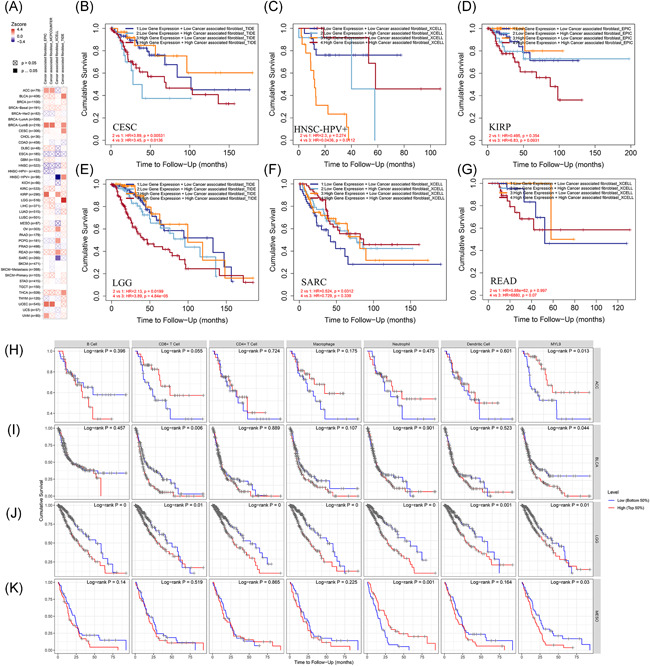
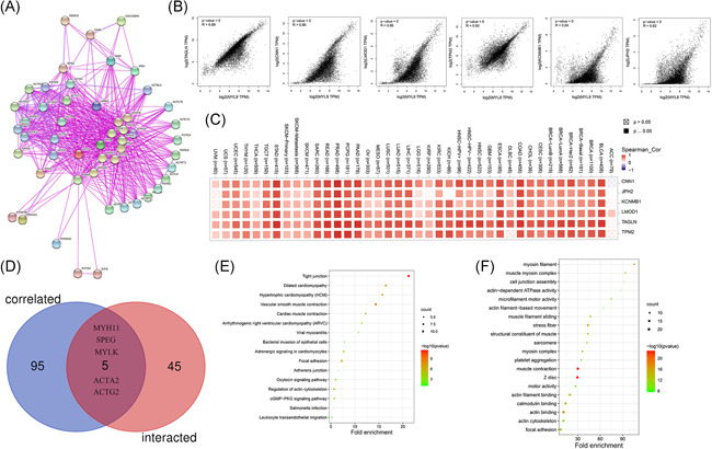
Methods: The expression pattern and prognostic value of MYL9 were analyzed across multiple public datasets in different cancer. The correlations between MYL9 expression and immune infiltration among multiple cancers were analyzed by using the TIMER2.0. The MYL9-related gene enrichment analysis was implemented by mainly using KEGG and GO datasets.
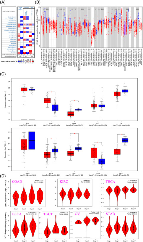
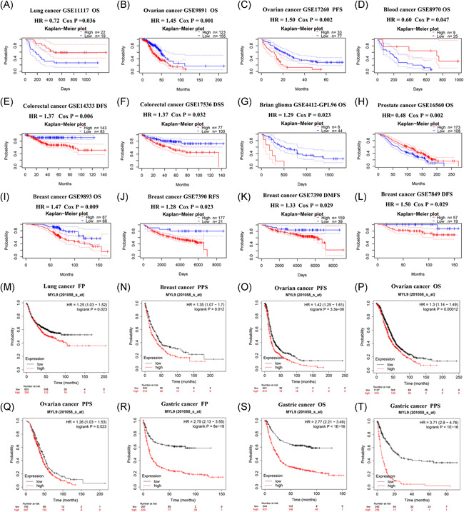
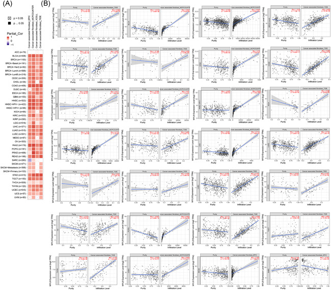
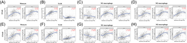
Results: MYL9 was lowly expressed in most cancers, such as breast cancer, lung adenocarcinoma and squamous cell carcinoma, and stomach adenocarcinoma; but it was highly expressed in several cancers, such as cholangiocarcinoma, head and neck squamous cell carcinoma, and liver hepatocellular carcinoma. Furthermore, MYL9 expression was distinctively associated with prognosis in adrenocortical carcinoma, colon adenocarcinoma, brain glioma, lung cancer, ovarian cancer, gastric cancer, breast cancer, blood cancer, and prostate cancer patients. The expressions of MYL9 were significantly associated with the infiltration of cancer-associated fibroblasts, B cell, CD8+ T cell, CD4+ T cell, macrophage, neutrophil, dendritic cell in different tumors as well as immune markers. In addition, we found that the functional mechanisms of MYL9 involved muscle contraction and focal adhesion.
Conclusion: MYL9 can serve as a prognostic signature in pan-cancer and is associated with immune infiltration. This pan-cancer study is the first to show a relatively comprehensive understanding of the prognostic and immunological roles of MYL9 across different cancers.
Keywords: MYL9; cancer; immune infiltration; prognosis.
© 2021 The Authors. Immunity, Inflammation and Disease published by John Wiley & Sons Ltd.
Figures
Similar articles
-
MYL5 as a Novel Prognostic Marker is Associated with Immune Infiltrating in Breast Cancer: A Preliminary Study.Breast J. 2023 Feb 17;2023:9508632. doi: 10.1155/2023/9508632. eCollection 2023. Breast J. 2023. PMID: 36846347 Free PMC article.
-
MYLK and MYL9 expression in non-small cell lung cancer identified by bioinformatics analysis of public expression data.Tumour Biol. 2014 Dec;35(12):12189-200. doi: 10.1007/s13277-014-2527-3. Epub 2014 Sep 2. Tumour Biol. 2014. PMID: 25179839
-
High Expression of MYL9 Indicates Poor Clinical Prognosis of Epithelial Ovarian Cancer.Recent Pat Anticancer Drug Discov. 2021;16(4):533-539. doi: 10.2174/1574891X16666210706153740. Recent Pat Anticancer Drug Discov. 2021. PMID: 34551701
-
A new therapeutic target: the CD69-Myl9 system in immune responses.Semin Immunopathol. 2019 May;41(3):349-358. doi: 10.1007/s00281-019-00734-7. Epub 2019 Apr 5. Semin Immunopathol. 2019. PMID: 30953160 Review.
-
CD4+ T cells in inflammatory diseases: pathogenic T-helper cells and the CD69-Myl9 system.Int Immunol. 2021 Nov 25;33(12):699-704. doi: 10.1093/intimm/dxab053. Int Immunol. 2021. PMID: 34427648 Review.
Cited by
-
Multi‑dimensional analysis reveals NCKAP5L is a promising biomarker for the diagnosis and prognosis of human cancers, especially colorectal cancer.Oncol Lett. 2023 Dec 12;27(2):53. doi: 10.3892/ol.2023.14186. eCollection 2024 Feb. Oncol Lett. 2023. PMID: 38192666 Free PMC article.
-
Comparison of differentially expressed genes in longissimus dorsi muscle of Diannan small ears, Wujin and landrace pigs using RNA-seq.Front Vet Sci. 2024 Jan 5;10:1296208. doi: 10.3389/fvets.2023.1296208. eCollection 2023. Front Vet Sci. 2024. PMID: 38249550 Free PMC article.
-
Epigenome-wide DNA methylation in leukocytes and toenail metals: The normative aging study.Environ Res. 2023 Jan 15;217:114797. doi: 10.1016/j.envres.2022.114797. Epub 2022 Nov 12. Environ Res. 2023. PMID: 36379232 Free PMC article.
-
A Transcriptomic Response to Lactiplantibacillus plantarum-KCC48 against High-Fat Diet-Induced Fatty Liver Diseases in Mice.Int J Mol Sci. 2022 Jun 17;23(12):6750. doi: 10.3390/ijms23126750. Int J Mol Sci. 2022. PMID: 35743193 Free PMC article.
-
PRPF19 facilitates colorectal cancer liver metastasis through activation of the Src-YAP1 pathway via K63-linked ubiquitination of MYL9.Cell Death Dis. 2023 Apr 8;14(4):258. doi: 10.1038/s41419-023-05776-2. Cell Death Dis. 2023. PMID: 37031206 Free PMC article.
References
-
- Blum A, Wang P, Zenklusen JC. SnapShot: TCGA‐analyzed tumors. Cell. 2018;173(2):530. - PubMed
-
- Wang Z, Jensen MA, Zenklusen JC. A Practical Guide to The Cancer Genome Atlas (TCGA). Methods Mol Biol. 2016;1418:111‐141. - PubMed
-
- Lu Q, Li J, Zhang M. Cargo recognition and cargo‐mediated regulation of unconventional myosins. Acc Chem Res. 2014;47(10):3061‐3070. - PubMed
MeSH terms
Substances
LinkOut - more resources
Full Text Sources
Research Materials
