Graphene oxide-ellagic acid nanocomposite as effective anticancer and antimicrobial agent
- PMID: 34694731
- PMCID: PMC8675783
- DOI: 10.1049/nbt2.12009
Graphene oxide-ellagic acid nanocomposite as effective anticancer and antimicrobial agent
Abstract
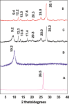
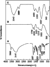
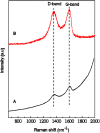
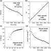
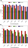
In this study, ellagic acid (ELA), a skin anticancer drug, is capped on the surface(s) of functionalised graphene oxide (GO) nano-sheets through electrostatic and π-π staking interactions. The prepared ELA-GO nanocomposite have been thoroughly characterised by using eight techniques: Fourier-transform infrared spectroscopy (FTIR), zeta potential, X-ray diffraction (XRD), thermogravimetric analysis (TGA), Raman spectroscopy, atomic force microscopy (AFM) topographic imaging, transmission electron microscopy (TEM), and surface morphology via scanning electron microscopy (SEM). Furthermore, ELA drug loading and release behaviours from ELA-GO nanocomposite were studied. The ELA-GO nanocomposite has a uniform size distribution averaging 88 nm and high drug loading capacity of 30 wt.%. The in vitro drug release behaviour of ELA from the nanocomposite was investigated by UV-Vis spectrometry at a wavelength of λmax 257 nm. The data confirmed prolonged ELA release over 5000 min at physiological pH (7.4). Finally, the IC50 of this ELA-GO nanocomposite was found to be 6.16 µg/ml against B16 cell line; ELA and GO did not show any cytotoxic effects up to 50 µg/ml on the same cell lines.
© 2021 The Authors. IET Nanobiotechnology published by John Wiley & Sons Ltd on behalf of The Institution of Engineering and Technology.
Figures




Similar articles
-
Design and Characterization of Chitosan-Graphene Oxide Nanocomposites for the Delivery of Proanthocyanidins.Int J Nanomedicine. 2020 Feb 20;15:1229-1238. doi: 10.2147/IJN.S240305. eCollection 2020. Int J Nanomedicine. 2020. PMID: 32110019 Free PMC article.
-
Carboxymethyl cellulose/graphene oxide bio-nanocomposite hydrogel beads as anticancer drug carrier agent.Carbohydr Polym. 2017 Jul 15;168:320-326. doi: 10.1016/j.carbpol.2017.03.014. Epub 2017 Mar 8. Carbohydr Polym. 2017. PMID: 28457456
-
Graphene oxide as a nanocarrier for controlled release and targeted delivery of an anticancer active agent, chlorogenic acid.Mater Sci Eng C Mater Biol Appl. 2017 May 1;74:177-185. doi: 10.1016/j.msec.2016.11.114. Epub 2016 Dec 5. Mater Sci Eng C Mater Biol Appl. 2017. PMID: 28254283
-
Surface modification of graphene oxide with stimuli-responsive polymer brush containing β-cyclodextrin as a pendant group: Preparation, characterization, and evaluation as controlled drug delivery agent.Colloids Surf B Biointerfaces. 2018 Dec 1;172:17-25. doi: 10.1016/j.colsurfb.2018.08.017. Epub 2018 Aug 13. Colloids Surf B Biointerfaces. 2018. PMID: 30121487
-
The Physicochemical Properties of Graphene Nanocomposites Influence the Anticancer Effect.J Oncol. 2019 Jul 3;2019:7254534. doi: 10.1155/2019/7254534. eCollection 2019. J Oncol. 2019. PMID: 31354821 Free PMC article. Review.
Cited by
-
Graphene Oxide Thin Films with Drug Delivery Function.Nanomaterials (Basel). 2022 Mar 30;12(7):1149. doi: 10.3390/nano12071149. Nanomaterials (Basel). 2022. PMID: 35407267 Free PMC article. Review.
-
Ellagic Acid Inclusion Complex-Loaded Hydrogels as an Efficient Controlled Release System: Design, Fabrication and In Vitro Evaluation.J Funct Biomater. 2023 May 16;14(5):278. doi: 10.3390/jfb14050278. J Funct Biomater. 2023. PMID: 37233388 Free PMC article.
-
Anti-microbial, anti-biofilm, and efflux pump inhibitory effects of ellagic acid-bonded magnetic nanoparticles against Escherichia coli isolates.Int Microbiol. 2024 Aug 6. doi: 10.1007/s10123-024-00560-4. Online ahead of print. Int Microbiol. 2024. PMID: 39105888
-
Preparation, Characterization, and Evaluation of Curcumin-Graphene Oxide Complex-Loaded Liposomes against Staphylococcus aureus in Topical Disease.ACS Omega. 2022 Nov 20;7(48):43499-43509. doi: 10.1021/acsomega.2c03940. eCollection 2022 Dec 6. ACS Omega. 2022. PMID: 36506117 Free PMC article.
-
The Green Synthesis of Reduced Graphene Oxide Using Ellagic Acid: Improving the Contrast-Enhancing Effect of Microbubbles in Ultrasound.Molecules. 2023 Nov 17;28(22):7646. doi: 10.3390/molecules28227646. Molecules. 2023. PMID: 38005368 Free PMC article.
References
-
- Strati, A. , et al.: Effect of ellagic acid on the expression of human telomerase reverse transcriptase (hTERT) α+ β+ transcript in estrogen receptor‐positive MCF‐7 breast cancer cells. Clin Biochem. 42(13‐14), 1358–1362 (2009) - PubMed
-
- Kim, S. , et al.: Development of chitosan–ellagic acid films as a local drug delivery system to induce apoptotic death of human melanoma cells. J. Biomed. Mater. Res. B Appl. Biomater. 90(1), 145–155 (2009) - PubMed
-
- Hussein‐Al‐Ali, S.H. , et al.: The in vitro therapeutic activity of ellagic acid‐alginate‐silver nanoparticles on breast cancer cells (MCF‐7) and normal fibroblast cells (3T3). Sci. Adv. Mater. 8(3), 545–553 (2016)
MeSH terms
Substances
Grants and funding
LinkOut - more resources
Full Text Sources
Research Materials
Miscellaneous

