. 2021 Sep 29:12:706240.
doi: 10.3389/fphar.2021.706240. eCollection 2021.

Lysophosphatidic Acid-Induced EGFR Transactivation Promotes Gastric Cancer Cell DNA Replication by Stabilizing Geminin in the S Phase

Affiliations

Lysophosphatidic Acid-Induced EGFR Transactivation Promotes Gastric Cancer Cell DNA Replication by Stabilizing Geminin in the S Phase

Haile Zhao et al. Front Pharmacol. .

Abstract

Geminin, an inhibitor of the DNA replication licensing factor, chromatin licensing and DNA replication factor (Cdt) 1, is essential for the maintenance of genomic integrity. As a multifunctional protein, geminin is also involved in tumor progression, but the molecular details are largely unknown. Here, we found that lysophosphatidic acid (LPA)-induced upregulation of geminin was specific to gastric cancer cells. LPA acted via LPA receptor (LPAR) 3 and matrix metalloproteinases (MMPs) signaling to transactivate epidermal growth factor receptor (EGFR) (Y1173) and thereby stabilize geminin expression level during the S phase. LPA also induced the expression of deubiquitinating protein (DUB) 3, which prevented geminin degradation. These results reveal a novel mechanism underlying gastric cancer progression that involves the regulation of geminin stability by LPA-induced EGFR transactivation and provide potential targets for the signaling pathway and tumor cell-specific inhibitors.

Keywords: DNA replication; EGFR; LPA; geminin; transactivation.


Conflict of interest statement

The authors declare that the research was conducted in the absence of any commercial or financial relationships that could be construed as a potential conflict of interest.

Figures
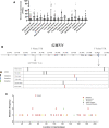
FIGURE 1
FIGURE 1
Mutation of geminin is non-predominant in human cancer. (A) RNA expression of geminin in 17 cancer types. (B) Location of mutation sites in GMNN gene. (C) Summary of geminin mutations across different tumor types from TCGA.
FIGURE 2
FIGURE 2
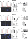
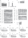
Depletion of geminin induces DNA re-replication in gastric cancer cells but not in normal gastric epithelial cells. Two gastric cancer cells (MKN45 and BGC-803) and normal epithelial cells (GES-1) were transfected with siGL or siGEM. After 48-h posttransfection, the cells were harvested and stained with PI to quantify DNA content by FACS analysis (A, D, G) or with DAPI to visualize nuclei by LSM (B, E, H). Geminin and tubulin were detected by Western blotting (C, F, I). Densitometory of three independent experiments was performed, normalized to tubulin, and expressed as a ratio of siGL, respectively, mean ± SD, *p < 0.05, **p < 0.01, ***p < 0.001 (T test).
FIGURE 3
FIGURE 3
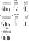
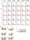
LPA selectively triggers the upregulation of geminin in gastric cancer cells. (A, D, G) G1/S–arrested cells were treated with LPA time gradient. (B, E) Protein level of geminin with or without 10 µM LPA treatment. (C, F) G1/S–arrested cells were under the treatment with or without 1% BSA. Densitometry of three independent experiments was performed, normalized to tubulin and expressed as a ratio of 0 h or 0 µM, respectively. Mean ± SD, *p < 0.05, **p < 0.01 (ANOVA test and Dunnett’s multiple comparisons test )
FIGURE 4
FIGURE 4
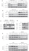
LPA potentiates geminin stability through DUB3 in gastric cancer cells. (A, B) Gastric cancer cells MKN45 (A) and BGC-803 (B) were transfected with the indicated vectors (empty vector; DUB3, a vector that contains Homo sapiens ubiquitin-specific peptidase 17-like family member 2 [USP17L2] mRNA [DUB3]), and incubated with cycloheximide (CHX, 50 µg/ml) for the indicated time points. Western blotting analysis with the indicated antibodies. (C) Two gastric cancer cells (MKN45 and BGC-803) were transfected with the indicated siRNA. Cell lysates were analyzed by Western blotting. (D) Two gastric cancer cells were treated with LPA time gradient, and cell lysates were analyzed by Western blotting. (E) Two gastric cancer cells (MKN45 and BGC-803) were transfected with the indicated siRNA, together with LPA time gradient treatment, and cell lysates were analyzed by Western blotting.
FIGURE 5
FIGURE 5
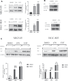
LPA stimulates EGFR transactivation in gastric cancer cells. (A–B) Tyrosine-phosphorylated EGFR ((A), PY) and Y1173-phosphorylated EGFR (B) level after treated with 0.1% DMSO (vehicle), 10 µM LPA, or 10 ng/ml EGF for 3 min. (C) Tyrosine-phosphorylated EGFR levels after pretreated with 0.1% DMSO or 10 µM BB94 for 30 min and stimulated with 10 µM LPA. Densitometry of three independent experiments was performed normalized to tubulin and expressed as a ratio of DMSO; mean ± SD, **p < 0.01 (ANOVA test and Dunnette’s multiple comparisons test).
FIGURE 6
FIGURE 6
LPA potentiates geminin stability through LPAR3/MMPS/EGFR/PI3K/mTOR signaling axis in MKN45 cells. (A) mRNA expression of geminin under 10 µM LPA time gradient. Densitometry of three independent experiments was performed normalized to actin and expressed as ratio of 0 h; mean ± SD (ANOVA test and Dunnett’s multiple comparisons test). (B) mRNA expression of LPARI-6 in MKN45 cells. (C) Geminin protein levels after transfected with or without sitrl/siLPAR3 for 6 h, and the cells were cultured for another 48 h in the total medium until harvested. (D–E) Gemimin protein level after pretreated with DMSO (vehicle, 0.1%), Kil6425 (10 µM), BB94 (10 µM), AG1478 (250 nM), LY294002 (10 µM), or rapamycin (100 nM) for 30 min, and stimulated with 10 µM LPA time gradient. In siRNA-silencing experiments, cells were transfected with either siGL or siGEM for 6 h and then cultured for another 48 h. (F–K) Quantification of geminin protein levels in (D) and (E). Densitometry of three independent experiments was performed normalized to tubulin and expressed as a ratio of control group; mean ± SD, *p < 0.05, **p < 0.01, ***p < 0.001 (ANOVA test and Dunnette’s multiple comparisons test). (L) Activation level of mTOR and p-mTOR (Ser2448) under 10 µM LPA treatment.
FIGURE 7
FIGURE 7
LPA induces S phase cell-cycle progression through LPAR3/MMPS/EGFR/PI3K/mTOR signaling axis in MKN45 cells. (A) Quiescent cells were pretreated with DMSO (vehicle 0.1%), Kil6425 (10 µM), BB94 (10 µM), AG1478 (250 nM), LY294002 (10 µM), or rapamycin (100 nM) for 30 min and stimulated with 10 µM LPA up to 4 h. After the treatment, the cells were harvested and stained with PI to analyze the quality of DNA content by FACS analysis. (B) Qualification of DNA content in (A), mean ± SD, n = 3. *p < 0.05, ANOVA test and Dunette’s multiple comparisons test were used for data analysis. (C) Expression of p27 after LPA time grade stimulation.
FIGURE 8
FIGURE 8
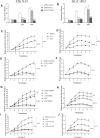
Kil6425, BB94, and AG1478 inhibit LPA-induced efficient DNA synthesis and cell proliferation in gastric cancer cells. (A–B) LPA-induced efficient DNA synthesis after pretreated with inhibitors for 30 min and grown in the ligands (0.1% DMSO, 10 µM LPA, or 10 ng/ml EGF) for 4 days. (C–D) Cell proliferation of gastric cancer cells with ligands. (E–J) Cell proliferation of gastric cancer cells after pretreated with inhibitors for 30 min and grown in the ligand. In qualifications, the cell growth were calculated using GraphPad Prism v8.0 software at each time point, and the fold is compared relative to the level at 1 day, respectively, mean ± SD, n=3, *p < 0.05, ***p < 0.001 (ANOVA test and Dunnette’s multiple comparisons test).
FIGURE 9
FIGURE 9
Signaling pathway of LPA-mediated DNA replication initiation. LPA work through LPAR3 to transactive EGFR by MMPs and to increase the expression of geminin protein level in the S phase through PI3K/mTOR signaling pathway. Meanwhile, LPA stimulation induces upregulation of DUB3 in a short time, inhibiting the ubiquitination degradation of geminin protein and enhancing its stability, and then positively regulating the DNA replication initiation of gastric cancer cells.

Similar articles

Cited by

References

    1. Alaeddini M., Etemad-Moghadam S. (2020). Cell Kinetic Markers in Cutaneous Squamous and Basal Cell Carcinoma of the Head and Neck. Braz. J. Otorhinolaryngol 10.1016/j.bjorl.2020.07.010 - DOI - PMC - PubMed
    1. Alsahafi E. N., Thavaraj S., Sarvestani N., Novoplansky O., Elkabets M., Ayaz B., et al. (2020). EGFR Overexpression Increases Radiotherapy Response in HPV-Positive Head and Neck Cancer through Inhibition of DNA Damage Repair and HPV E6 Downregulation. Cancer Lett. 498, 80–97. 10.1016/j.canlet.2020.10.035 - DOI - PubMed
    1. Ballabeni A., Melixetian M., Zamponi R., Masiero L., Marinoni F., Helin K. (2004). Human Geminin Promotes Pre-RC Formation and DNA Replication by Stabilizing CDT1 in Mitosis. EMBO J. 23, 3122–3132. 10.1038/sj.emboj.7600314 - DOI - PMC - PubMed
    1. Ballabeni A., Zamponi R., Moore J. K., Helin K., Kirschner M. W. (2013). Geminin Deploys Multiple Mechanisms to Regulate Cdt1 before Cell Division Thus Ensuring the Proper Execution of DNA Replication. Proc. Natl. Acad. Sci. U S A. 110, E2848–E2853. 10.1073/pnas.1310677110 - DOI - PMC - PubMed
    1. Champeris Tsaniras S., Villiou M., Giannou A. D., Nikou S., Petropoulos M., Pateras I. S., et al. (2018). Geminin Ablation In Vivo Enhances Tumorigenesis through Increased Genomic Instability. J. Pathol. 246, 134–140. 10.1002/path.5128 - DOI - PubMed