. 2021 Dec 1;321(6):H1083-H1095.
doi: 10.1152/ajpheart.00065.2021. Epub 2021 Oct 15.

TNF-α-activated eNOS signaling increases leukocyte adhesion through the S-nitrosylation pathway

Affiliations

TNF-α-activated eNOS signaling increases leukocyte adhesion through the S-nitrosylation pathway

Gaynor Aguilar et al. Am J Physiol Heart Circ Physiol. .

Abstract

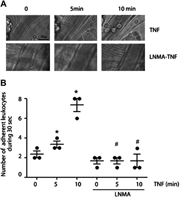
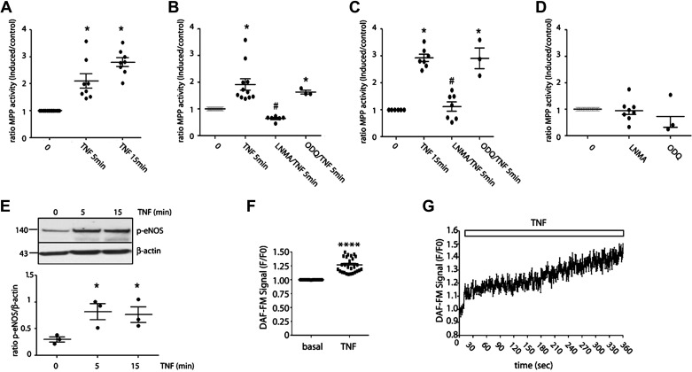
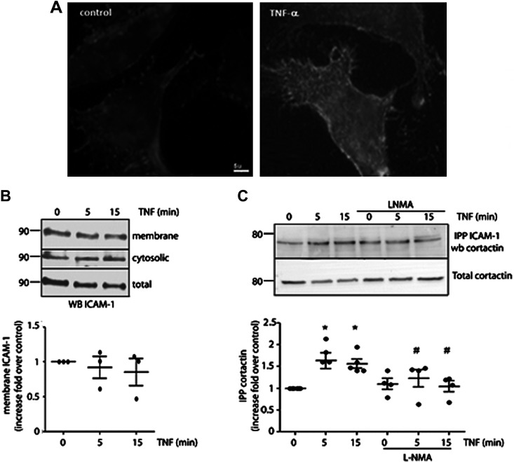
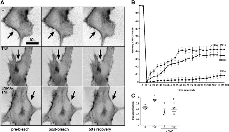
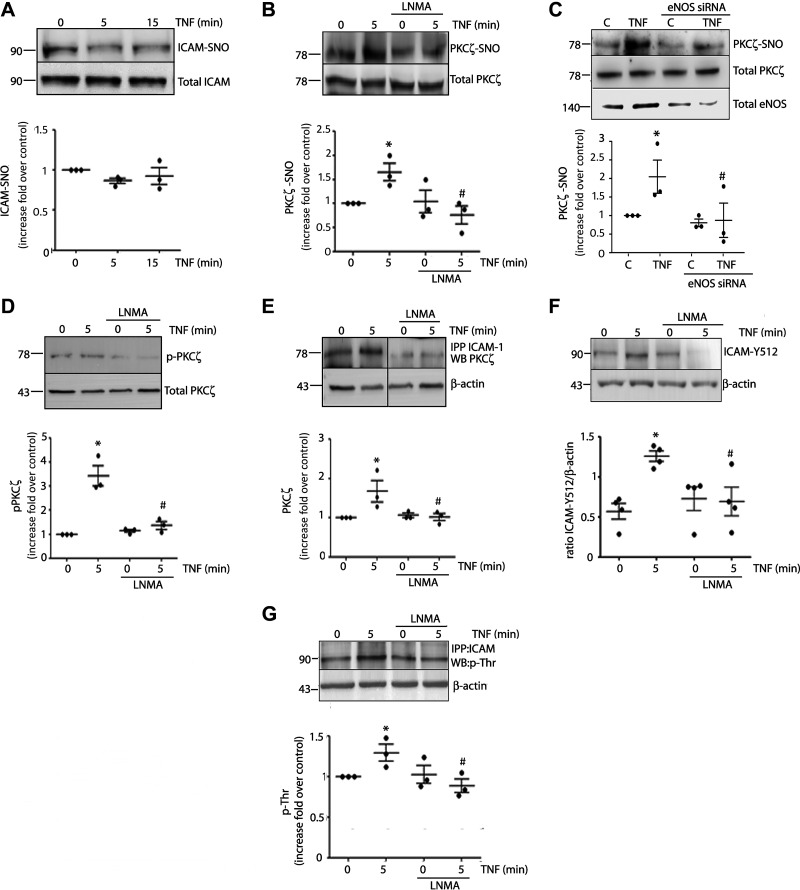
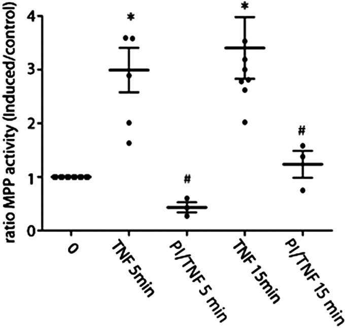
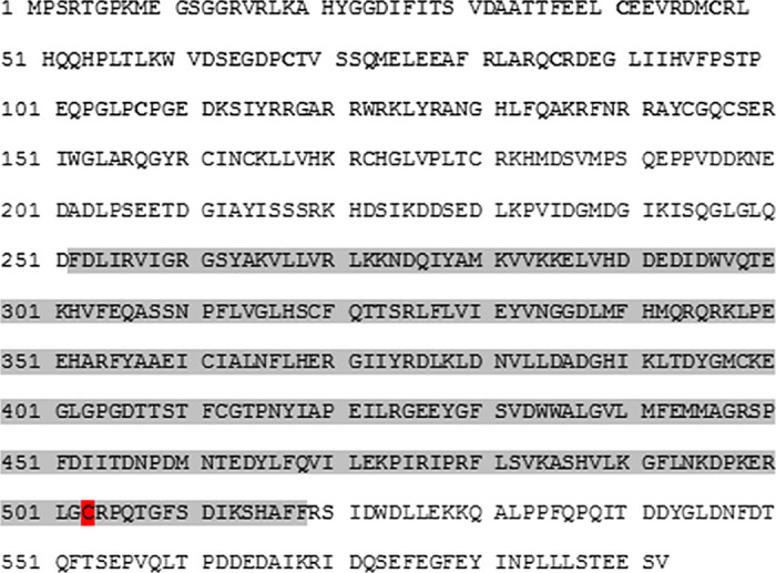
Nitric oxide (NO) is a key factor in inflammation. Endothelial nitric oxide synthase (eNOS), whose activity increases after stimulation with proinflammatory cytokines, produces NO in endothelium. NO activates two pathways: 1) soluble guanylate cyclase-protein kinase G and 2) S-nitrosylation (NO-induced modification of free-thiol cysteines in proteins). S-nitrosylation affects phosphorylation, localization, and protein interactions. NO is classically described as a negative regulator of leukocyte adhesion to endothelial cells. However, agonists activating NO production induce a fast leukocyte adhesion, which suggests that NO might positively regulate leukocyte adhesion. We tested the hypothesis that eNOS-induced NO promotes leukocyte adhesion through the S-nitrosylation pathway. We stimulated leukocyte adhesion to endothelium in vitro and in vivo using tumor necrosis factor-α (TNF-α) as proinflammatory agonist. ICAM-1 changes were evaluated by immunofluorescence, subcellular fractionation, immunoprecipitation, and fluorescence recovery after photobleaching (FRAP). Protein kinase Cζ (PKCζ) activity and S-nitrosylation were evaluated by Western blot analysis and biotin switch method, respectively. TNF-α, at short times of stimulation, activated the eNOS S-nitrosylation pathway and caused leukocyte adhesion to endothelial cells in vivo and in vitro. TNF-α-induced NO led to changes in ICAM-1 at the cell surface, which are characteristic of clustering. TNF-α-induced NO also produced S-nitrosylation and phosphorylation of PKCζ, association of PKCζ with ICAM-1, and ICAM-1 phosphorylation. The inhibition of PKCζ blocked leukocyte adhesion induced by TNF-α. Mass spectrometry analysis of purified PKCζ identified cysteine 503 as the only S-nitrosylated residue in the kinase domain of the protein. Our results reveal a new eNOS S-nitrosylation-dependent mechanism that induces leukocyte adhesion and suggests that S-nitrosylation of PKCζ may be an important regulatory step in early leukocyte adhesion in inflammation.NEW & NOTEWORTHY Contrary to the well-established inhibitory role of NO in leukocyte adhesion, we demonstrate a positive role of nitric oxide in this process. We demonstrate that NO induced by eNOS after TNF-α treatment induces early leukocyte adhesion activating the S-nitrosylation pathway. Our data suggest that PKCζ S-nitrosylation may be a key step in this process.

Keywords: S-nitrosylation; leukocyte adhesion; nitric oxide; protein kinase.


Conflict of interest statement

No conflicts of interest, financial or otherwise, are declared by the authors.

Figures

Figure 1.
Figure 1.
NO signaling regulates leukocyte adhesion in vivo. A: intravital microscopy of cremaster showing leukocyte adhesion at 5 and 10 min after treatment with 1 nM TNF-α. Pretreatment with l-NMA (50 mg/kg iv) inhibits TNF-α-induced leukocyte adhesion. B: statistical analysis of leukocyte adhesion. Two-way ANOVA and Tukey test, *P < 0.05 compared with time 0 of treatment with TNF-α; #P < 0.05 in comparison to the treatment with TNF-α. n = 3 different animals. l-NMA, NG-methyl-l-arginine; NO, nitric oxide; TNF-α, tumor necrosis factor-α.
Figure 2.
Figure 2.
S-nitrosylation regulates early PMN adhesion induced by TNF-α in vitro. A: confluent monolayers of EAhy926 cells were incubated with 1 nM TNF-α for 5 and 15 min. Then, isolated human preactivated PMNs were added to the monolayers and allowed to adhere for 30 min at 37°C. Nonadherent PMN were washed away and MPO activity was assayed in the cell lysates. One-way ANOVA and Tukey’s multiple comparison test. *P < 0.05 compared with time 0. n = 8 independent experiments. Confluent monolayers of EAhy926 cells were preincubated with 300 µM l-NMA for 45 min or 10 µM ODQ for 10 min before TNF-α treatment for 5 (B) and 15 min (C). Increase in leukocyte adhesion induced by TNF-α was blocked by inhibition of eNOS with l-NMA but not by inhibition of GC-1-PKG with ODQ. One-way ANOVA and Bonferroni’s multiple comparison test. *P < 0.05 compared with time 0; #P < 0.05 compared with TNF-α treatment. n = 6 independent experiments. D: confluent monolayers of EAhy926 cells were preincubated with 300 µM l-NMA for 45 min or 10 µM ODQ for 10 min. Neither agent caused leukocyte adhesion. One-way ANOVA and Bonferroni’s multiple comparison test. n = 8 independent experiments. All data were normalized to adhesion seen at time 0. E: TNF-α significantly increases phosphorylation of eNOS at Ser1177 at 5 and 15 min. One-way ANOVA and Bonferroni’s multiple comparison test. *P < 0.05; n = 3 independent experiments. F: real-time detection of NO induced by TNF-α in live EAHy926 cells. Paired t test. ****P < 0.0001; n = 5 independent experiments. G: representative image of one experiment showing the increases in NO levels in the time after TNF-α application. eNOS, endothelial nitric oxide synthase; l-NMA, NG-methyl-l-arginine; NO, nitric oxide; ODQ, 1H-[1,2, 4]oxadiazolo[4,3-a]quinoxalin-1-one; PMN, polymorphonuclear leukocyte; TNF-α, tumor necrosis factor-α.
Figure 3.
Figure 3.
TNF-α induces ICAM-1 clustering dependent on NO signaling. A: confluent monolayers of EAhy926 cells were incubated with 1 nM TNF-α for 15 min and processed by immunofluorescence to detect ICAM-1 in the cell surface. The images are representative of 3 independent experiments. B: confluent monolayers of EAhy926 cells were incubated with 1 nM TNF-α for different times and membrane and cytosolic fractions were isolated, separated by PAGE-SDS, and probed with anti-ICAM antibodies. One-way ANOVA and Dunnett’s multiple comparison test, n = 3 independent experiments. C: confluent monolayers of EAhy926 cells were incubated with 1 nM TNF-α for different times in the presence or absence of l-NMA; ICAM-1 was immunoprecipitated and the interaction with cortactin evaluated by Western blot analysis. Two-way ANOVA and Tukey’s test, *P < 0.05 in comparison to time 0, #P < 0.05 in comparison to the treatment with TNF-α; n = 4 independent experiments. Data were normalized to band intensity seen at time 0. ICAM-1, intercellular adhesion molecule 1; l-NMA, NG-methyl-l-arginine; NO, nitric oxide; TNF-α, tumor necrosis factor-α.
Figure 4.
Figure 4.
TNF-α-induced NO increases ICAM-1 immobile fraction. A: ICAM-1-GFP (grayscale, inverted contrast) was expressed in EAhy926 cells, and FRAP was recorded. Arrow depicts the FRAP region. Top shows FRAP without treatment applied. Middle shows ICAM-1-GFP FRAP after treatment with 1 nM TNF-α for 5 min. Bottom shows ICAM-1-GFP FRAP after pretreatment of the cells with l-NMA before TNF-α application for 5 min. B: recovery ICAM-GFP fluorescence curves after different treatments. C: quantification of the FRAP data show increase in the immobile fraction of ICAM-1-GFP following TNF-α treatment and inhibition of this effect by application of l-NMA. Two-way ANOVA and Bonferroni t test. *P < 0.05 in comparison to time 0, #P < 0.05 in comparison to the treatment with TNF-α; n = 5 independent experiments. FRAP, fluorescence recovery after photobleaching; ICAM-1, intercellular adhesion molecule 1; l-NMA, NG-methyl-l-arginine; NO, nitric oxide; TNF-α, tumor necrosis factor-α.
Figure 5.
Figure 5.
TNF-α leads to PKCζ S-nitrosylation, PKCζ activation, association with ICAM-1, and ICAM-1 phosphorylation. A: confluent monolayers of EAhy926 cells were incubated with 1 nM TNF-α for different times. Cell lysates were obtained and processed by biotin switch to detect S-nitrosylated ICAM-1. One-way ANOVA and Tukey’s test, n = 3 independent experiments. B: confluent monolayers of EAhy926 cells were incubated with 1 nM TNF-α for 5 min in the presence or absence of 300 µM l-NMA for 1 h. Cell lysates were obtained and processed to detect S-nitrosylated PKCζ by biotin switch. C: depletion of eNOS in EAHy926 cells abrogates PKCζ S-nitrosylation induced by TNF-α. EAHy926 cells transfected with scrambled siRNA served as a control. Western blot for eNOS confirms protein depletion. One-way ANOVA and Dunnett’s multiple comparison test, n = 3 independent experiments; *P < 0.05 in comparison to control (C), #P < 0.05 in comparison to TNF-α treatment in the absence of eNOS siRNA. Data were normalized at band intensity seen at control without eNOS siRNA treatment. Confluent monolayers of EAhy926 cells were incubated with 1 nM TNF-α for 5 min in the presence or absence of 300 µM l-NMA for 1 h. Cell lysates were obtained and processed to detect phosphorylated p-PKCζ by Western blot analysis (D) and interaction between ICAM-1 and PKCζ by immunoprecipitation (E). Two-way ANOVA and Holm–Sidak test, *P < 0.05 in comparison to time 0, #P < 0.05 in comparison to the treatment with TNF-α; n = 3 independent experiments. All data were normalized at band intensity seen at time 0. F: ICAM-Y512 by Western blot analysis. Two-way ANOVA and Holm–Sidak test, *P < 0.05 in comparison to time 0, #P < 0.05 in comparison to the treatment with TNF-α; n = 4 independent experiments. G: ICAM-1 Thr phosphorylation by immunoprecipitation with anti ICAM-1 antibody, followed by Western blot analysis against p-Thr. Two-way ANOVA and Holm–Sidak test, *P < 0.05 in comparison to time 0, #P < 0.05 in comparison to the treatment with TNF-α; n = 3 independent experiments. Data were normalized at band intensity seen at time 0. eNOS, endothelial nitric oxide synthase; ICAM-1, intercellular adhesion molecule 1; l-NMA, NG-methyl-l-arginine; PKCζ, protein kinase Cζ; TNF-α, tumor necrosis factor-α.
Figure 6.
Figure 6.
PKCζ regulates leukocyte adhesion. Confluent monolayers of EAhy926 cells were preincubated with 1 µM inhibitor peptide for 20 min before TNF-α treatment for 5 and 15 min. Then, isolated human preactivated PMNs were added to the monolayers and allowed to adhere for 30 min at 37°C. Nonadherent PMNs were washed away and MPO activity was assayed in the cell lysates. Two-way ANOVA and Holm–Sidak test, *P < 0.05 in comparison to time 0, #P < 0.05 in comparison to the treatment with TNF-α; n = 3 independent experiments. Data were normalized at adhesion seen at time 0. MPO, myeloperoxidase; PKCζ, protein kinase Cζ; PMN, polymorphonuclear leukocyte; TNF-α, tumor necrosis factor-α.
Figure 7.
Figure 7.
Identification of S-nitrosylated Cys 503 in PKCζ. Protein sequence from human PKCζ. Purified PKCζ was S-nitrosylated with GSNO for 30 min, subjected to biotin switch assay followed by in-solution trypsin digestion. The kinase domain of PKCζ is shown in gray background. Cysteine 503 (red background) was the only S-nitrosylated cysteine according to mass spectrometry. GSNO, S-nitrosoglutathione; PKCζ, protein kinase Cζ.

Similar articles

Cited by

References

    1. Ma XL, Weyrich AS, Lefer DJ, Lefer AM. Diminished basal nitric oxide release after myocardial ischemia and reperfusion promotes neutrophil adherence to coronary endothelium. Circ Res 72: 403–412, 1993. doi:10.1161/01.RES.72.2.403. - DOI - PubMed
    1. Furchgott RF, Vanhoutte PM. Endothelium-derived relaxing and contracting factors. FASEB J 3: 2007–2018, 1989. - PubMed
    1. Ignarro LJ. Biological actions and properties of endothelium-derived nitric oxide formed and released from artery and vein. Circ Res 65: 1–21, 1989. doi:10.1161/01.RES.65.1.1. - DOI - PubMed
    1. Kubes P, Suzuki M, Granger DN. Nitric oxide: an endogenous modulator of leukocyte adhesion. Proc Natl Acad Sci USA 88: 4651–4655, 1991. doi:10.1073/pnas.88.11.4651. - DOI - PMC - PubMed
    1. Moncada S. The 1991 Ulf von Euler lecture. The l-arginine: nitric oxide pathway. Acta Physiol Scand 145: 201–227, 1992. doi:10.1111/j.1748-1716.1992.tb09359.x. - DOI - PubMed

Publication types

MeSH terms

LinkOut - more resources