Review
. 2021 Oct 1;22(19):10661.
doi: 10.3390/ijms221910661.

The Keratinocyte as a Crucial Cell in the Predisposition, Onset, Progression, Therapy and Study of the Atopic Dermatitis

Affiliations
Review

The Keratinocyte as a Crucial Cell in the Predisposition, Onset, Progression, Therapy and Study of the Atopic Dermatitis

Pamela Gallegos-Alcalá et al. Int J Mol Sci. .

Abstract

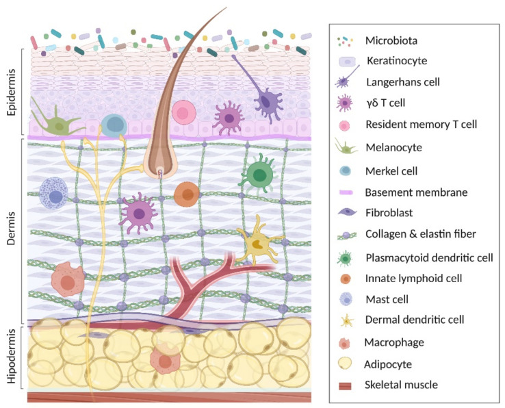
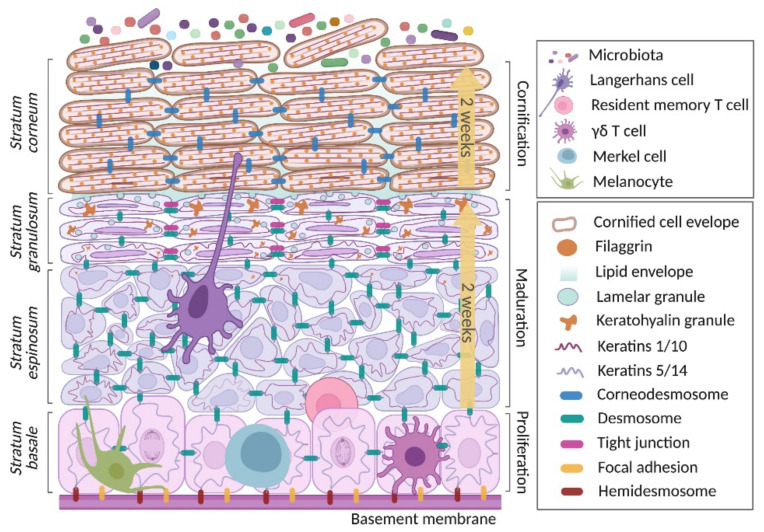
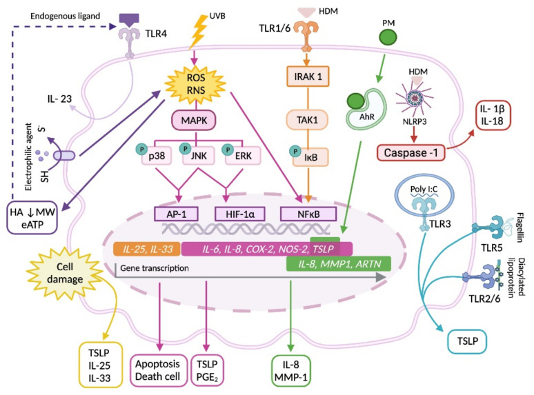
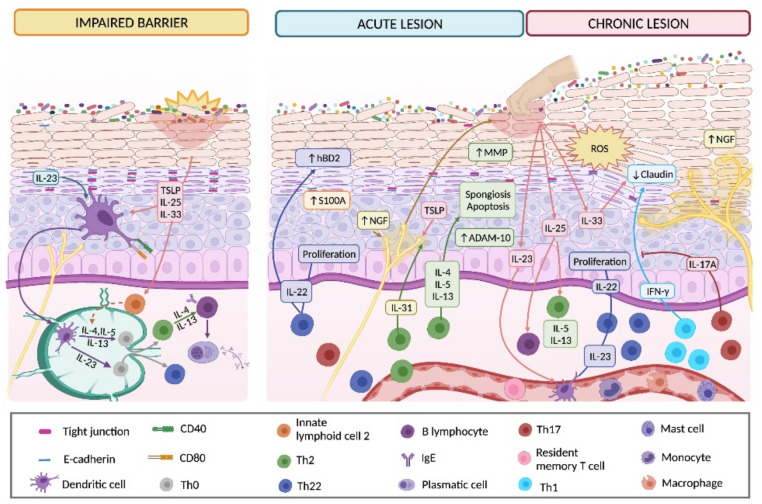
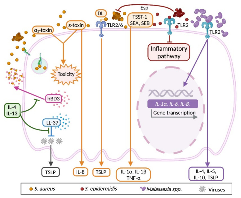
The keratinocyte (KC) is the main functional and structural component of the epidermis, the most external layer of the skin that is highly specialized in defense against external agents, prevention of leakage of body fluids and retention of internal water within the cells. Altered epidermal barrier and aberrant KC differentiation are involved in the pathophysiology of several skin diseases, such as atopic dermatitis (AD). AD is a chronic inflammatory disease characterized by cutaneous and systemic immune dysregulation and skin microbiota dysbiosis. Nevertheless, the pathological mechanisms of this complex disease remain largely unknown. In this review, we summarize current knowledge about the participation of the KC in different aspects of the AD. We provide an overview of the genetic predisposing and environmental factors, inflammatory molecules and signaling pathways of the KC that participate in the physiopathology of the AD. We also analyze the link among the KC, the microbiota and the inflammatory response underlying acute and chronic skin AD lesions.

Keywords: allergic inflammatory response; atopic dermatitis; in vitro atopic dermatitis models; keratinocyte; keratinocyte differentiation; pharmacological therapy; skin microbiome.


Conflict of interest statement

The authors declare no conflict of interest.

Figures

Figure 1
Figure 1
Anatomy of the skin. This image represents the three main layers of the skin, including the most abundant cellular populations in each layer, together with the immune cells present in each anatomical region. Created with BioRender.com (access date: 26 August 2021).
Figure 2
Figure 2
Keratinocyte life cycle. This begins when the keratinocyte (KC) proliferates in the stratum basale. Later, in a course of two weeks, it matures and migrates through the suprabasal layers (spinosum and granulosum stratums) until reaching the top part of the skin, the stratum corneum. Here, the KC acquires the highest degree of maturity and gradually loses viability. Finally, in another two weeks, it moves through the cornified layer to be eliminated by flaking. Created with BioRender.com (access date: 26 August 2021).
Figure 3
Figure 3
Keratinocyte response to environmental factors. In response to UVB radiation, allergens, endogenous ligands, electrophilic agents, particulate matter present in air pollutants, or ligands of pathogen pattern receptors, KCs activate different signaling pathways that induce apoptosis and cellular death, gene transcription and the release of de novo synthesized type-2 response promoting cytokines and inflammatory mediators, or caspase-1 activation and the subsequent maturation of inflammatory cytokines pro-IL-1β and pro-IL-18. Abbreviations: AhR, aryl hydrocarbon receptor; COX-2, cyclooxygenase-2; eATP, extracellular ATP; HA, hyaluronic acid; HDM, house dust mite; IL, interleukin; MMP-1, matrix metalloproteinase-1; MW, molecular weight; NOS-2, nitric oxide synthase -2; PGE2, prostaglandin E2; TSLP, thymic stromal lymphopoietin. Created with BioRender.com (access date: 26 August 2021).
Figure 4
Figure 4
Keratinocyte participation in the onset, development and chronification of atopic dermatitis lesions. Due to an impaired cutaneous barrier, KC-derived cytokines (mainly TSLP, IL-23, IL-25 and IL-33) induce dendritic cell activation and mobilization to nearby lymphatic nodes where they active naïve CD4+ T cell (Th0) and promote Th2 and Th22 polarization. Th2-derived cytokines trigger antibody isotype switching in B lymphocytes to produce IgE. Cytokines produced by Th2 and Th22 cells increase KC proliferation and S100A expression, prompt KC apoptosis and induce cutaneous spongiosis due to a decrease in E-cadherin levels mediated by increased enzymatic activity. These changes are clinically manifested in skin as acute lesions. In this stage, IL-22 and hBD2 mutually enhance their production, perpetuating the inflammatory response associated with AD. Pruritic mediators released by Th2 cells (IL-31) or KCs (NGF and TSLP) increase itching sensation which triggers scratching and worsening of skin lesions. Lesions becomes chronic due to the intensification of the pre-existing Th2 and Th22 inflammatory response enhanced by Th1 and Th17 cytokines. Augmented production of IL-23, IL-25 and IL-33 by KCs maintains Th22 differentiation, up-regulates Th2 cytokine production and diminishes claudin expression, respectively. The decrease in tight junctions, which is enhanced by IFN-γ production, together with the increased levels of NGF, favors epidermal hyperinnervation. Altogether, this inflammatory environment exacerbates the remodeling processes of the epidermis and hyperplasia, while IL-17 down-regulates the IFN-γ effect on claudin expression. Abbreviations: hBD2, human beta-defensin 2; MMP, matrix metalloproteinase; NGF, nerve growth factor. Created with BioRender.com (access date: 26 August 2021).
Figure 5
Figure 5
Keratinocyte response to microbiota stimuli under the type 2 immune milieu. KCs recognize bacteria, fungi, and viruses from skin microbiota through specific receptors. Colonization with Staphylococcus aureus is present in the skin of AD patients, and the bacteria or their components can activate KCs to secrete proallergic (TSLP) and proinflammatory (IL-1α, IL-1β, IL-8, TNF-α) cytokines, exacerbating the underlying immune response. Exotoxins from S. aureus can also damage the cell. Chronic infection of S. aureus is partly due to its internalization into KCs through extracellular adherence protein recognition. Th2 cytokines (IL-4, IL-13) avoid S. aureus destruction by KC-derived hBD3. As Staphylococcus epidermidis/S. aureus ratio is diminished in AD skin, the anti-inflammatory and protective effects, and the anti-microbial properties of S. epidermidis on KCs and S. aureus, respectively, are impaired. In response to Malassezia colonization, KCs express proinflammatory IL-1α, IL-6, and IL-8, and release type 2 (IL-4, IL-5, IL-10, TSLP) cytokines. Levels of LL-37 in KCs are down-regulated by type 2 cytokines, increasing viral replication and dissemination of vaccinia, herpes simplex and human papilloma viruses, which at the same time promotes type 2 inflammation through an induction of TSLP expression by the cell. Abbreviations: DL, diacylated lipopeptide; Esp, serine protease; hBD3, human β-defensin 3; LL-37, cathelicidin LL-37; SEA, staphylococcal enterotoxin A; SEB, staphylococcal enterotoxin B; TNF, tumoral necrosis factor; TSST-1, toxic shock syndrome toxin. Created with BioRender.com (access date: 30 August 2021).

Similar articles

Cited by

References

    1. Boniol M., Verriest J.-P., Pedeux R., Doré J.-F. Proportion of Skin Surface Area of Children and Young Adults from 2 to 18 Years Old. J. Investig. Dermatol. 2008;128:461–464. doi: 10.1038/sj.jid.5701032. - DOI - PubMed
    1. Gallo R.L. Human Skin Is the Largest Epithelial Surface for Interaction with Microbes. J. Investig. Dermatol. 2017;137:1213–1214. doi: 10.1016/j.jid.2016.11.045. - DOI - PMC - PubMed
    1. Wong R., Geyer S., Weninger W.J., Guimberteau J.-C., Wong J.K. The dynamic anatomy and patterning of skin. Exp. Dermatol. 2015;25:92–98. doi: 10.1111/exd.12832. - DOI - PubMed
    1. Lancerotto L., Stecco C., Macchi V., Porzionato A., Stecco A., De Caro R. Layers of the abdominal wall: Anatomical investigation of subcutaneous tissue and superficial fascia. Surg. Radiol. Anat. 2011;33:835–842. doi: 10.1007/s00276-010-0772-8. - DOI - PubMed
    1. Baroni A., Buommino E., De Gregorio V., Ruocco E., Ruocco V., Wolf R. Structure and function of the epidermis related to barrier properties. Clin. Dermatol. 2012;30:257–262. doi: 10.1016/j.clindermatol.2011.08.007. - DOI - PubMed

MeSH terms

LinkOut - more resources