. 2021 Oct 26;12(5):e0241021.
doi: 10.1128/mBio.02410-21. Epub 2021 Oct 5.

Citrobacter amalonaticus Inhibits the Growth of Citrobacter rodentium in the Gut Lumen

Affiliations

Citrobacter amalonaticus Inhibits the Growth of Citrobacter rodentium in the Gut Lumen

Caroline Mullineaux-Sanders et al. mBio. .

Abstract

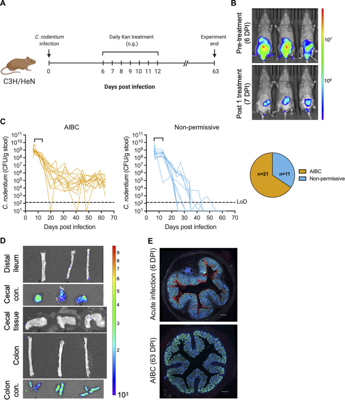
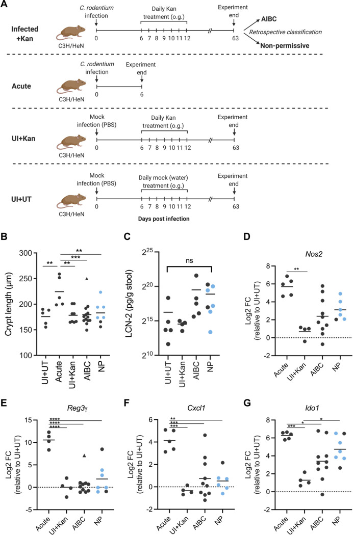
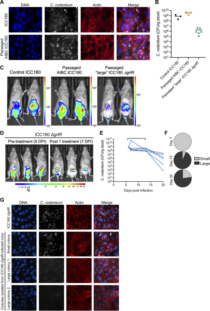
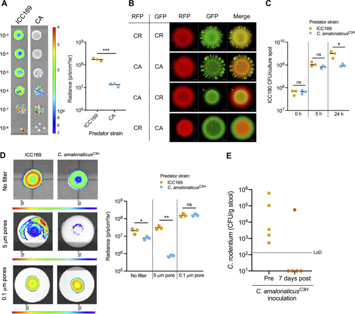
The gut microbiota plays a crucial role in susceptibility to enteric pathogens, including Citrobacter rodentium, a model extracellular mouse pathogen that colonizes the colonic mucosa. C. rodentium infection outcomes vary between mouse strains, with C57BL/6 and C3H/HeN mice clearing and succumbing to the infection, respectively. Kanamycin (Kan) treatment at the peak of C57BL/6 mouse infection with Kan-resistant C. rodentium resulted in relocalization of the pathogen from the colonic mucosa and cecum to solely the cecal luminal contents; cessation of the Kan treatment resulted in rapid clearance of the pathogen. We now show that in C3H/HeN mice, following Kan-induced displacement of C. rodentium to the cecum, the pathogen stably colonizes the cecal lumens of 65% of the mice in the absence of continued antibiotic treatment, a phenomenon that we term antibiotic-induced bacterial commensalization (AIBC). AIBC C. rodentium was well tolerated by the host, which showed few signs of inflammation; passaged AIBC C. rodentium robustly infected naive C3H/HeN mice, suggesting that the AIBC state is transient and did not select for genetically avirulent C. rodentium mutants. Following withdrawal of antibiotic treatment, 35% of C3H/HeN mice were able to prevent C. rodentium commensalization in the gut lumen. These mice presented a bloom of a commensal species, Citrobacter amalonaticus, which inhibited the growth of C. rodentium in vitro in a contact-dependent manner and the luminal growth of AIBC C. rodentium in vivo. Overall, our data suggest that commensal species can confer colonization resistance to closely related pathogenic species. IMPORTANCE Gut bacterial infections involve three-way interactions between virulence factors, the host immune responses, and the microbiome. While the microbiome erects colonization resistance barriers, pathogens employ virulence factors to overcome them. Treating mice infected with kanamycin-resistant Citrobacter rodentium with kanamycin caused displacement of the pathogen from the colonic mucosa to the cecal lumen. Following withdrawal of the kanamycin treatment, 65% of the mice were persistently colonized by C. rodentium, which seemed to downregulate virulence factor expression. In this model of luminal gut colonization, 35% of mice were refractory to stable C. rodentium colonization, suggesting that their microbiotas were able to confer colonization resistance. We identify a commensal bacterium of the Citrobacter genus, C. amalonaticus, which inhibits C. rodentium growth in vitro and in vivo. These results show that the line separating commensal and pathogenic lifestyles is thin and multifactorial and that commensals may play a major role in combating enteric infection.

Keywords: Citrobacter; colonization resistance; gastrointestinal infection.


Figures

FIG 1
FIG 1
Kan treatment of ICC180-infected C3H mice induces AIBC (A) Schematic of the experimental timeline. o.g., oral gavage. (B) Representative in vivo BLIs of mice at 6 DPI prior to Kan treatment (top) and at 7 DPI 24 h after the first Kan treatment (bottom). (C) C. rodentium (strain ICC180) colonization of mice designated AIBC (left) or nonpermissive (middle). Brackets on the line graphs denote the Kan treatment period. Each line represents an individual mouse. Only AIBC mice monitored until 63 DPI are shown (24 mice from five biological repeats). (Right, pie chart) The ratio of AIBC to nonpermissive designations of all mice used in this study (32 mice from six biological repeats). LoD, limit of detection. (D) Ex vivo BLIs of organs from AIBC mice at 63 DPI. Images are representative of 4 mice from one biological repeat. con., contents. In panels B and D, color scale bar indicates radiance (photons per second per square centimeter per surface radiance). (E) Immunofluorescence staining of the distal colons of mice acutely infected with C. rodentium (6 DPI with strain ICC169) or mice harboring AIBC C. rodentium at 63 DPI (AIBC), demonstrating no staining of C. rodentium on the colonic mucosa in the AIBC group. C. rodentium, red; DNA, blue; E-cadherin, green. Images are representative of 8 mice from three biological repeats (AIBC) or 5 mice from one biological repeat (acute infection). Scale bar = 200 μm.
FIG 2
FIG 2
AIBC C. rodentium is not inflammatory. (A) Schematic of the experimental timeline of the AIBC, nonpermissive, Acute, UI+Kan, and UI+UT groups of mice. AIBC and nonpermissive mice are the same mice shown in Fig. 1C. (B) Colonic crypt lengths of mice from the indicated groups. Each point represents the average crypt length of an individual mouse. Data are from one (UI+UT, UI+Kan, Acute), two (nonpermissive), or four (AIBC) biological repeats. (C) Stool LCN-2 concentrations in mice from the indicated groups at 63 DPI. Each point represents an individual mouse. Data are from one (UI+UT, UI+Kan) or two (AIBC, nonpermissive) biological repeats. ns, not significant (P < 0.05 for all comparisons, as determined by a one-way ANOVA with Tukey’s posttest for multiple comparisons between all groups). (D to G) qRT-PCR analysis of the indicated mRNA isolated from IECs of mice in the Acute, AIBC, nonpermissive, or UI+Kan group. All are shown relative to the mean of results from the UI+UT group. Each point represents an individual mouse. Data are from one (UI+UT, UI+Kan, and Acute), two (NP), or three (AIBC) biological repeats. (B, E to G) *, P < 0.05; **, P < 0.01; ***, P < 0.001; ****, P < 0.0001 (as determined by a one-way ANOVA with Tukey’s posttest for multiple comparisons between all groups). The triangle points in panels B and E indicate data points identified as an outlier and not included in statistical analyses. (D) Nos2 data did not pass a normality test and were analyzed by a Kruskal-Wallis test with Dunn’s posttest for multiple comparisons between all groups (**, P < 0.01). (B to G) Blue points indicate nonpermissive (NP) mice harboring C. amalonaticusC3H.
FIG 3
FIG 3
C. rodentium T3SS repression is favored in the AIBC state. (A) CMT-93 cells infected with passaged C. rodentium isolated from mice with AIBC C. rodentium at 63 DPI (mice shown in Fig. 1C). The image is representative of infections with 22 colonies from 6 mice. Scale bar = 10 μm. (B) C. rodentium colonization of mice at 6 DPI with the indicated C. rodentium strain. Each point represents an individual mouse. Data are from one (ICC180 and passaged AIBC ICC180) or two (passaged large ICC180 ΔgrlR) biological repeats. (C) Representative in vivo BLI images of mice shown in panel B at 6 DPI. (D) In vivo BLI images of ICC180 ΔgrlR-infected mice at 6 DPI, prior to Kan treatment (left) and at 7 DPI, 24 h after the first Kan treatment (right). (C, D) Color scale bars indicate radiance (photons per second per square centimeter per surface radiance). (E) C. rodentium colonization of mice infected with ICC180 ΔgrlR and treated daily with Kan from 6 to 12 DPI inclusive (denoted by the bracket on the graph). Each line represents an individual mouse, and data are from two biological repeats. (F) Percentages of isolated ICC180 ΔgrlR colonies which were assigned as having a large- or small-colony morphology at the indicated DPI. Pie charts show the average percentages of all mice shown in panel E. (G) CMT-93 cells infected with control ICC180 ΔgrlR or passaged small or large ICC180 ΔgrlR colonies isolated from stool samples of mice shown in panel E at 11 to 20 DPI. Immunofluorescence images are representative of 9 colonies from 5 mice (large colonies) and 8 colonies from 4 mice (small colonies). Scale bar = 10 μm.
FIG 4
FIG 4
Commensal Citrobacter blooms in nonpermissive mice. (A) Non-C. rodentium bacteria assigned to the Citrobacter genus in stool samples from AIBC and nonpermissive mice collected from the same mice prior to C. rodentium infection (0 DPI) and at 34 or 41 DPI. Each point represents an individual mouse. Data are from three biological repeats. LoD, limit of detection. (B) The 16S rRNA gene of C. amalonaticusC3H was compared to selected Citrobacter species using a maximum likelihood model. The percentage of trees in which the associated taxa cluster together in the bootstrap test (1,000 replicates) is shown next to each branch. The tree is drawn to scale, with branch lengths measured in numbers of substitutions per site. Klebsiella pneumoniae is used as an outgroup. The red arrow denotes the C. amalonaticusC3H strain isolated in this study. (C) 16S rRNA gene taxonomic analysis of the stool microbiomes from AIBC and nonpermissive (NP) mice at 0 and 45 DPI. Each bar represents a mouse; stool samples from the same mice were analyzed at 0 and 45 DPI. p, phylum; c, class; o, order; f, family.
FIG 5
FIG 5
C. amalonaticusC3H outcompetes C. rodentium. (A, left) Representative BLI of cocultures of ICC180 and ICC169 or C. amalonaticusC3H (CA). The number on the left of the cultures indicates the dilution factor. (Right) Graph showing the quantification of BL from the 10−3 culture spot of the indicated coculture from three biological repeats. (B) Fluorescence microscopy of cocultures of C. rodentium (ICC169; CR) and C. amalonaticusC3H (CA) expressing RFP or GFP from a plasmid as indicated after 24 h of growth on LB agar. Scale bar = 1,000 μm. (C) Quantification of ICC180 CFU at 0, 5, and 24 h after coincubation with ICC169 or C. amalonaticusC3H (D, left) Representative BLI of ICC180 grown with ICC169 or C. amalonaticusC3H separated by a filter with 5-μm or 0.1-μm pores or not separated by a filter. (Right) Graph showing the quantification of BL from the culture spots shown on the left from three biological repeats. (A, C, D) *, P < 0.05; **, P < 0.01; ***, P < 0.001 (as determined by Student’s unpaired two-tailed t test). (A, D) Color scale bars indicate radiance (photons per second per square centimeter per surface radiance). (E) C. rodentium colonization of AIBC mice before (Pre) C. amalonaticusC3H inoculation (42 DPI) and 7 days after (post) the first C. amalonaticusC3H inoculation (53 DPI). Five mice from one biological repeat were used. LoD, limit of detection.

Similar articles

Cited by

References

    1. Brestoff JR, Artis D. 2013. Commensal bacteria at the interface of host metabolism and the immune system. Nat Immunol 14:676–684. doi:10.1038/ni.2640. - DOI - PMC - PubMed
    1. Baümler AJ, Sperandio V. 2016. Interactions between the microbiota and pathogenic bacteria in the gut. Nature 535:85–93. doi:10.1038/nature18849. - DOI - PMC - PubMed
    1. Thaiss CA, Zmora N, Levy M, Elinav E. 2016. The microbiome and innate immunity. Nature 535:65–74. doi:10.1038/nature18847. - DOI - PubMed
    1. Curtis MM, Hu Z, Klimko C, Narayanan S, Deberardinis R, Sperandio V. 2014. The gut commensal Bacteroides thetaiotaomicron exacerbates enteric infection through modification of the metabolic landscape. Cell Host Microbe 16:759–769. doi:10.1016/j.chom.2014.11.005. - DOI - PMC - PubMed
    1. Mullineaux-Sanders C, Sanchez-Garrido J, Hopkins EGD, Shenoy AR, Barry R, Frankel G. 2019. Citrobacter rodentium-host-microbiota interactions: immunity, bioenergetics and metabolism. Nat Rev Microbiol 17:701–715. doi:10.1038/s41579-019-0252-z. - DOI - PubMed

Publication types

Supplementary concepts

LinkOut - more resources