Oklahoma Nathan Shock Aging Center - assessing the basic biology of aging from genetics to protein and function
- PMID: 34606039
- PMCID: PMC8599778
- DOI: 10.1007/s11357-021-00454-7
Oklahoma Nathan Shock Aging Center - assessing the basic biology of aging from genetics to protein and function
Abstract
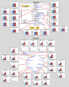
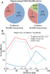
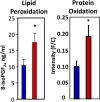
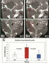
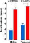
The Oklahoma Shock Nathan Shock Center is designed to deliver unique, innovative services that are not currently available at most institutions. The focus of the Center is on geroscience and the development of careers of young investigators. Pilot grants are provided through the Research Development Core to junior investigators studying aging/geroscience throughout the USA. However, the services of our Center are available to the entire research community studying aging and geroscience. The Oklahoma Nathan Shock Center provides researchers with unique services through four research cores. The Multiplexing Protein Analysis Core uses the latest mass spectrometry technology to simultaneously measure the levels, synthesis, and turnover of hundreds of proteins associated with pathways of importance to aging, e.g., metabolism, antioxidant defense system, proteostasis, and mitochondria function. The Genomic Sciences Core uses novel next-generation sequencing that allows investigators to study the effect of age, or anti-aging manipulations, on DNA methylation, mitochondrial genome heteroplasmy, and the transcriptome of single cells. The Geroscience Redox Biology Core provides investigators with a comprehensive state-of-the-art assessment of the oxidative stress status of a cell, e.g., measures of oxidative damage and redox couples, which are important in aging as well as many major age-related diseases as well as assays of mitochondrial function. The GeroInformatics Core provides investigators assistance with data analysis, which includes both statistical support as well as analysis of large datasets. The Core also has developed number of unique software packages to help with interpretation of results and discovery of new leads relevant to aging. In addition, the Geropathology Research Resource in the Program Enhancement Core provides investigators with pathological assessments of mice using the recently developed Geropathology Grading Platform.
Keywords: Genomic Sciences Core; Geropathology Grading Platform; Geroscience.
© 2021. American Aging Association.
Figures





Similar articles
-
University of Washington Nathan Shock Center: innovation to advance aging research.Geroscience. 2021 Oct;43(5):2161-2165. doi: 10.1007/s11357-021-00413-2. Epub 2021 Jul 7. Geroscience. 2021. PMID: 34232461 Free PMC article.
-
San Antonio Nathan Shock Center: your one-stop shop for aging research.Geroscience. 2021 Oct;43(5):2105-2118. doi: 10.1007/s11357-021-00417-y. Epub 2021 Jul 9. Geroscience. 2021. PMID: 34240333 Free PMC article.
-
Einstein-Nathan Shock Center: translating the hallmarks of aging to extend human health span.Geroscience. 2021 Oct;43(5):2167-2182. doi: 10.1007/s11357-021-00428-9. Epub 2021 Aug 31. Geroscience. 2021. PMID: 34463901 Free PMC article.
-
University of Alabama at Birmingham Nathan Shock Center: comparative energetics of aging.Geroscience. 2021 Oct;43(5):2149-2160. doi: 10.1007/s11357-021-00414-1. Epub 2021 Jul 25. Geroscience. 2021. PMID: 34304389 Free PMC article. Review.
-
The Role of the National Institute on Aging in the Development of the Field of Geroscience.Cold Spring Harb Perspect Med. 2023 Oct 3;13(10):a041211. doi: 10.1101/cshperspect.a041211. Cold Spring Harb Perspect Med. 2023. PMID: 36878648 Review.
Cited by
-
Geroscience and pathology: a new frontier in understanding age-related diseases.Pathol Oncol Res. 2024 Feb 23;30:1611623. doi: 10.3389/pore.2024.1611623. eCollection 2024. Pathol Oncol Res. 2024. PMID: 38463143 Free PMC article. Review.
-
Longitudinal Fragility Phenotyping Predicts Lifespan and Age-Associated Morbidity in C57BL/6 and Diversity Outbred Mice.bioRxiv [Preprint]. 2024 Feb 8:2024.02.06.579096. doi: 10.1101/2024.02.06.579096. bioRxiv. 2024. Update in: Geroscience. 2024 Oct;46(5):4937-4954. doi: 10.1007/s11357-024-01226-9 PMID: 38370707 Free PMC article. Updated. Preprint.
-
Longitudinal fragility phenotyping contributes to the prediction of lifespan and age-associated morbidity in C57BL/6 and Diversity Outbred mice.Geroscience. 2024 Oct;46(5):4937-4954. doi: 10.1007/s11357-024-01226-9. Epub 2024 Jun 27. Geroscience. 2024. PMID: 38935230 Free PMC article.
References
Publication types
MeSH terms
Grants and funding
LinkOut - more resources
Full Text Sources
Medical

