Observational Study
. 2021 Sep 27;11(1):19092.
doi: 10.1038/s41598-021-98560-1.

Detailed characterization of the transcriptome of single B cells in mantle cell lymphoma suggesting a potential use for SOX4

Affiliations
Observational Study

Detailed characterization of the transcriptome of single B cells in mantle cell lymphoma suggesting a potential use for SOX4

Simone Valentin Hansen et al. Sci Rep. .

Abstract

Mantle cell lymphoma (MCL) is a malignancy arising from naive B lymphocytes with common bone marrow (BM) involvement. Although t(11;14) is a primary event in MCL development, the highly diverse molecular etiology and causal genomic events are still being explored. We investigated the transcriptome of CD19+ BM cells from eight MCL patients at single-cell level. The transcriptomes revealed marked heterogeneity across patients, while general homogeneity and clonal continuity was observed within the patients with no clear evidence of subclonal involvement. All patients were SOX11+CCND1+CD20+. Despite monotypic surface immunoglobulin (Ig) κ or λ protein expression in MCL, 10.9% of the SOX11 + malignant cells expressed both light chain transcripts. The early lymphocyte transcription factor SOX4 was expressed in a fraction of SOX11 + cells in two patients and co-expressed with the precursor lymphoblastic marker, FAT1, in a blastoid case, suggesting a potential prognostic role. Additionally, SOX4 was found to identify non-malignant SOX11- pro-/pre-B cell populations. Altogether, the observed expression of markers such as SOX4, CD27, IgA and IgG in the SOX11+ MCL cells, may suggest that the malignant cells are not fixed in the differentiation state of naïve mature B cells, but instead the patients carry B lymphocytes of different differentiation stages.


Conflict of interest statement

The authors declare no competing interests.

Figures

Figure 1
Figure 1
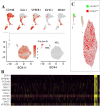
Clustering of combined single-cell transcriptomes, expression and correlation of molecular pathology MCL markers. The MCL cohort displayed heterogeneous expression profiles, forming distinct clusters of single-cell transcriptomes, except for co-located sample 7 and 8. Cells are colored and numbered according to patient origin (A). A highly significant positive correlation (red) was found between the percentage of SOX11expressing cells and cells expressing frequently used molecular pathology markers in MCL (B) including CCND1, LDHA/B, PAX5, CD20, immunoglobulin κ/λ light chain (IGKC/IGLC) (p = 0.003). The fraction of SOX11 positive cells also showed a positive correlation with the percentage of B cells in the bone marrow samples known from the clinical flow cytometry analysis (BM B cells, p = 0.003). The rest of the listed markers showed strong multicollinearity and were hence excluded in the linear regression. The percentage of SOX4+ cells was negatively correlated (green), with other markers and associated with residual non-malignant pro-/pre-B cells (see also Fig. 2b). The percentage of cells expressing molecular pathology markers were calculated for the individual patients (bars) and from the total single cell cohort (numbers) as described in the methods section (C). The total single cell population (numbers) and the cells from individual patients (bars) were markedly positive for CCND1, CD20 and κ/λ (IGKC/IGLC), while merely 37.5% of the total sequenced cells were SOX11+, varying from 9.8 to 64.6%. 10.8% of the combined cohort was found to harbor dual expression of κ and λ light chains.
Figure 2
Figure 2
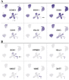
Global expression of molecular pathology markers frequently applied in diagnosis of mantle cell lymphoma (A) and markers associated with immature B cells (B) in combined mantle cell lymphoma (MCL) single cell transcriptomes. The resolved representations of cells from each MCL patient (pt.) were in general agreement with molecular pathology markers of MCL (A), although CD19 was only positive in a fraction of the single cells at the transcriptional level (12.2%). The dominating Ig κ and λ light chain restriction could be transcriptionally identified, with pt. 2, 3, 5, 6, 7, 8 being κ, and 1 and 4 being λ, the average double-positive was 7%, ranging from 0 (pt. 1) to 24% (pt. 4). CCND1 and CD20 were the most widely expressed molecular pathology markers. Markers of immature pre-pro B cells and pro-B cells were expressed in SOX11- areas containing cells from multiple patients (B). Three patients (pt. 1, 3 and 7) had a substantial cell fraction expressing SOX4 co-localized with SOX11+. Purple indicates positive expression, and the intensity of the color reflects increased expression. MME: also known as CD10.
Figure 3
Figure 3
Cluster trio of patient 2. The resolved subpopulations could be identified as a large cohort of lymphoma cells (MCL), a minor subpopulation assumed to reflect monoclonal B cell lymphocytosis (MBL), and a relatively small cluster of cells expressing (red) IGLL1 with a possible role in pro-B cell to pre-B cell differentiation, with little evidence of isotype-switched B cells (Pro/pre B) (A). The profile of this cluster was significant for immature B cells of either pro- or pre-B cells (yellow indicates positive expression) (B). Importantly, immature SOX11+ cells was discernible. The minor population assumed to represent an MBL clone, resolved from flow cytometry, was positive for both immunoglobulin light chain λ (green) and κ (red) genes (IGLC, IGKC) as was a fraction of immature cells (C), while the MCL clone was λ negative. Also, the MBL cluster was enriched in CD23 and markers of isotype-switched B cells (IgA and IgG) (not shown).
Figure 4
Figure 4
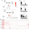
Expression signature of the blastoid mantle cell lymphoma case. Patient 1 had highly specific FAT1 expression (red, upper) among SOX11+ cells, while SOX4 expression (red, lower) was also found in pt. 3, 4, and 7 as well as in a small fraction of pro-/pre-B cells (A). A large fraction of these cells was also significant for an AHR+, CHD3+, DST+, SOX4+ expression (red) signature (B), providing potential evidence of a more immature cell type based on previous findings in lymphoblastic leukemia/lymphoma. While the blastoid patient had a very high fraction of malignant SOX4+ cells, comprising 19.9% of the SOX11+ population and no evidence of healthy pro-/pre-B cells, it was not specific for SOX4+ cells in general. Violin plots show the expression (y-axis) of genes in each patient (x-axis) (C).

Similar articles

Cited by

References

    1. Jain P, Wang M. Mantle cell lymphoma: 2019 update on the diagnosis, pathogenesis, prognostication, and management. Am. J. Hematol. 2019;94(6):710–725. doi: 10.1002/ajh.25487. - DOI - PubMed
    1. Veloza L, Ribera-Cortada I, Campo E. Mantle cell lymphoma pathology update in the 2016 WHO classification. Ann. Lymphoma. 2019;3:1–17. doi: 10.21037/aol.2019.03.01. - DOI
    1. Tsujimoto Y, Jaffe E, Cossman J, Gorham J, Nowell PC, Croce CM. Clustering of breakpoints on chromosome 11 in human B-cell neoplasms with the t(11;14) chromosome translocation. Nature. 1985;315(6017):340–343. doi: 10.1038/315340a0. - DOI - PubMed
    1. Jares P, Colomer D, Campo E. Genetic and molecular pathogenesis of mantle cell lymphoma: Perspectives for new targeted therapeutics. Nat. Rev. Cancer. 2007;7(10):750–762. doi: 10.1038/nrc2230. - DOI - PubMed
    1. Salaverria I, Royo C, Carvajal-Cuenca A, Clot G, Navarro A, Valera A, et al. CCND2 rearrangements are the most frequent genetic events in cyclin D1(-) mantle cell lymphoma. Blood. 2013;121(8):1394–1402. doi: 10.1182/blood-2012-08-452284. - DOI - PMC - PubMed

Publication types

Substances