. 2021 Sep 17;12(9):1434.
doi: 10.3390/genes12091434.

Meiotic Behavior of Achiasmate Sex Chromosomes in the African Pygmy Mouse Mus mattheyi Offers New Insights into the Evolution of Sex Chromosome Pairing and Segregation in Mammals

Affiliations

Meiotic Behavior of Achiasmate Sex Chromosomes in the African Pygmy Mouse Mus mattheyi Offers New Insights into the Evolution of Sex Chromosome Pairing and Segregation in Mammals

Ana Gil-Fernández et al. Genes (Basel). .

Abstract

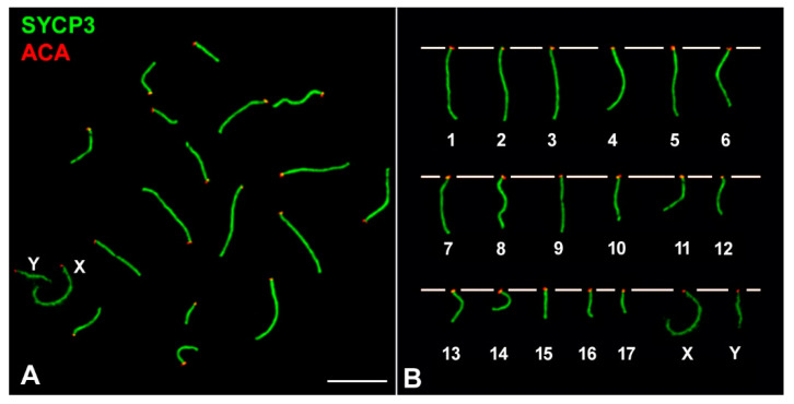
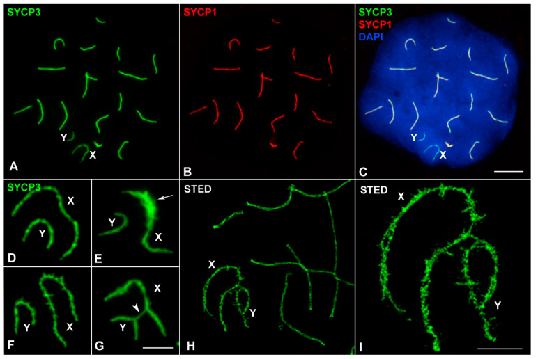
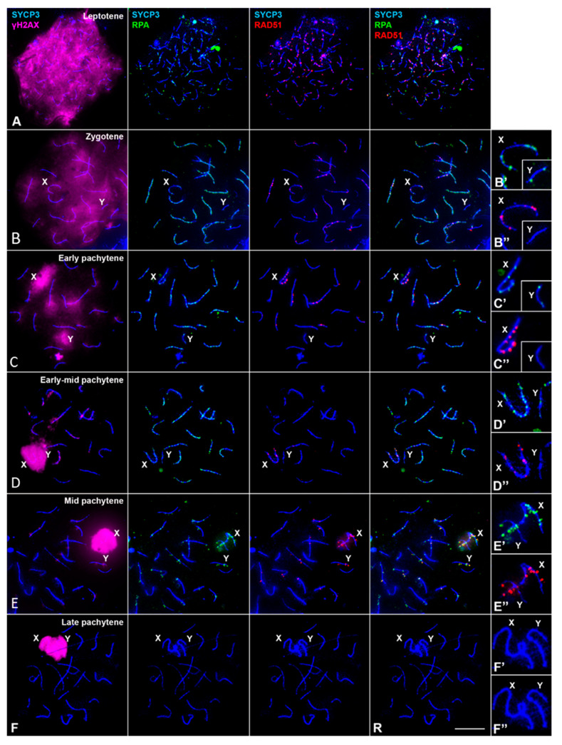
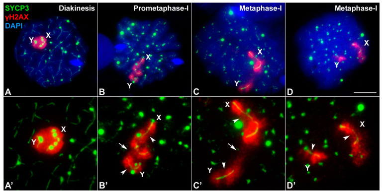
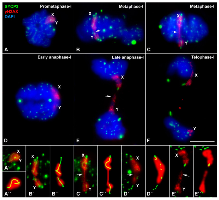
X and Y chromosomes in mammals are different in size and gene content due to an evolutionary process of differentiation and degeneration of the Y chromosome. Nevertheless, these chromosomes usually share a small region of homology, the pseudoautosomal region (PAR), which allows them to perform a partial synapsis and undergo reciprocal recombination during meiosis, which ensures their segregation. However, in some mammalian species the PAR has been lost, which challenges the pairing and segregation of sex chromosomes in meiosis. The African pygmy mouse Mus mattheyi shows completely differentiated sex chromosomes, representing an uncommon evolutionary situation among mouse species. We have performed a detailed analysis of the location of proteins involved in synaptonemal complex assembly (SYCP3), recombination (RPA, RAD51 and MLH1) and sex chromosome inactivation (γH2AX) in this species. We found that neither synapsis nor chiasmata are found between sex chromosomes and their pairing is notably delayed compared to autosomes. Interestingly, the Y chromosome only incorporates RPA and RAD51 in a reduced fraction of spermatocytes, indicating a particular DNA repair dynamic on this chromosome. The analysis of segregation revealed that sex chromosomes are associated until metaphase-I just by a chromatin contact. Unexpectedly, both sex chromosomes remain labelled with γH2AX during first meiotic division. This chromatin contact is probably enough to maintain sex chromosome association up to anaphase-I and, therefore, could be relevant to ensure their reductional segregation. The results presented suggest that the regulation of both DNA repair and epigenetic modifications in the sex chromosomes can have a great impact on the divergence of sex chromosomes and their proper transmission, widening our understanding on the relationship between meiosis and the evolution of sex chromosomes in mammals.

Keywords: Mus mattheyi; evolution; meiosis; pygmy mouse; sex chromosomes.


Conflict of interest statement

The authors declare no conflict of interest. The funders had no role in the design of the study; in the collection, analyses, or interpretation of data; in the writing of the manuscript, or in the decision to publish the results.

Figures

Figure 1
Figure 1
Chromosomes of Mus mattheyi (A) Spread spermatocyte at pachytene labelled with antibodies against SYCP3 (green) and centromeres (red). (B) Meiotic karyotype. Bivalents are arranged according to their length. The sex chromosomes (X, Y) are indicated. Scale bar: 10 µm.
Figure 2
Figure 2
Prophase I progression in M. mattheyi spermatocytes. SYCP3 (green) and γH2AX (red). (A) Leptotene. Axial elements (AEs) appear as short filaments and γH2AX labelling appears as large foci scattered over the nucleus. (B) Early zygotene. AEs associate in some regions. γH2AX signal occupies the entire nucleus. (C) Late zygotene. Most autosomes have completed synapsis. γH2AX labelling still covers most of the nucleus. Sex chromosomes (X, Y) are distinguishable from the rest of chromosomes as thinner filaments. The Y chromosome has a single focus of γH2AX while the X is intensely labelled. (D) Early pachytene. All autosomes have completed synapsis, but large γH2AX foci remain associated with many of them. The AEs of sex chromosomes remain apart and surrounded by a large γH2AX signal. (E) Early pachytene. Sex chromosomes are completely separated from each other. An intense and irregular γH2AX signal surrounds each sex chromosome. (F) Early-mid pachytene. γH2AX signal in the autosomes appears as small foci closely associated to the SCs. The AE of the X chromosome is bent and the signal of γH2AX is more concentrated around the AE in both the X and the Y chromosomes. (G) Mid pachytene. γH2AX remains in many autosomes as small foci. Sex chromosomes appear closely associated inside a single γH2AX signal, which shows a compact appearance and a well-defined outline. No contact between their AEs is observed. (H) Late pachytene. Sex chromosomes remain associated but without contact of their AEs. γH2AX is only observed in the sex body. (I) Diplotene. Autosomes desynapse and remain associated only in some regions. Sex chromosomes remain associated inside a sex body intensely labelled with γH2AX. Scale bar: 10 µm.
Figure 3
Figure 3
Structural organization of sex chromosomes in prophase-I. (AC) Mid pachytene spermatocyte labelled with SYCP3 (green), SYCP1 (red) and DAPI (blue). SYCP1 is absent in the sex chromosomes. A well-defined sex body is observed with DAPI staining. (DG) Details of the AEs of the sex chromosomes. (D) AEs without obvious modifications. (E) The AE of the X chromosome is thickened in an interstitial region (arrow). (F) Lateral excrescences are observed in both sex chromosomes. (G) Bridges of SYCP3 are occasionally observed between the AEs of sex chromosomes (arrowhead). (H) A pachytene spermatocyte observed with super-resolution STED confocal microscopy. The two LEs of autosomes are distinguishable. (I) Enlarged image showing sex chromosomes. The AEs of both chromosomes are irregular and show profuse excrescences and splitting into two filaments along their trajectory. Scale bars: 10 µm in (AC); 5 µm in (DI).
Figure 4
Figure 4
Distribution of RPA and RAD51 during prophase-I in spermatocyte spreads. First column on the left: SYCP3 (blue) and γH2AX (magenta); second column: SYCP3 (blue) and RPA (green); third column: SYCP3 (blue) and RAD51 (red); fourth column: SYCP3 (blue), RPA (green) and RAD51 (red); right column: enlarged details of sex chromosomes for each row. Staging was determined according to SYCP3 and γH2AX labelling patterns, in agreement with Figure 2. (A) Leptotene: RPA and RAD51 foci are observed throughout the nucleus associated to forming AEs. (B) Zygotene: RPA foci are distributed along synapsed and unsynapsed regions of chromosomes. RAD51 foci are less abundant and mainly localize to unsynapsed regions. Some foci overlap while others do not. Sex chromosomes are distinguishable. The X chromosome shows abundant RPA and RAD51 foci, but the Y chromosome only shows a few RPA foci and rarely accumulates RAD51 foci. (C) Early pachytene. The number of RPA and RAD51 foci decreases in the autosomes and the X chromosome. (D) Early-mid pachytene. The number of RPA foci is similar to the previous stage, but RAD51 is almost undetectable, except in the X chromosome. (E) Mid pachytene. The number of RPA foci further decreases in the autosomes. However, the number of RAD51 foci is conspicuously higher than in the previous stage. Many of these foci appear located out of the SCs in some autosomes. Many RPA foci are observed in the X chromosome, while only a few are detectable in the Y chromosome. Most RPA and RAD51 foci do not colocalize. (F) Late Pachytene. RPA and RAD512 are not detected in either the autosomes or the sex chromosomes. Scale bar: 10 µm.
Figure 5
Figure 5
Distribution of MLH1 in a pachytene spermatocyte. SYCP3 (blue), MLH1 (green) and centromeres (red). All autosomes show at least one MLH1 focus. Sex chromosomes do not have any focus. Scale bar: 10 µm.
Figure 6
Figure 6
Association of sex chromosomes during late stages of first meiotic division. Spread spermatocytes labelled with antibodies against SYCP3 (green) and γH2AX (red) and counterstained with DAPI (blue). (A′D′) shows enlarged images of the sex chromosomes presented in (AD). (A,A′) Diakinesis: SYCP3 appears as thin lines along each bivalent. Some protein aggregates are observed, particularly in the sex chromosomes. γH2AX signal covers the sex chromosomes, which are closely associated. (B,B′) Prometaphase-I. SYCP3 is observed as a discontinuous line inside each bivalent, and abundant SYCP3 aggregates decorate the nucleus. SYCP3 is also observed running inside the X and Y chromosome. The X chromosome shows a conspicuous bending, adopting a hook-like shape. The γH2AX signal in the sex chromosomes becomes discernible for each chromosome, although both chromosomes are still closely associated (arrow). Some regions along sex chromosomes have a weaker labelling with γH2AX (arrowheads). (C) Metaphase-I. SYCP3 signal becomes weaker inside the autosomes and preferentially accumulates in the centromeric regions. Sex chromosomes are clearly more separated than in previous stages, but there is a γH2AX bridge connecting them (arrow). The X chromosome still has a hook-like configuration. (D) Metaphase-I. SYCP3 signal is barely detectable inside the autosomes and mostly accumulates in the centromeric regions. Sex chromosomes have apparently lost their contact. γH2AX weaker regions are still differentiated in the sex chromosomes (arrowheads). Scale bar: 10 µm.
Figure 7
Figure 7
Segregation of sex chromosomes during first meiotic division. Squashed spermatocytes labelled with antibodies against SYCP3 (green) and γH2AX (red) and counterstained with DAPI (blue). Lower row shows enlarged images of the sex chromosomes presented in (AE). (A,A′) Prometaphase-I. SYCP3 signal is observed in the autosomes, while γH2AX marks sex chromosomes, which are tightly associated at the nucleus periphery. (B,B′) Metaphase-I. Bivalents appear aligned at the metaphase plate. SYCP3 in autosomes accumulates especially in centromeric regions and in some large scattered foci. Sex chromosomes are oriented to opposite poles and appear associated and labelled with γH2AX, which is continuous between both chromosomes (arrow). The hook-like configuration of the X chromosome is discernible. (C,C′) Metaphase-I. Autosomal bivalents are aligned at the cell equator, while the sex chromosomes appear further apart. A γH2AX filament bridges from the X to the Y chromosome (arrow). (D,D) Early anaphase-I. Autosomes start migration. SYCP3 is observed as aggregates in the centromeric regions and as large accumulation in the cytoplasm. Sex chromosomes initiate their segregation, but a γH2AX contact is still observed (arrow). (E,E′) Late anaphase-I. Two chromatin masses appear clearly separated. A chromatin connection between these two chromosome groups is observed (arrow), most probably representing sex chromosomes. (F) Telophase-I. Chromosomes reach the cell poles. A γH2AX signal is detected in each pole, but without connection between them. (A″E″) shows enlarged images of the sex chromosomes presented in (A′E′). Scale bar: 10 µm.

Similar articles

Cited by

References

    1. John B. Meiosis. Cambridge University Press; Cambridge, MA, USA: 1990.
    1. Page S.L., Hawley R.S. The genetics and molecular biology of the synaptonemal complex. Annu. Rev. Cell Dev. Biol. 2004;20:525–558. doi: 10.1146/annurev.cellbio.19.111301.155141. - DOI - PubMed
    1. von Wettstein D., Rasmussen S.W., Holm P.B. The synaptonemal complex in genetic segregation. Annu. Rev. Genet. 1984;18:331–413. doi: 10.1146/annurev.ge.18.120184.001555. - DOI - PubMed
    1. Keeney S., Neale M.J. Initiation of meiotic recombination by formation of DNA double-strand breaks: Mechanism and regulation. Biochem. Soc. Trans. 2006;34:523–525. doi: 10.1042/BST0340523. - DOI - PubMed
    1. Grey C., de Massy B. Chromosome Organization in Early Meiotic Prophase. Front. Cell Dev. Biol. 2021;9:688878. doi: 10.3389/fcell.2021.688878. - DOI - PMC - PubMed

Publication types