. 2021 Sep 17;10(9):2448.
doi: 10.3390/cells10092448.

Dynamic Expression of Membrane Type 1-Matrix Metalloproteinase (Mt1-mmp/Mmp14) in the Mouse Embryo

Affiliations

Dynamic Expression of Membrane Type 1-Matrix Metalloproteinase (Mt1-mmp/Mmp14) in the Mouse Embryo

Emma Muñoz-Sáez et al. Cells. .

Abstract

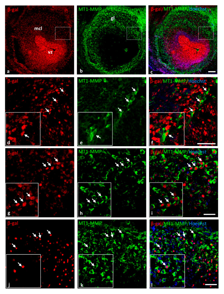
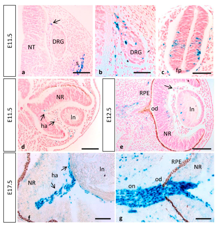
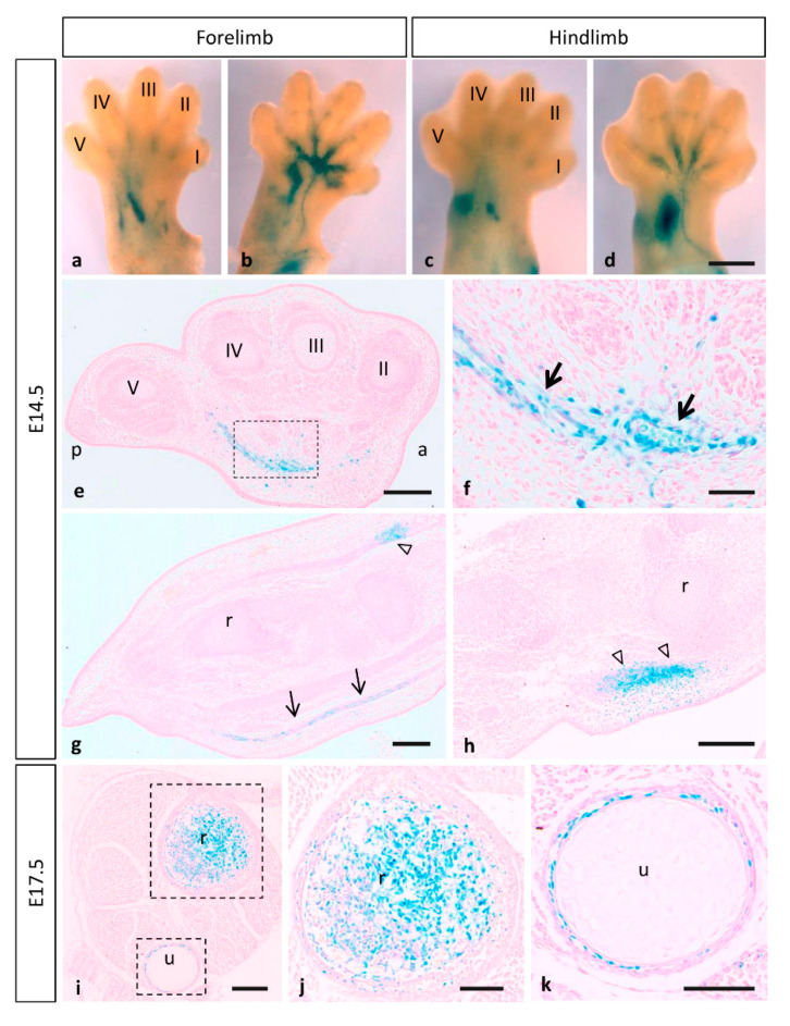
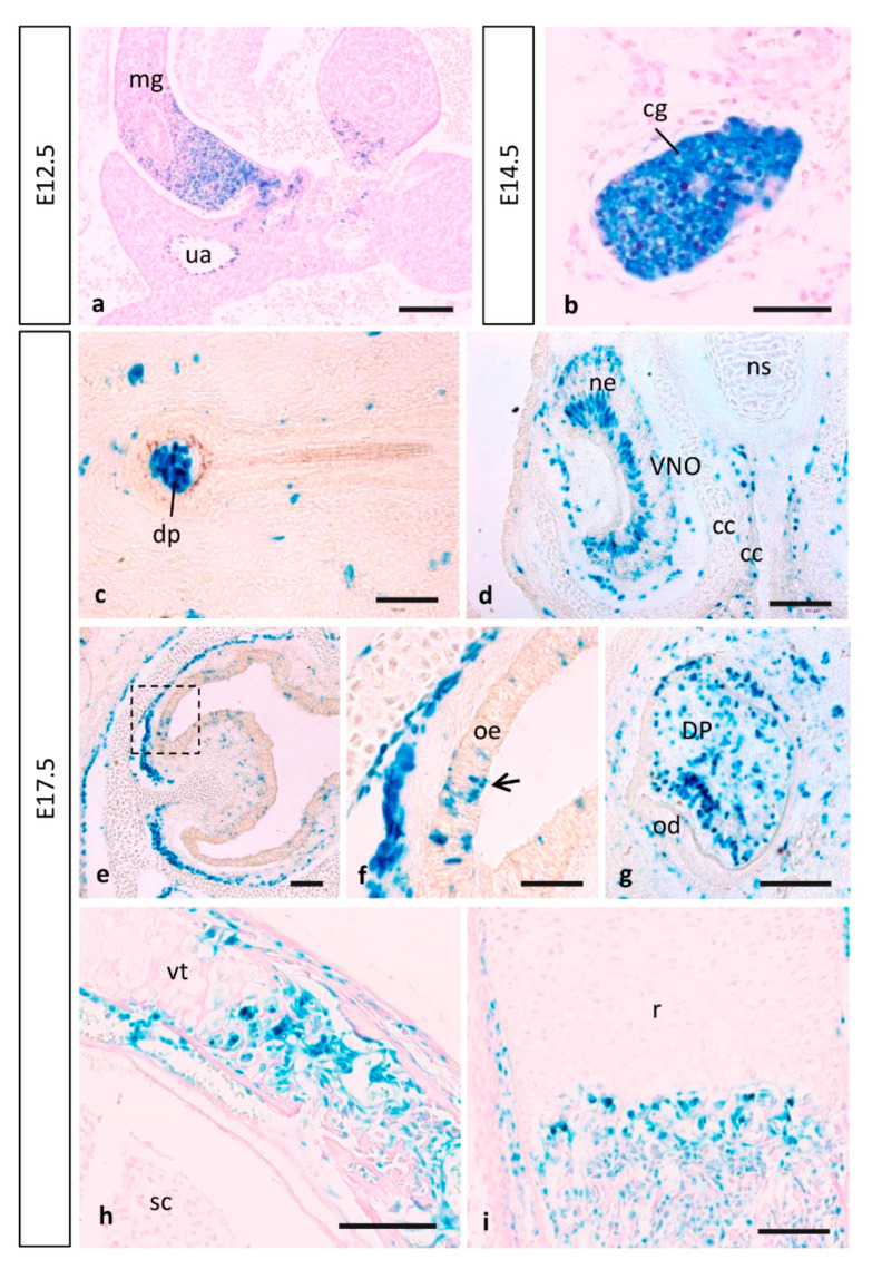
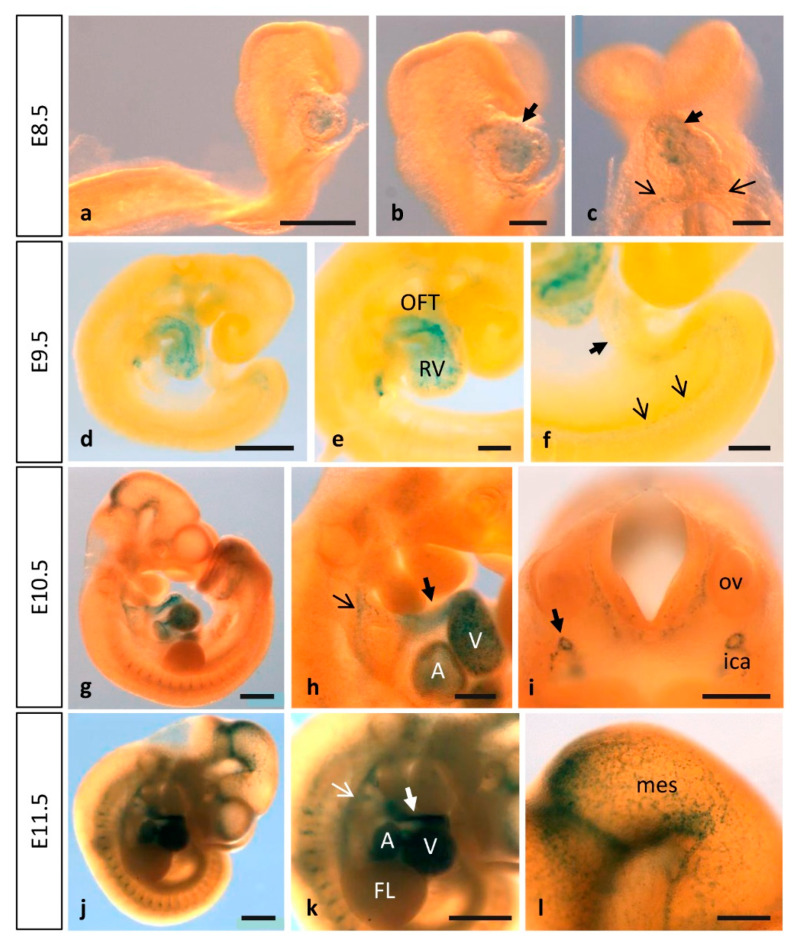
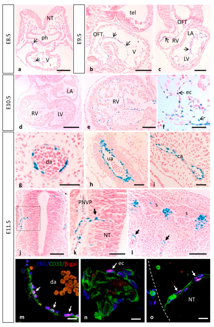
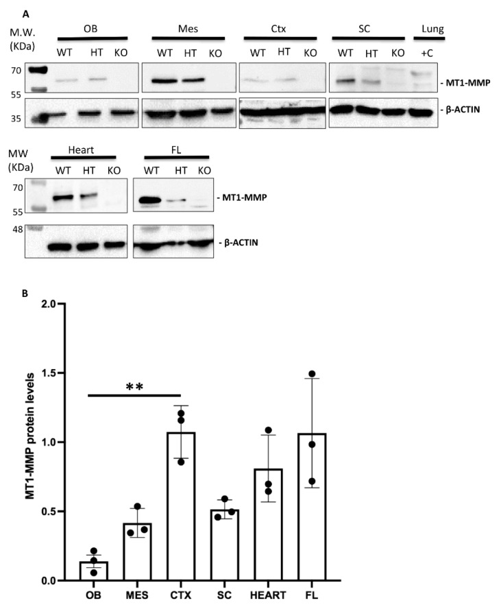
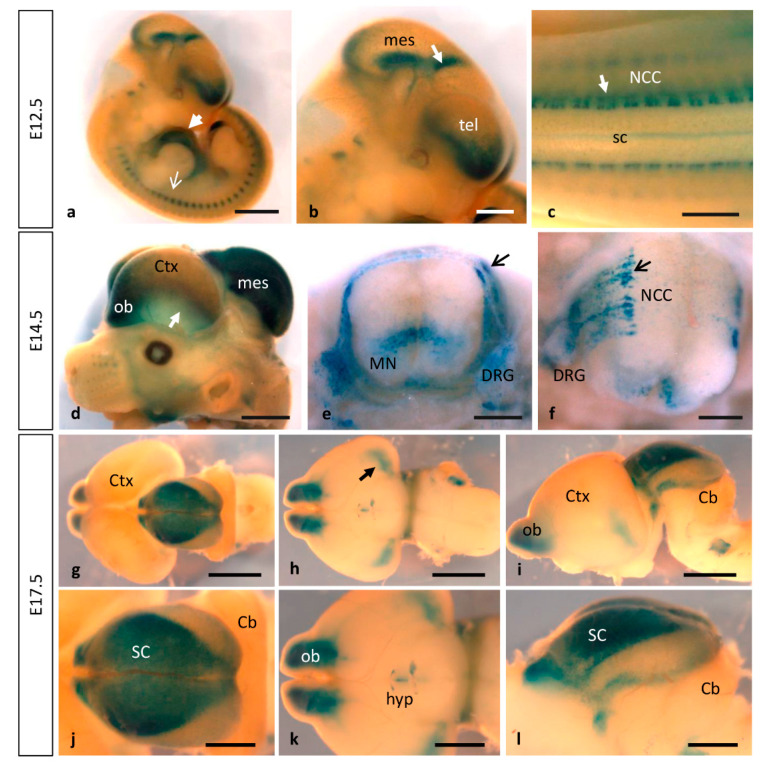
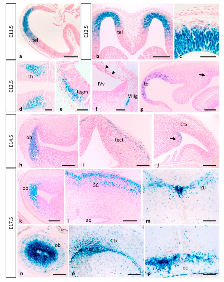
MT1-MMP/MMP14 belongs to a subgroup of the matrix metalloproteinases family that presents a transmembrane domain, with a cytosolic tail and the catalytic site exposed to the extracellular space. Deficient mice for this enzyme result in early postnatal death and display severe defects in skeletal, muscle and lung development. By using a transgenic line expressing the LacZ reporter under the control of the endogenous Mt1-mmp promoter, we reported a dynamic spatiotemporal expression pattern for Mt1-mmp from early embryonic to perinatal stages during cardiovascular development and brain formation. Thus, Mt1-mmp shows expression in the endocardium of the heart and the truncus arteriosus by E8.5, and is also strongly detected during vascular system development as well as in endothelial cells. In the brain, LacZ reporter expression was detected in the olfactory bulb, the rostral cerebral cortex and the caudal mesencephalic tectum. LacZ-positive cells were observed in neural progenitors of the spinal cord, neural crest cells and the intersomitic region. In the limb, Mt1-mmp expression was restricted to blood vessels, cartilage primordium and muscles. Detection of the enzyme was confirmed by Western blot and immunohistochemical analysis. We suggest novel functions for this metalloproteinase in angiogenesis, endocardial formation and vascularization during organogenesis. Moreover, Mt1-mmp expression revealed that the enzyme may contribute to heart, muscle and brain throughout development.

Keywords: MMP14; Mt1-mmp; angiogenesis; brain; cardiovascular development; dorsal aorta; embryo development; expression pattern; limb; metalloproteinases.


Conflict of interest statement

The authors declare no conflict of interest.

Figures

Figure 6
Figure 6
MT1-MMP is expressed in the olfactory bulb, the cerebral cortex and the superior colliculus of E14.5 HT embryos. Double-labeling immunohistochemistry for β-gal (in red) and MT1-MMP (in green) confirmed the expression of the enzyme and the reporter β-gal (white arrows) in the olfactory bulb (af), the cerebral cortex (gi) and the superior colliculus (jl). Abbreviations: gl: glomerular layer; mcl: mitral cell layer; vz: ventricular zone. Scale bars: 100 µm (ac) and 50 µm (dl).
Figure 7
Figure 7
MT1-MMP expression in the embryonic spinal cord and eye. (a,b) MT1-MMP expression was detected in the spinal cord particularly in neural crest cells, somites, the dorsal nerve root and the dorsal root ganglia at E11.5. Reporter expression was also observed in neural precursors of the spinal cord and the floor plate (c). (d,e) In the developing eye, β-gal positive cells localize in the hyaloid artery at E11.5 (arrows in (b)) and E12.5 (arrow in (e)). Further, scattered cells were detected in the lens and the neural retina by E12.5 (e). Later in development, MT1-MMP expression increases in the hyaloid artery and is present in cells surrounding the optic nerve and at the level of the optic disc and optic nerve head, possibly corresponding to glial cells (f,g). Abbreviations: DRG, dorsal root ganglia; fp, floor plate; ha, hyaloid artery; od, optic disc; on, optic nerve; ln, lens; NR, neural retina; NT, neural tube; RPE, retinal pigmented epithelium. Scale bars: 100 µm (ag).
Figure 8
Figure 8
Mt1-mmpLacZ/+ distribution during mouse limb development. Whole mount forelimbs ((a), dorsal side; (b), ventral side) and hindlimbs ((c), dorsal side; (d), ventral side) stained with β-gal show MT1-MMP expression restricted to the vasculature and muscles of the limb at E14.5. (eh) Paraffin cross (e,f) and longitudinal sections (g,h) through the hindlimb showed β-gal positive cells in endothelial cells of blood vessels entering the limbs (arrows in (f,g)) and dorsally in the earliest muscle blocks (open arrowheads in (g,h)) at E14.5. (ik) Limb crossed sections at the level of proximal radius and ulna revealed LacZ expressing cells related to the ossification within the cartilage primordium of these bones at E17.5. Abbreviations: a, anterior; p, posterior; r, radius; u, ulna; digits I, II, III, IV and V. Scale bars: 500 µm (ad), 200 µm (e,gi), 100 µm (j,k), 50 µm (f).
Figure 9
Figure 9
MT1-MMP expression in other mouse embryonic structures. Reporter expression in Mt1-mmpLacZ/+ early embryos was detected in the midgut loop and the cervical sensory ganglia. At E17.5, β-gal positive cells localized in the dermal papilla of the hair follicle (c), in sensory neurons of the vomeronasal organ (d) and the olfactory epithelium (e,f), and also in the condensing dental mesenchyme. Notably, MT1-MMP expression was also detected during endochondral ossification in the vertebra (h) and the radius of the hindlimb (i) by this stage. Abbreviations: cc, cartilaginous capsule; cg, cervical sensory ganglia; dp, dermal papillae; DP, dental pulp; mg, midgut loop; ne, neurosensory epithelium; ns, nasal septum; od, odontoblast; oe, olfactory epithelium; r, radius; sc, spinal cord; ua, umbilical artery; VNO, vomeronasal organ; vt, vertebra. Scale bars: 100 mm (a,d,e,gi), 50 mm (b,c,f).
Figure 1
Figure 1
MT1-MMP is expressed in the early mouse embryo. Whole-mount β-gal staining in Mt1-mmpLacZ/+ embryos from E8.5 to E11.5. Staining is observed in the endocardium of the developing heart tube (ac) as early as E8.5 embryonic stage. At later stages of development, this expression persists and the strongest labeling is observed in the atrium, the ventricle, the cardiac outflow tract (black and white thick arrows in (h,k)), the aortic arch arteries (black and white thin arrows in (h,k)) and the dorsal aorta at E9.5 (df), E10.5 (gi) and E11.5 (jl). Expression in the central nervous system was first detected by E10.5 (g) and E11.5 (j) in the caudal midbrain and the rostral rhombencephalon (l). Furthermore, β-gal positive cells were detected in the perineural vascular plexus and the first blood vessels entering the brain (i,l). Somites also show β-gal positive cells at these embryonic stages (g,j,k). Abbreviations: A, atrium; FL, forelimb; ica, internal carotid artery; mes, mesencephalon; OFT, outflow tract; ov, otic vesicle; RV, right ventricle; V, ventricle. Scale bars: 1 mm (g,j,k), 500 µm (a,d,h,i,l), 200 µm (b,c,e,f).
Figure 2
Figure 2
MT1-MMP is expressed at early embryonic stages in the developing cardiovascular system. (af) β-gal staining of paraffin sections from E8.5 to E10.5 Mt1-mmpLacZ/+ embryos revealed expression of MT1-MMP in the endocardial tissue lining the primitive heart tube at early embryonic stages (arrows in (a)), and the endocardium of the atrium, the ventricle and the cardiac outflow tract at E9.5 (arrows in (b), sagittal section; arrows in (c), coronal section) and at E10.5 (df). (gl) Mt1-mmpLacZ/+ expression was detected in large blood vessels such as the dorsal aorta (g), the umbilical artery (h) and the carotid artery (i) of E11.5 embryos. At this embryonic stage, β-gal positive cells were detected in the perineural vascular plexus that surrounds the neural tube (j,k) and in somites (l), in a pattern compatible with intersomitic arteries (arrows in (l)). Note the β-gal labeling in endothelial cells of a blood vessel sprouting into the neural tube (arrow in (k)). (mo) Confocal images of immunohistochemistry for the endothelial transcription factor ERG (blue), the membrane marker for endothelial cells CD31 (green) and the reporter β-gal (red), demonstrate MT1-MMP expression in endothelial cells of the dorsal aorta (m) and the endocardial endothelium of the primitive heart tube (n) of E11.5 embryos. MT1-MMP expression was also demonstrated in the endothelial cells from the first blood vessels entering the brain parenchyma (o). See images from split channels in Figure S1. Abbreviations: ca, carotid artery; da, dorsal aorta; ec, endothelial cell; OFT, outflow tract; LA, left atrium; LV, left ventricle; NT, neural tube; ph, pharyngeal region; PNVP, perineural vascular plexus; RV, right ventricle; s, somite; tel, telencephalon; V, ventricle; ua, umbilical artery. Scale bars: 250 µm (d), 100 µm (ac,e,j,l), 50 µm (f,g,k,mo), 20 µm (h,i).
Figure 3
Figure 3
(A) Western blot analysis confirms MT1-MMP protein expression in distinct embryonic tissues. (A) Protein detection by Western blot of E14.5 embryonic tissues revealed MT1-MMP expression in the olfactory bulb (OB), mesencephalon (MES), cerebral cortex (CTX), spinal cord (SC), heart and forelimbs (FL). Specific bands correspond to the mature form of MT1-MMP (57 kDa) and β-actin (42 kDa). Adult lungs were used as a positive control (+C lane) due to the high expression of the protein in this tissue. As expected, MT1-MMP protein levels were significantly decreased in the heterozygous (HT) compared to the wild type (WT), and absent in the knock-out (KO) embryonic tissues. (B) Quantification of Western blot in WT embryonic tissues demonstrates higher levels of the active MT1-MMP in the cerebral cortex, heart and the forelimbs compared to the olfactory bulb, mesencephalon and spinal cord (** indicates significant differences at p < 0.01) (n = 3 per tissue). MT1-MMP protein levels were expressed as arbitrary units/50 µg protein.
Figure 4
Figure 4
Dynamic expression of MT1-MMP at later stages of brain and embryo development. As development proceeds, stronger pattern of MT1-MMP expression persists in the cardiovascular system and the brain. (ac) E12.5 Mt1-mmpLacZ/+ embryos show reporter expression in the developing heart (white arrowhead in a), somites (white arrow in (a)) and at distinct regions of the brain: the rostral telencephalon (the prospective olfactory bulb), zona limitans intrathalamica (white arrow in (b)), the floor plate and the caudal mesencephalon and neural crest cells (white arrow in (c)). (df) At E14.5, MT1-MMP expression increases in the olfactory bulb and the caudal cerebral cortex (arrow in a), the dorsal part of the mesencephalon and in neural progenitors and motoneurons of the spinal cord (e,f). Further, β-gal positive cells were located in streams of migrating neural crest cells (arrow in (e,f)), the dorsal nerve root and the dorsal root ganglia. (gl) Later in development, LacZ staining persists in the brain at E17.5: in the olfactory bulb, the amygdala (arrow in (h,k)), the hypothalamic region and the superior colliculus ((g,j), dorsal view; (h,k) ventral view; (i,l), lateral view of the brain). Abbreviations: Cb, cerebellum; Ctx, cerebral cortex; DRG, dorsal root ganglia; hyp, hypothalamic region; mes, mesencephalon; MN, motoneurons; NCC, neural crest cell; ob, olfactory bulb; sc, spinal cord; SC, superior colliculus; tel, telencephalon. Scale bars: 2 mm (a,d,gi), 1 mm (b,c,jl), 500 µm (e,f).
Figure 5
Figure 5
Expression of MT1-MMP in the developing mouse brain. β-gal staining of horizontal (ac,g,h,k) and coronal sections (n) from E11.5 to E17.5 Mt1-mmpLacZ/+ embryos revealed high expression of MT1-MMP in the ventricular zone of the rostral telencephalon at early embryonic stages (ac,g) that will give rise to the olfactory bulb later in development (h,k,n). In addition, β-gal positive cells were detected in the thalamus (d), the mesencephalon (e), the VIII nerve ganglia (f) and the meninges (arrowheads in f). At this embryonic stage, β-gal stained cells appear in the caudal portion of the telencephalon (arrow in g) where the amygdala will develop later (arrow in j) at E14.5. LacZ-positive cells were also located in the mesencephalic tectum of E14.5 embryos (i) and restricted to the superficial layers of the superior colliculus by E17.5 (l). By this perinatal stage, strong LacZ staining localized in the periventricular zone of the III ventricle and the zona limitans intrathalamica (m) and the optic chiasm region (p). Streams of β-gal positive cells entered the cerebral cortex by E17.5 (o). Abbreviations: aq, cerebral aqueduct; Ctx, cerebral cortex; ob, olfactory bulb; oc, optic chiasm; tect, mesencephalic tectum; tegm, mesencephalic tegmentum; SC, superior colliculus; tel, telencephalon; th, thalamus; vz, ventricular zone; IVv, IV ventricle; VIIIg, VIII nerve ganglia; ZLI, zona limitans intrathalamica. Scale bars: 250 µm (a,b), 200 µm (fp), 100 µm (d,e), 50 µm (c).

Similar articles

Cited by

References

    1. Seiki M. Membrane-type 1 matrix metalloproteinase: A key enzyme for tumor invasion. Cancer Lett. 2003;194:1–11. doi: 10.1016/S0304-3835(02)00699-7. - DOI - PubMed
    1. Itoh Y., Seiki M. MT1-MMP: A potent modifier of pericellular microenvironment. J. Cell. Physiol. 2006;206:1–8. doi: 10.1002/jcp.20431. - DOI - PubMed
    1. Itoh Y. Membrane-type matrix metalloproteinases: Their functions and regulations. Matrix Biol. 2015;44:207–223. doi: 10.1016/j.matbio.2015.03.004. - DOI - PubMed
    1. Seiki M. Membrane-type matrix metalloproteinases. APMIS. 1999;107:137–143. doi: 10.1111/j.1699-0463.1999.tb01536.x. - DOI - PubMed
    1. Moracho N., Learte A.I.R., Muñoz-Sáez E., Marchena M.A., Cid M.A., Arroyo A.G., Sánchez-Camacho C. Emerging roles of MT-MMPs in embryonic development. Dev. Dyn. 2021 doi: 10.1002/dvdy.398. Epub ahead of print. - DOI - PubMed

Publication types

Substances