. 2021 Nov 23;95(24):e0052921.
doi: 10.1128/JVI.00529-21. Epub 2021 Sep 22.

Asymmetric Structures and Conformational Plasticity of the Uncleaved Full-Length Human Immunodeficiency Virus Envelope Glycoprotein Trimer

Affiliations

Asymmetric Structures and Conformational Plasticity of the Uncleaved Full-Length Human Immunodeficiency Virus Envelope Glycoprotein Trimer

Shijian Zhang et al. J Virol. .

Abstract

The functional human immunodeficiency virus (HIV-1) envelope glycoprotein (Env) trimer [(gp120/gp41)3] is produced by cleavage of a conformationally flexible gp160 precursor. gp160 cleavage or the binding of BMS-806, an entry inhibitor, stabilizes the pretriggered, "closed" (state 1) conformation recognized by rarely elicited broadly neutralizing antibodies. Poorly neutralizing antibodies (pNAbs) elicited at high titers during natural infection recognize more "open" Env conformations (states 2 and 3) induced by binding the receptor, CD4. We found that BMS-806 treatment and cross-linking decreased the exposure of pNAb epitopes on cell surface gp160; however, after detergent solubilization, cross-linked and BMS-806-treated gp160 sampled non-state-1 conformations that could be recognized by pNAbs. Cryo-electron microscopy of the purified BMS-806-bound gp160 revealed two hitherto unknown asymmetric trimer conformations, providing insights into the allosteric coupling between trimer opening and structural variation in the gp41 HR1N region. The individual protomer structures in the asymmetric gp160 trimers resemble those of other genetically modified or antibody-bound cleaved HIV-1 Env trimers, which have been suggested to assume state-2-like conformations. Asymmetry of the uncleaved Env potentially exposes surfaces of the trimer to pNAbs. To evaluate the effect of stabilizing a state-1-like conformation of the membrane Env precursor, we treated cells expressing wild-type HIV-1 Env with BMS-806. BMS-806 treatment decreased both gp160 cleavage and the addition of complex glycans, implying that gp160 conformational flexibility contributes to the efficiency of these processes. Selective pressure to maintain flexibility in the precursor of functional Env allows the uncleaved Env to sample asymmetric conformations that potentially skew host antibody responses toward pNAbs. IMPORTANCE The envelope glycoprotein (Env) trimers on the surface of human immunodeficiency virus (HIV-1) mediate the entry of the virus into host cells and serve as targets for neutralizing antibodies. The functional Env trimer is produced by cleavage of the gp160 precursor in the infected cell. We found that the HIV-1 Env precursor is highly plastic, allowing it to assume different asymmetric shapes. This conformational plasticity is potentially important for Env cleavage and proper modification by sugars. Having a flexible, asymmetric Env precursor that can misdirect host antibody responses without compromising virus infectivity would be an advantage for a persistent virus like HIV-1.

Keywords: Env; antibody; asymmetry; cleavage; conformation; cryo-electron microscopy; furin; processing; structure.


Figures

FIG 1
FIG 1
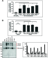
Antibody recognition of cleaved and uncleaved HIV-1 Envs on the cell surface. (A) HOS cells transiently expressing the wild-type HIV-1JR-FL Env, a fraction of which is cleaved in these cells, were incubated with the indicated broadly neutralizing antibodies or poorly neutralizing antibodies. After washing and lysis of the cells, the antibody-Env complexes were purified with protein A-Sepharose beads and analyzed by Western blotting with a goat anti-gp120 polyclonal serum. (B) The effect of cross-linking with BS3 and/or BMS-806 treatment on antibody binding to HIV-1JR-FL Env(−) on the surface of CHO cells was evaluated by cell-based ELISA. BMS-806 (10 μM) was added to the CHO cells at the time of induction of Env(−) expression with doxycycline. All values were normalized against 2G12 antibody binding and were derived from at least three independent experiments. Note that the HIV-1JR-FL Env(−) glycoprotein is not recognized by the PGT145 V2 quaternary antibody, which serves as a negative control.
FIG 2
FIG 2
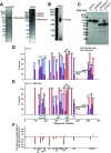
Characterization of the full-length HIV-1JR-FL Env(−) glycoprotein in CHO cell lysates and in detergent-solubilized purified forms. (A) Purified HIV-1JR-FL Env(−) without and with cross-linking by BS3 was run on a NUPAGE 4-to-12% BT gel stained by Coomassie blue. (B) Purified HIV-1JR-FL Env(−) cross-linked by BS3 was run on a native PAGE 4-to-16% Bis-Tris gel and subjected to Western blotting with an HRP-conjugated anti-HIV-1 gp120 antibody. (C to E) To evaluate the effect of BMS-806 on the glycosylation of the synthesized Env(−) glycoprotein, BMS-806 (10 μM) was added to the CHO cells at the time of doxycycline induction. (C) The effect of BMS-806 on HIV-1JR-FL Env(−) glycosylation was evaluated by Western blotting after digestion with glycosidases (sialidase, peptide-N-glycosidase F [PNGase F], and endoglycosidase H [Endo H]). The purified HIV-1JR-FL Env(−) glycoproteins were digested with the indicated glycosidase, run on a NUPAGE 4-to-12% Bis-Tris gel, and subjected to Western blotting with a goat anti-HIV-1 gp120 antiserum. The results shown are representative of those obtained in three independent experiments. Note that BMS-806 treatment decreases Env(−) heterogeneity by reducing the levels of sialidase-sensitive and Endo H-resistant glycoforms. (D and E) The bar graphs show the glycan profiles at each glycosylation site of HIV-1JR-FL Env(−) purified from untreated CHO cells (D) or CHO cells treated with 10 μM BMS-806 (E), as determined by mass spectrometry. The glycan composition (in percent) was broadly characterized as high-mannose (red bars) or processed (complex plus hybrid) glycans (blue bars). (F) The results in panels D and E were used to calculate the change in the percentage of processed glycans after BMS-806 treatment, which is shown for each N-linked glycosylation site.
FIG 3
FIG 3
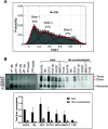
Conformations of purified HIV-1JR-FL Env(−) treated with BMS-806 and cross-linked with BS3. (A) HIV-1JR-FL Env(−) with V1 and V4 labeling tags was purified from 293T cell membranes using a protocol identical to that used for preparation of Env(−) for cryo-EM imaging. The purified Env(−) was labeled and analyzed by smFRET. FRET trajectories were compiled into a population FRET histogram and fit to the Gaussian distributions associated with each conformational state, according to a hidden Markov model (42). The percentage of the population that occupies each state as well as the number of molecules analyzed (N) is shown. The error bars represent the standard deviation from three independent data sets. (B) Membranes from BMS-806-treated CHO cells expressing HIV-1JR-FL Env(−) were cross-linked with BS3 and then solubilized in Cymal-5 detergent. The lysate was successively incubated with the 19b anti-gp120 (V3) antibody and protein A-Sepharose beads. The Env(−) glycoproteins precipitated by the 19b antibody or by the negative-control protein A-Sepharose beads during the indicated rounds of immunoprecipitation were analyzed by SDS-PAGE and Western blotting (upper left panel). The Env(−) glycoproteins in the initial cell membrane lysate (input) and those glycoproteins remaining after four rounds of 19b counterselection were precipitated with Ni-NTA beads or the indicated antibodies; the precipitated Env(−) glycoproteins were analyzed by SDS-PAGE and Western blotting (upper right panel). The total amounts of Env(−) glycoprotein in the input and after 19b counterselection, normalized to the input Env(−) glycoprotein amount precipitated by the Ni-NTA beads, are shown in the bar graph (lower panel). Means and standard deviations derived from two independent experiments are shown.
FIG 4
FIG 4
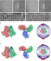
Cryo-EM analysis of the full-length HIV-1JR-FL Env(−) trimer. (A) A typical cryo-EM micrograph of Env(−) trimers taken with a Gatan K2 direct electron detector at 0 degrees of tilt. (B) Fourier transform of the image in panel A. Left panel, simulated logarithmic amplitude spectra in Gctf (134); upper right panel, background-subtracted logarithmic amplitude spectra; lower right panel, equiphase average in Gctf. (C) Unsupervised 2D class averages for nontilt particles. (D) A typical cryo-EM micrograph of Env(−) trimers taken with a Gatan K2 direct electron detector at 45 degrees of tilt. (E) Fourier transform of the image in panel D. Left panel, simulated logarithmic amplitude spectra in Gctf (134); upper right panel, background-subtracted logarithmic amplitude spectra; lower right panel, equiphase average in Gctf. (F) Unsupervised 2D class averages for tilted particles. (G) Final refined cryo-EM density map for the state-U1 Env(−) trimers. Left, side view, with gp120 at the bottom of the figure and gp41 at the top. Middle, top view from the gp120 side. The right inset shows the orientation distribution of the particles used for reconstruction of the final state-U1 map. (H) Final refined cryo-EM density map for the state-U2 Env(−) trimers. Left, side view, with gp120 at the bottom of the figure and gp41 at the top. Middle, top view from the gp120 side. The right inset shows the orientation distribution of the particles used for reconstruction of the final state-U2 map.
FIG 5
FIG 5
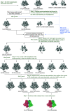
Cryo-EM classification workflow. The diagram illustrates the major steps of our classification strategy for the 300-kV data set. Iterated steps with the same parameters were omitted for clarity.
FIG 6
FIG 6
Cryo-EM density maps of the state-U1 and state-U2 Env(−) trimers. (A) Masks used for the FSC calculation. Masks for state-U1 and state-U2 maps were both generated in RELION 3.0 (136), using unmasked maps low-pass filtered to 10 Å. (B) Local resolution measurement of the state-U1 and state-U2 maps, as measured by ResMap (142). The maps are colored according to the local resolution, indicated by the color gradient (units in angstroms). Side views of the Env(−) maps are shown, with gp120 at the bottom of the figure and gp41 at the top. (C) Gold standard FSC plots of the state-U1 and state-U2 cryo-EM maps. The “map vs model” FSC curve was calculated with protein models without glycans.
FIG 7
FIG 7
Cross-linking solubilized and cell-surface Env(−) glycoproteins with BS3. (A) The percentage of solubilized BMS-806-treated Env(−) that cross-linked with BS3 into gel-stable trimers was calculated from the experiments shown in Fig. 3B. The values are reported for the Env(−) subset that was precipitated by the 19b pNAb and for the 19b-counterselected Env(−) subset precipitated by Ni-NTA beads or the indicated bNAbs. Differences between the values associated with the Env(−) subsets were evaluated by Student's t test (*, P < 0.05; **, P < 0.01). (B) BMS-806-treated CHO cells were induced to express HIV-1JR-FL Env(−) and incubated with BS3 cross-linker. The cells were washed and incubated with the indicated pNAbs or bNAbs. After washing, the cells were lysed. Cell lysates were incubated with protein A-agarose beads, and the precipitated Envs were analyzed by SDS-PAGE and Western blotting with a goat anti-gp120 antibody. In the lower left panel, the results of a typical experiment are shown. The Env(−) monomers (m), dimers (d), and trimers (t) are indicated. The percentage of monomers, dimers, and trimers recognized by each antibody was calculated from the results of two independent experiments (lower right panel). Means and standard deviations are shown. In the upper panel, the percentage of the total Env(−) that was cross-linked into gel-stable trimers is shown for each antibody. Statistical comparisons were made using Student's t test (*, P < 0.05; **, P < 0.01; ns, not significant).
FIG 8
FIG 8
Comparison of U1 and U2 Env(−) structures. (A) Protomer 2 of the state-U1 and state-U2 models are superposed, showing that protomer 1 and protomer 3 are rotated 4.0° and 2.8°, respectively. (B) Three protomers of the state-U1 model are superposed. (C) Three protomers of the state-U2 model are superposed. (D) With protomer 2 of the state-U1 and state-U2 models superposed, the Cα distances between the same residues on the U1 and U2 structures are measured for four residues (from i to iv, T90, D230, S481, and N392). In the side views of Env(−) shown in panels B to D, gp120 is at the bottom of the figure and gp41 is at the top.
FIG 9
FIG 9
Comparison of Env(−) structures with those of cleaved HIV-1 Envs. (A) Left, protomer 1 of the state-U1 trimer is superposed on the unliganded HIV-1BG505 sgp140 SOSIP.664 trimer (PDB ID 4ZMJ) (101), demonstrating how the other two protomers in state U1 are rotated toward each other. Right, side views of the superposed protomers, with red parts representing the major areas of difference between the two protomers. (B) Left, protomer 1 of the state-U1 trimer is superposed on the HIV-1JR-FL EnvΔCT trimer complexed with PGT151 Fabs (PDB ID 5FUU) (102), indicating that binding of the PGT151 antibodies introduces asymmetry into the Env trimer that differs from that of U1. Right, side views of the superposed protomers, with red parts representing the major areas of difference between the two protomers. In the side views of the Env protomers shown on the right in panels A and B, gp120 is at the bottom of the figure and gp41 at the top.
FIG 10
FIG 10
Comparison of Env trimer geometry among Env(−) trimers and mature Env trimers. (A) The interprotomer distances (in Å) between selected atoms of gp120 and gp41 are shown for different Env structures: the smaller, inner triangle depicts distances measured between gp41 residues W628 and I635; the larger, outer triangle depicts distances measured between gp120 residues A336 and Q352. The U1 and U2 structures are compared with those of the unliganded sgp140 SOSIP.664 trimer (PDB ID 4ZMJ) (101) and the PGT151-bound HIV-1JR-FL and HIV-1AMC011 EnvΔCT trimers (PDB IDs 5FUU and 6OLP, respectively) (102, 105). For 5FUU and 6OLP, the sides of the Env trimer that are bound by the PGT151 Fabs are marked. (B) The three gp120 subunits of four Env trimer atomic structures were superposed with the gp120 subunits of the state-U1 Env(−) trimer. Each protomer was aligned separately. After gp120 alignment, the relative positions of the gp41 HR1C helixes are jointly shown here. In each case, the U1 HR1C helices are colored cyan. With gp120 aligned, the gp41 in state U1 is displaced compared with the other structures. Upper row, top views of 3-helix bundles; bottom row, side views of 3-helix bundles. 5FYK is the structure of an HIV-1JR-FL sgp140 SOSIP.664 trimer complexed with several neutralizing antibody Fabs (63).
FIG 11
FIG 11
Relationship between HR1N helicity and the opening angle of the trimer. (A) Sequences of the gp41 HR1N region from three U1 protomers are shown, with residues in α-helices highlighted in red. (B) The relationship between HR1N helicity and the opening angle of three asymmetric HIV-1 Env trimers (U1 and two PGT151-Fab-bound EnvΔCT trimers (PDB ID 5FUU and 6OLP]) is shown. The x axis represents the opening angle for each of three sides, measured using the “angle_between_domains” command in PyMOL (141). The y axis represents the number of residues in an α-helical conformation for the HR1N region of that side. (C) The HR1N and HR1C regions from the three indicated atomic models are superposed. (D) The HR1N regions from the three protomers in state U1 are shown.
FIG 12
FIG 12
HIV-1JR-FL Env(−) glycan structure. Glycans on state-U1 trimers are colored according to the following scheme: glycans that exhibit significant decreases in the addition of processed glycans as a result of BMS-806 treatment are colored purple, high-mannose glycans are colored yellow, and the remaining mixed or processed glycans that are not affected by BMS-806 binding are colored green.
FIG 13
FIG 13
BMS-806 binding site. The BMS-806 binding sites within three protomers of the state-U1 structure (cyan) are compared with those in the BMS-806-bound sgp140 SOSIP.664 trimer (PDB 5U70) (green) (115).
FIG 14
FIG 14
Effect of BMS-806 on the synthesis, processing, and glycosylation of wild-type HIV-1AD8 Env. A549-Gag/Env cells were treated with BMS-806 (10 μM) or mock treated during doxycycline induction of Gag/Env expression. Lysates were prepared from cells (A) and supernatants containing virus-like particles (VLPs) (B) and were treated with peptide-N-glycosidase F (PNGase F) or endoglycosidase Hf (Endo Hf) or were mock treated (no Rx). The Envs were run on reducing SDS-polyacrylamide gels and analyzed by Western blotting. The deglycosylated gp160, gp120, and gp41 proteins (dgp160, dgp120, and dgp41, respectively) are indicated by arrows (red, PNGase F-treated sample; green, Endo Hf-treated sample). Data in this figure are representative of those obtained in two independent experiments.

Similar articles

Cited by

References

    1. Wyatt R, Sodroski J. 1998. The HIV-1 envelope glycoproteins: fusogens, antigens, and immunogens. Science 280:1884–1888. 10.1126/science.280.5371.1884. - DOI - PubMed
    1. Allan JS, Coligan JE, Barin F, McLane MF, Sodroski JG, Rosen CA, Haseltine WA, Lee TH, Essex M. 1985. Major glycoprotein antigens that induce antibodies in AIDS patients are encoded by HTLV-III. Science 228:1091–1094. 10.1126/science.2986290. - DOI - PubMed
    1. Robey WG, Safai B, Oroszlan S, Arthur LO, Gonda MA, Gallo RC, Fischinger PJ. 1985. Characterization of envelope and core structural gene products of HTLV-III with sera from AIDS patients. Science 228:593–595. 10.1126/science.2984774. - DOI - PubMed
    1. Klatzmann D, Champagne E, Chamaret S, Gruest J, Guetard D, Hercend T, Gluckman JC, Montagnier L. 1984. T-lymphocyte T4 molecule behaves as the receptor for human retrovirus LAV. Nature 312:767–768. 10.1038/312767a0. - DOI - PubMed
    1. Dalgleish AG, Beverley PC, Clapham PR, Crawford DH, Greaves MF, Weiss RA. 1984. The CD4 (T4) antigen is an essential component of the receptor for the AIDS retrovirus. Nature 312:763–767. 10.1038/312763a0. - DOI - PubMed

Publication types

Substances