

Molecular characterization of a complex of apoptosis-inducing factor 1 with cytochrome c oxidase of the mitochondrial respiratory chain
- PMID: 34548399
- PMCID: PMC8488679
- DOI: 10.1073/pnas.2106950118
Molecular characterization of a complex of apoptosis-inducing factor 1 with cytochrome c oxidase of the mitochondrial respiratory chain
Abstract
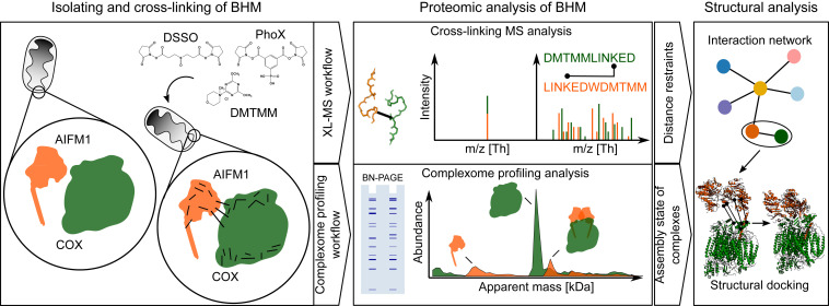
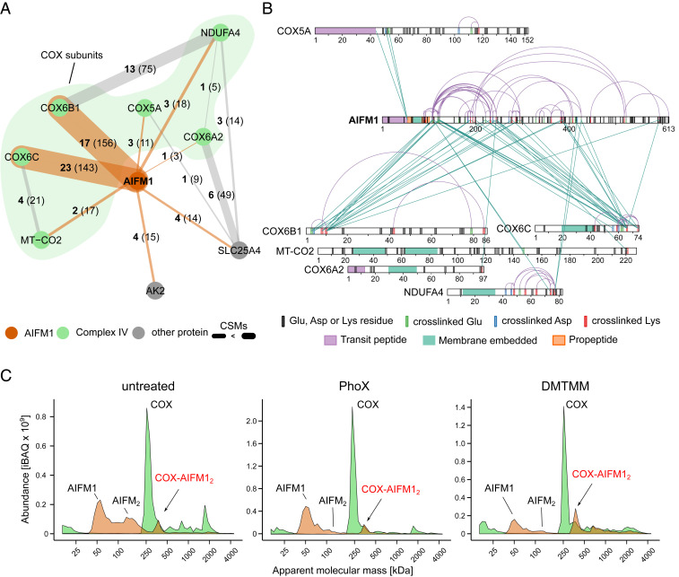
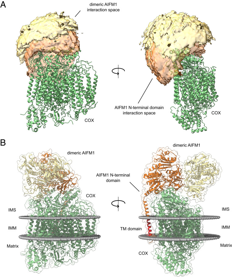
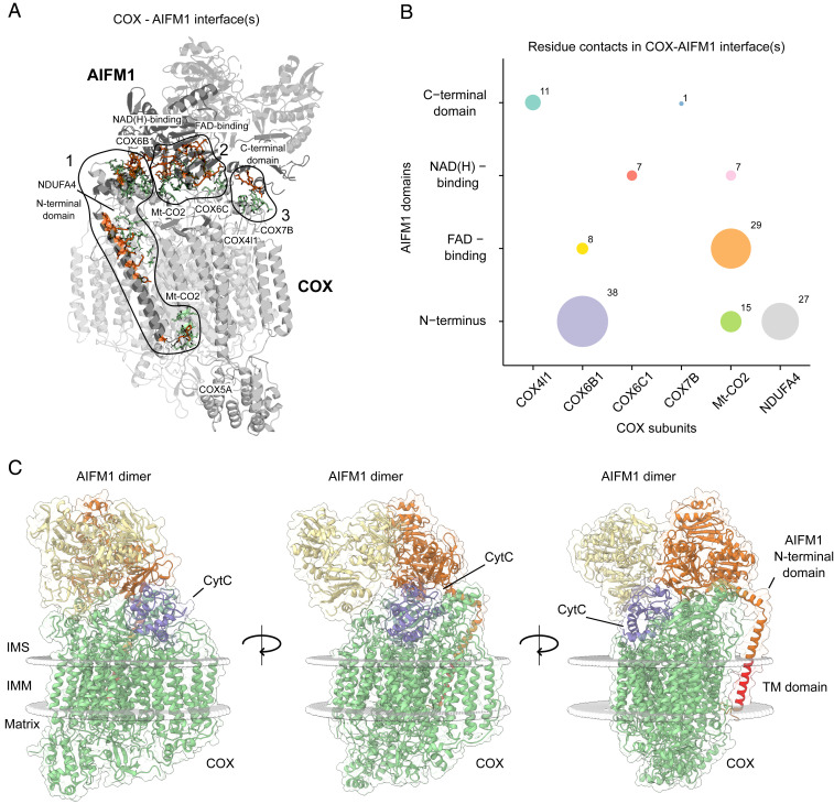
Combining mass spectrometry-based chemical cross-linking and complexome profiling, we analyzed the interactome of heart mitochondria. We focused on complexes of oxidative phosphorylation and found that dimeric apoptosis-inducing factor 1 (AIFM1) forms a defined complex with ∼10% of monomeric cytochrome c oxidase (COX) but hardly interacts with respiratory chain supercomplexes. Multiple AIFM1 intercross-links engaging six different COX subunits provided structural restraints to build a detailed atomic model of the COX-AIFM12 complex (PDBDEV_00000092). An application of two complementary proteomic approaches thus provided unexpected insight into the macromolecular organization of the mitochondrial complexome. Our structural model excludes direct electron transfer between AIFM1 and COX. Notably, however, the binding site of cytochrome c remains accessible, allowing formation of a ternary complex. The discovery of the previously overlooked COX-AIFM12 complex and clues provided by the structural model hint at potential roles of AIFM1 in oxidative phosphorylation biogenesis and in programmed cell death.
Keywords: AIFM1; COX; complexome profiling; cross-linking mass spectrometry; mitochondria.
Copyright © 2021 the Author(s). Published by PNAS.
Conflict of interest statement
The authors declare no competing interest.
Figures
Similar articles
-
COX7A2L Is a Mitochondrial Complex III Binding Protein that Stabilizes the III2+IV Supercomplex without Affecting Respirasome Formation.Cell Rep. 2016 Aug 30;16(9):2387-98. doi: 10.1016/j.celrep.2016.07.081. Epub 2016 Aug 18. Cell Rep. 2016. PMID: 27545886 Free PMC article.
-
Initiation of electron transport chain activity in the embryonic heart coincides with the activation of mitochondrial complex 1 and the formation of supercomplexes.PLoS One. 2014 Nov 26;9(11):e113330. doi: 10.1371/journal.pone.0113330. eCollection 2014. PLoS One. 2014. PMID: 25427064 Free PMC article.
-
Arrangement of electron transport chain components in bovine mitochondrial supercomplex I1III2IV1.EMBO J. 2011 Sep 9;30(22):4652-64. doi: 10.1038/emboj.2011.324. EMBO J. 2011. PMID: 21909073 Free PMC article.
-
AIFM1 beyond cell death: An overview of this OXPHOS-inducing factor in mitochondrial diseases.EBioMedicine. 2022 Sep;83:104231. doi: 10.1016/j.ebiom.2022.104231. Epub 2022 Aug 19. EBioMedicine. 2022. PMID: 35994922 Free PMC article. Review.
-
The road to the structure of the mitochondrial respiratory chain supercomplex.Biochem Soc Trans. 2020 Apr 29;48(2):621-629. doi: 10.1042/BST20190930. Biochem Soc Trans. 2020. PMID: 32311046 Free PMC article. Review.
Cited by
-
Chemical cross-linking and mass spectrometry enabled systems-level structural biology.Curr Opin Struct Biol. 2024 Aug;87:102872. doi: 10.1016/j.sbi.2024.102872. Epub 2024 Jun 26. Curr Opin Struct Biol. 2024. PMID: 38936319 Review.
-
MRPS36 provides a structural link in the eukaryotic 2-oxoglutarate dehydrogenase complex.Open Biol. 2023 Mar;13(3):220363. doi: 10.1098/rsob.220363. Epub 2023 Mar 1. Open Biol. 2023. PMID: 36854377 Free PMC article.
-
Unbiased complexome profiling and global proteomics analysis reveals mitochondrial impairment and potential changes at the intercalated disk in presymptomatic R14Δ/+ mice hearts.PLoS One. 2024 Oct 24;19(10):e0311203. doi: 10.1371/journal.pone.0311203. eCollection 2024. PLoS One. 2024. PMID: 39446877 Free PMC article.
-
Mechanisms of Survival of Cytomegalovirus-Infected Tumor Cells.Mol Biol. 2022;56(5):668-683. doi: 10.1134/S0026893322050132. Epub 2022 Oct 5. Mol Biol. 2022. PMID: 36217337 Free PMC article.
-
Comparative Clustering (CompaCt) of eukaryote complexomes identifies novel interactions and sheds light on protein complex evolution.PLoS Comput Biol. 2023 Aug 7;19(8):e1011090. doi: 10.1371/journal.pcbi.1011090. eCollection 2023 Aug. PLoS Comput Biol. 2023. PMID: 37549177 Free PMC article.
References
Publication types
MeSH terms
Substances
LinkOut - more resources
Full Text Sources
