Review
. 2021 Aug 26:8:741718.
doi: 10.3389/fmolb.2021.741718. eCollection 2021.

Single-Molecule Insights Into the Dynamics of Replicative Helicases

Affiliations
Review

Single-Molecule Insights Into the Dynamics of Replicative Helicases

Richard R Spinks et al. Front Mol Biosci. .

Abstract

Helicases are molecular motors that translocate along single-stranded DNA and unwind duplex DNA. They rely on the consumption of chemical energy from nucleotide hydrolysis to drive their translocation. Specialized helicases play a critically important role in DNA replication by unwinding DNA at the front of the replication fork. The replicative helicases of the model systems bacteriophages T4 and T7, Escherichia coli and Saccharomyces cerevisiae have been extensively studied and characterized using biochemical methods. While powerful, their averaging over ensembles of molecules and reactions makes it challenging to uncover information related to intermediate states in the unwinding process and the dynamic helicase interactions within the replisome. Here, we describe single-molecule methods that have been developed in the last few decades and discuss the new details that these methods have revealed about replicative helicases. Applying methods such as FRET and optical and magnetic tweezers to individual helicases have made it possible to access the mechanistic aspects of unwinding. It is from these methods that we understand that the replicative helicases studied so far actively translocate and then passively unwind DNA, and that these hexameric enzymes must efficiently coordinate the stepping action of their subunits to achieve unwinding, where the size of each step is prone to variation. Single-molecule fluorescence microscopy methods have made it possible to visualize replicative helicases acting at replication forks and quantify their dynamics using multi-color colocalization, FRAP and FLIP. These fluorescence methods have made it possible to visualize helicases in replication initiation and dissect this intricate protein-assembly process. In a similar manner, single-molecule visualization of fluorescent replicative helicases acting in replication identified that, in contrast to the replicative polymerases, the helicase does not exchange. Instead, the replicative helicase acts as the stable component that serves to anchor the other replication factors to the replisome.

Keywords: DNA replication; dynamics; fluorescence; helicases; multi-protein complexes; replisome; single-molecule.


Conflict of interest statement

The authors declare that the research was conducted in the absence of any commercial or financial relationships that could be construed as a potential conflict of interest.

Figures

FIGURE 1
FIGURE 1
Single-molecule methods used to study replicative helicases. (A) An optical trap uses a laser to apply a pulling force (0.5–100 pN) to a dielectric bead, which can in turn apply force to a DNA template and measure changes in DNA length during helicase unwinding. (B) A magnetic trap uses a magnetic field to apply force (10–100 pN) and torque to a paramagnetic bead and thus measure DNA length changes during helicase unwinding. (C) Single-molecule FRET can measure helicase unwinding through the changes in proximity of the donor and acceptor fluorophores positioned within the DNA template. (Inset) The relationship between FRET efficiency and distance between the fluorophores. (D) The simple technique of multi-color colocalization is effective at detecting helicase interactions in complex reactions. (E) Single-molecule fluorescence recovery after photobleaching (FRAP) method can be used to quantify protein exchange in the form of recovered fluorescence following a deliberate bleaching event. (F) Single-molecule fluorescence loss in photobleaching (FLIP) method can also detect protein exchange when comparing different fluorescence lifetimes between conditions.
FIGURE 2
FIGURE 2
Translocation states of the replicative helicases of interest. T7 gp4 (A) and E. coli DnaB (B) are homo-hexamers that move along ssDNA in the 5′–3′ direction (i). Both of these prokaryotic, homologous helicases are thought to translocate in a spiral staircase conformation, where each subunit contacts 2 nt and moves sequentially along DNA (ii); gp4: PDB ID: 6N7N (note: the gp4 N-terminal primase domain in the structure has been omitted); DnaB: PDB ID: 4ESV (note: the last orange subunit is semi-transparent). The top view of these structures (iii) shows that each subunit binds a nucleoside triphosphate molecule (red) at the subunit interface, except at the opening at the first (pink) and last (orange) subunit. Note, structures of the T4 gp41 helicase are currently not available but are expected to be similar to gp4 (Mueser et al., 2010). The eukaryotic CMG replicative helicase (C) is a hetero-hexamer that moves along ssDNA in the 3′–5′ direction (i). The MCM2–7 hexamer of CMG is expected to translocate in a bridged spiral conformation with the final subunits closing the gap (ii); CMG: PDB ID: 5U8T (note: Cdc45 and GINS are omitted here, and the last orange subunit is semi-transparent). The top view of CMG (iii) shows only two subunits binding nucleotides (red), even though all subunits have binding pockets.
FIGURE 3
FIGURE 3
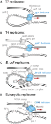
The best-studied replisomes and their helicases. (A) The T7 phage replicative helicase, gp4 (blue), translocates in the 5′–3′ direction on the lagging strand and contacts the leading and lagging strand polymerases (Scherr et al., 2018). (B) The T4 phage replicative helicase, gp41 (blue), translocates in the 5′–3′ direction on the lagging strand and contacts only the gp61 primase (Benkovic and Spiering, 2017). (C) The E. coli replicative helicase, DnaB (blue), translocates in the 5′–3′ direction on the lagging strand and contacts the DnaG primase and the τ subunit of the leading- and lagging-strand arms of the clamp loader complex (Lewis et al., 2016). (D) The S. cerevisiae replicative helicase, CMG (Cdc45 in green; MCM2–7 in blue, GINS in dark blue), translocates in the 3′–5′ direction on the leading strand and contacts the leading strand polymerase Pol ε, as well as the primase Pol α, firing factor Mcm10, organizing factor Ctf1, and MTC accessory factor (omitted) (Lewis et al., 2020).
FIGURE 4
FIGURE 4
Single-molecule manipulation of replicative helicase unwinding events. (A) Magnetic trap measurement of a single T4 gp41 helicase unwinding and rezipping a hairpin DNA template. (B) A single CMG unwinding event as measured by a magnetic trap shows more sporadic unwinding, but no rezipping. (C) Gp41 unwinding and rezipping rates measured at different forces. Rezipping is independent of force, but equivalent to the ssDNA translocation rate (<v ss> = 409 ± 16 bp/s) and unwinding rate increases with force (Ribeck and Saleh, 2013). (D) CMG unwinding rate also increases with force but with a less obvious trend (Burnham et al., 2019).
FIGURE 5
FIGURE 5
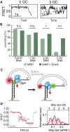
Detection of replicative helicase stepping using single-molecule FRET. (A) Typical FRET unwinding traces for G40P, the DnaB-like helicase (Schlierf et al., 2019). On templates containing no GC base pairs, G40P unwinding exhibits a rapid decrease in FRET. On templates containing 3 GC base pairs, G40P frequently stalls (see arrows), slips backwards and then re-attempts unwinding. (B) The percentage of FRET templates unwound by G40P decreases with increasing GC DNA content, but a higher percentage can be recovered with the inclusion of the DnaG primase. (C) A nanotensioner applied to the single-molecule FRET unwinding assay stabilizes the overhangs of the unwound DNA strands and thus improves the resolution in FRET signal to <1 bp (Lin et al., 2017). (D) Typical FRET unwinding traces for the gp4 helicase, where a nanotensioner is incorporated into the DNA template. (Ma et al., 2020). A histogram of gp4 step sizes (right) shows the helicase can sample a hierarchy of steps with 2 nt/step being the most common size.
FIGURE 6
FIGURE 6
Single-molecule visualization of CMG assembly during S. cerevisiae replication initiation. (A) A single-molecule assay where two-color colocalization is used to detect CMG loading in vitro. (B) Fluorescently labeled MCM2–7 (red) binds simultaneously with Cdt1 (green), with multiple hexamers appearing to bind sequentially (Ticau et al., 2015). (C) A single-molecule assay where FRET is used to detect MCM2–7 ring opening and closing. (D) Observation of both donor and acceptor emission identifies when MCM2–7 binds DNA. The FRET efficiency indicates the hexameric ring state, where high FRET denotes a closed ring and low FRET denotes an open ring. Instances of low FRET (black lines) correlate with MCM2–7 binding, indicating that the ring briefly opens as it encircles DNA during loading (Ticau et al., 2017).
FIGURE 7
FIGURE 7
Single-molecule fluorescence visualization of replisome stability. (A) In the E. coli replisome, the polymerase holoenzyme (polymerase core and clamp loader complex) has been shown to exchange rapidly during replication (Beattie et al., 2017; Lewis et al., 2017), while the DnaB helicase remains stably associated (Spinks et al., 2021). (B) In the S. cerevisiae replisome, each of the polymerases demonstrates some degree of exchange, while the CMG helicase shows a complete lack of exchange (Kapadia et al., 2020; Lewis et al., 2020). (C) In vivo FRAP applied to fluorescent DnaB-YPet shows no recovery (yellow arrow) after the FRAP pulse (red arrow) and thus no exchange (Beattie et al., 2017). (D) In vivo FRAP applied to fluorescent Mcm4-mNG as part of CMG shows no recovery (yellow arrow) in the area bleached by the FRAP pulse (red arrow) (Kapadia et al., 2020). Therefore, CMG does not exchange. (E) In vitro FLIP applied to fluorescent DnaB-a647 within single-molecule rolling-circle replication. In this example kymograph the DnaB signal persists even though it is challenged with extra unlabeled DnaB, and therefore is not exchanging (Spinks et al., 2021). (F) In vitro FLIP applied to fluorescent CMG-LD650 during single-molecule replication of a linear DNA template. The CMG signal persists even though it is challenged with excess unlabeled CMG, and thus does not exchange (Lewis et al., 2020).

Similar articles

Cited by

References

    1. Abid Ali F., Renault L., Gannon J., Gahlon H. L., Kotecha A., Zhou J. C., et al. (2016). Cryo-EM Structures of the Eukaryotic Replicative Helicase Bound to a Translocation Substrate. Nat. Commun. 7, 10708. 10.1038/ncomms10708 - DOI - PMC - PubMed
    1. Alberts B. M., Barry J., Bedinger P., Formosa T., Jongeneel C. V., Kreuzer K. N. (1983). Studies on DNA Replication in the Bacteriophage T4 a--.Gif System. Cold Spring Harbor Symposia Quantitative Biol. 47 (Pt 2), 655–668. 10.1101/sqb.1983.047.01.077 - DOI - PubMed
    1. Arias-Palomo E., Puri N., O’Shea Murray V. L., Yan Q., Berger J. M. (2019). Physical Basis for the Loading of a Bacterial Replicative Helicase onto DNA. Mol. Cell. 74, 173–184. 10.1016/j.molcel.2019.01.023 - DOI - PMC - PubMed
    1. Beattie T. R., Kapadia N., Nicolas E., Uphoff S., Wollman A. J., Leake M. C., et al. (2017). Frequent Exchange of the DNA Polymerase during Bacterial Chromosome Replication. eLife 6, 21763. 10.7554/eLife.21763 - DOI - PMC - PubMed
    1. Beattie T. R., Reyes-Lamothe R. (2015). A Replisome's Journey through the Bacterial Chromosome. Front. Microbiol. 6, 562. 10.3389/fmicb.2015.00562 - DOI - PMC - PubMed