. 2021 Nov 1;32(21):ar22.
doi: 10.1091/mbc.E21-05-0279. Epub 2021 Sep 8.

Slk19 enhances cross-linking of microtubules by Ase1 and Stu1

Affiliations

Slk19 enhances cross-linking of microtubules by Ase1 and Stu1

Sarina Norell et al. Mol Biol Cell. .

Abstract

The Saccharomyces cerevisiae protein Slk19 has been shown to localize to kinetochores throughout mitosis and to the spindle midzone in anaphase. However, Slk19 clearly also has an important role for spindle formation and stabilization in prometaphase and metaphase, albeit this role is unresolved. Here we show that Slk19's localization to metaphase spindles in vivo and to microtubules (MTs) in vitro depends on the MT cross-linking protein Ase1 and the MT cross-linking and stabilizing protein Stu1. By analyzing a slk19 mutant that specifically fails to localize to spindles and MTs, we surprisingly found that the presence of Slk19 amplified the amount of Ase1 strongly and that of Stu1 moderately at the metaphase spindle in vivo and at MTs in vitro. Furthermore, Slk19 markedly enhanced the cross-linking of MTs in vitro when added together with Ase1 or Stu1. We therefore suggest that Slk19 recruits additional Ase1 and Stu1 to the interpolar MTs (ipMTs) of metaphase spindles and thus increases their cross-linking and stabilization. This is in agreement with our observation that cells with defective Slk19 localization exhibit shorter metaphase spindles, an increased number of unaligned nuclear MTs, and most likely reduced ipMT overlaps.


Figures

FIGURE 1:
FIGURE 1:
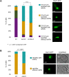
Slk19 localizes to metaphase spindles. (A, B) Statistics see Supplemental Table S4. (A) Cells were arrested in metaphase by Cdc20 depletion (GAL1 promoter shutoff). Subsequent Spc105 depletion (‘∆spc105’) was performed as indicated (AID-system) to interfere with KT localization of Slk19. The percentage of cells revealing the depicted phenotype is indicated. Genotypes of the strains with the indicated fusion proteins are listed in Supplemental Table S1. Bars, 2 µm. (B) Cells were treated as in A. Mean distribution of the indicated proteins along the horizontal spindle axes (longitudinal blots in Materials and Methods). Vertical bars indicate the SD of two independent experiments. (C) Domain organization of Stu1 as shown in (Kolenda et al., 2018).
FIGURE 2:
FIGURE 2:
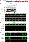
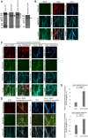
Slk19 deletion analysis. (A) Putative structural organization of Slk19 according to (Havens et al., 2010) and additional secondary structure analysis (PSIPRED). GD, globular domain; cc, coiled-coil. (B–E) Genotypes of the strains with the indicated fusion proteins are listed in Supplemental Table S1. All SLK19 deletion constructs were expressed from the endogenous SLK19 promoter. (B) cc6+cc7 confers Slk19–Slk19 interaction. FLAG-tagged Slk19 constructs were coexpressed in S. cerevisiae with Slk19-GFP and affinity purified. Proteins were detected by Western analysis. Arrowheads indicate the different Slk19 deletion constructs. The asterisk indicates a background band. (C–E) The percentage of cells revealing the depicted phenotype is indicated. Statistics see Supplemental Table S4. Bars, 2 µm. (C, D) Cells were arrested in metaphase by Cdc20 depletion (GAL1 promoter shutoff). (C) cc6+7 is essential for KT and spindle localization of Slk19 in metaphase. (D) cc1 is essential for spindle localization in metaphase. Spc105 depletion (‘∆spc105’) was used to interfere with KT localization of Slk19 (AID-system). (E) cc1 is essential for midzone localization of Slk19 and cc6+7 is essential for midzone and KT localization of Slk19 in anaphase. Cells were analyzed 2–2.5 h after release from a G1 arrest.
FIGURE 3:
FIGURE 3:
∆slk19 and slk19∆cc1 cells exhibit a highly increased number of short spindles and not-aligned nuclear MTs. (A and B) Nuclear MTs were visualized via Dad1-GFP localization. nrMTs, nuclear random MTs. n.s., not significant. Statistics see Supplemental Table S4. Bars, 2 µm. (A) Cells were arrested in metaphase by Cdc20 depletion (MET25 promoter shutoff). Slk19∆cc1 was expressed from the endogenous SLK19 promoter. (B) Cells were analyzed ∼90 min after release from G1. Only cells with bud diameters ranging from 1.9–2.8 µm were included in the quantification.
FIGURE 4:
FIGURE 4:
Slk19 localizes to metaphase spindles via Ase1 and the CL domain of Stu1. (A-C) Statistics see Supplemental Table S4. (A) Cells were arrested in metaphase by Cdc20 depletion (MET25 or GAL1 promoter shutoff). Spc105 depletion (‘∆spc105’) was used to interfere with KT localization of Slk19 (AID-system). The ‘∆spc105’ data from Figure 1A were included for better comparison. Genotypes of the strains with the indicated fusion proteins are listed in Supplemental Table S1. The percentage of cells revealing the depicted phenotype is indicated. Bars, 2 µm. (B) Quantification of Slk19 at metaphase spindles in indicated cells. The SD of two independent experiments are shown. (C) Slk19-Stu1 interaction in metaphase requires the CL domain of Stu1. Western analysis of affinity purified Stu1 constructs and copurifying Slk19. Black arrowheads mark full-length Slk19; green arrowheads mark a naturally occurring cleavage product of Slk19 (Sullivan et al., 2001) and red arrowheads indicate FLAG-Stu1 or FLAG-Stu1∆CL. The asterisk indicates a background band.
FIGURE 5:
FIGURE 5:
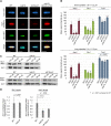
Slk19 amplifies the localization of Ase1 and Stu1 to metaphase spindles. (A–D) Genotypes of the strains with the indicated fusion proteins are listed in Supplemental Table S1. SLK19 constructs were expressed from the endogenous SLK19 promoter. (A) Example micrographs. Cells were arrested in metaphase by Cdc20 depletion (MET25 promoter shutoff). Bars, 2µm. (B, D) The SD of two independent experiments is shown. Statistics see Supplemental Table S4. (B) Quantification of Ase1, Stu1, and Tub1 at metaphase spindles as shown in A; n.s., not significant. (C) Cellular levels of Ase1 and Stu1. Western analysis of WCEs with the indicated amounts of total protein. Detection with anti-Myc or anti-Arc1 antibody. (D) Quantification of Western results shown in C. WT values were set to “1.”
FIGURE 6:
FIGURE 6:
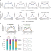
Slk19 synchronizes the overlap of ipMTs at the center of metaphase spindles. (A–D) Genotypes of the strains with the indicated fusion proteins are listed in Supplemental Table S1. Statistics see Supplemental Table S4. Cells were arrested in metaphase by Cdc20 depletion (MET25 promoter shutoff). (A, B) SLK19 constructs were expressed from the endogenous SLK19 promoter. (A) Mean tubulin distribution along the metaphase spindle axes (see longitudinal blots in Materials and Methods). The vertical bars represent the SD of two independent experiments. The WT result is super­imposed in the other blots for better comparison. (B) Mean tubulin distribution perpendicular (y-axes) to the spindle axes (x-axes) at the spindle center (see cross-section blots in Materials and Methods). The vertical bars represent the SD of two independent experiments. The WT result is superimposed in the other blots for better comparison. (C) Single cell examples of the distribution of Tub1, Ase1 and Stu1 along the spindle axes. Arrowheads indicate defined maxima and double headed arrows the lack of defined maxima. Note that for each protein, the maximal value was normalized to 1 so that the Ase1 distribution of the ∆slk19 cells can be presented in one blot together with the distribution of Stu1 and Tub1. (D) Quantification of the distribution phenotypes of Ase1 and Stu1 in individual cells, as depicted in the cartoons.
FIGURE 7:
FIGURE 7:
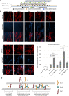
Slk19 amplifies the MT binding of Ase1 and Stu1 in vitro. (A) Purified proteins used in the in vitro experiment, as revealed by SDS–PAGE and Coomassie staining. Black arrowheads mark the full-length proteins, the green arrowhead a naturally occurring cleavage product of Slk19 (Sullivan et al., 2001) and red arrowheads Ase1 carrying two or one mCherry domains. An asterisk indicates IgG. (B–D) Stabilized and immobilized MTs were incubated with purified proteins and analyzed as indicated. Bars, 10 µm. (B) Slk19 does not bind to MTs in vitro. (C) Ase1 and Stu1 confer MT binding of Slk19 in vitro. MTs preloaded with Ase1 or Stu1 were subsequently incubated with Slk19 proteins. (D) Slk19 increases the binding of Ase1 and Stu1 to MTs in vitro. Where indicated, MTs were incubated with premixed Ase1 and Slk19 or Stu1 and Slk19. (E) Quantification of MT binding of Ase1 and Stu1. Statistics see Supplemental Table S4. The SD of two independent experiments is shown.
FIGURE 8:
FIGURE 8:
Slk19 enhances the cross-linking of MTs by Ase1 or Stu1 in vitro. (A) Experimental setup, modified from (Funk et al., 2014). (B) As illustrated in A, stabilized MTs were immobilized, incubated with the indicated proteins (Figure 7A), and subsequently incubated with mobile MTs. Where indicated, Slk19 constructs were applied premixed with Stu1 or Ase1. Bars, 10 µm. (C) Quantification of colocalization (overlaps) of immobile and mobile MTs (cross-linking efficiency). The SD of two independent experiments is shown; n.s., not significant. Statistics see Supplemental Table S4. (D) Model depicting how Slk19 may support the cross-linking and stability of metaphase ipMTs. See Discussion for further explanation.

Similar articles

Cited by

References

    1. Alberti S, Gladfelter A, Mittag T (2019). Considerations and challenges in studying liquid-liquid phase separation and biomolecular condensates. Cell 176, 419–434. - PMC - PubMed
    1. Alberti S, Saha S, Woodruff JB, Franzmann TM, Wang J, Hyman AA (2018). A user’s guide for phase separation assays with purified proteins. J Mol Biol 430, 4806–4820. - PMC - PubMed
    1. Bieling P, Telley IA, Surrey T (2010). A minimal midzone protein module controls formation and length of antiparallel microtubule overlaps. Cell 142, 420–432. - PubMed
    1. De Wulf P, McAinsh AD, Sorger PK (2003). Hierarchical assembly of the budding yeast kinetochore from multiple subcomplexes. Genes Dev 17, 2902–2921. - PMC - PubMed
    1. Faust AM, Wong CC, Yates JR 3rd, Drubin DG, Barnes G (2013). The FEAR protein Slk19 restricts Cdc14 phosphatase to the nucleus until the end of anaphase, regulating its participation in mitotic exit in Saccharomyces cerevisiae. PLoS One 8, e73194. - PMC - PubMed

Publication types

MeSH terms

Substances

LinkOut - more resources