. 2021 Sep 7;19(1):194.
doi: 10.1186/s12915-021-01137-7.

ACBD3 modulates KDEL receptor interaction with PKA for its trafficking via tubulovesicular carrier

Affiliations

ACBD3 modulates KDEL receptor interaction with PKA for its trafficking via tubulovesicular carrier

Xihua Yue et al. BMC Biol. .

Abstract

Background: KDEL receptor helps establish cellular equilibrium in the early secretory pathway by recycling leaked ER-chaperones to the ER during secretion of newly synthesized proteins. Studies have also shown that KDEL receptor may function as a signaling protein that orchestrates membrane flux through the secretory pathway. We have recently shown that KDEL receptor is also a cell surface receptor, which undergoes highly complex itinerary between trans-Golgi network and the plasma membranes via clathrin-mediated transport carriers. Ironically, however, it is still largely unknown how KDEL receptor is distributed to the Golgi at steady state, since its initial discovery in late 1980s.

Results: We used a proximity-based in vivo tagging strategy to further dissect mechanisms of KDEL receptor trafficking. Our new results reveal that ACBD3 may be a key protein that regulates KDEL receptor trafficking via modulation of Arf1-dependent tubule formation. We demonstrate that ACBD3 directly interact with KDEL receptor and form a functionally distinct protein complex in ArfGAPs-independent manner. Depletion of ACBD3 results in re-localization of KDEL receptor to the ER by inducing accelerated retrograde trafficking of KDEL receptor. Importantly, this is caused by specifically altering KDEL receptor interaction with Protein Kinase A and Arf1/ArfGAP1, eventually leading to increased Arf1-GTP-dependent tubular carrier formation at the Golgi.

Conclusions: These results suggest that ACBD3 may function as a negative regulator of PKA activity on KDEL receptor, thereby restricting its retrograde trafficking in the absence of KDEL ligand binding. Since ACBD3 was originally identified as PAP7, a PBR/PKA-interacting protein at the Golgi/mitochondria, we propose that Golgi-localization of KDEL receptor is likely to be controlled by its interaction with ACBD3/PKA complex at steady state, providing a novel insight for establishment of cellular homeostasis in the early secretory pathway.

Keywords: ACBD3; Arf1-GTP; ArfGAPs; Golgi; KDEL receptor; Protein Kinase A.


Conflict of interest statement

The authors declare no competing interests.

Figures
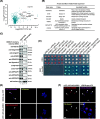
Fig. 1
Fig. 1
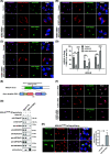
ACBD3 is a novel binding partner of KDELR1. A Volcano plot representing results of the label-free BioID of KDELR1. The logarithmic ratio of protein intensities in the KDELR1-BioID/KDELR1-myc were plotted against negative logarithmic p-values of the t test performed from the triplicates. Annotated Golgi proteins are candidate interaction partners of KDELR1 with a p-value of ratio significance <0.05 and marked with red dots. B Table summarizing annotated function of the Golgi proteins identified in KDELR1-BioID experiments. C Several Golgi-related proteins co-immunoprecipitate with KDELR1-mCherry. The protein extracts from HeLa cells transfected with mCherry tagged Sorting Nexin 3 (mCherry-SNX3 as a control) or KDELR1-mCherry were immunoprecipitated with anti-RFP agarose beads. These lysates and the immunoprecipitates (anti-RFP IPs) were analyzed by western blotting using anti-mCherry antibody and antibodies against the indicated Golgi proteins. D Split-ubiquitin-based membrane yeast two-hybrid assays suggest that KDELR1 interact with ACBD3 and ARFGAP1/3, but not with ARFGAP2 and Golgin97. Growth of yeast cells expressing KDELR1-Cub-LexA-VP16 bait or a pBT3-SUC bait vector with the following NubG-HA-preys: ACBD3, ARFGAP1, ARFGAP2, ARFGAP3, Golgin97, pPR3-N (prey vector as a negative control), and Ost1-NubI (positive control) were challenged on agar plates depleted of tryptophan and leucine (DDO plates, upper panel) or depleted of tryptophan, leucine, histidine, and adenine (QDO plates, bottom panel), plus 40 μg/ml X-α-Gal by spotting three independent transformants on different plates. The bait pTSU2-APP and the prey pNubG-Fe65 were used as positive controls in the MYTH assay. Experiments were repeated twice. D, E Interaction between ACBD3 and KDELR1 detected by split-YFP assays. HeLa cells were transfected with nYFP-3xFLAG-ACBD3 and KDELR1-myc-cYFP for 18 h before cells were fixed and observed by confocal microscopy (E). Single plasmid (nYFP-3xFLAG-ACBD3 or KDELR1-myc-cYFP) transfection was used as negative controls (D). White arrowheads indicate co-transfected cells, whereas white asterisks indicate cells transfected with a single plasmid, serving as a negative control. Scale bars = 10 μm
Fig. 2
Fig. 2
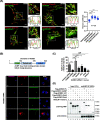
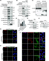
ACBD3 contributes significantly to Golgi localization of KDELR at steady state. AD Confocal micrographs of HeLa cells expressing KDELR1-mCherry (A), KDELR2-mCherry (B), and KDELR3A-mCherry (C), showing that depletion of ACBD3 results in significant re-distribution of all three isoforms of KDELRs from the Golgi to the ER in vivo, which could be restored by exogenous expression of RNAi-resistant EGFP-ACBD3. For quantification (D), the percentage of fluorescent intensity of Golgi-localized KDELRs over the total KDELR-mCherry were quantified and plotted onto the histogram as average ratio with s.d (n = 20 ~ 25) (ACBD3-KD vs Control, **p < 0.001; ACBD3-KD/rescued vs ACBD3-KD, ##p < 0.01; ###p < 0.001). E Schematic illustration of CRISPR-Cas9-mediated insertion of 3xFlag-mCherry tagging at the C-terminal end of endogenous KDELR1. F Confocal micrographs of KDELR1endo-3xFlag-mCherry in HeLa cells showing that depletion of ACBD3 results in re-distribution of KDELR1endo-3xFlag to the ER in vivo. G Endogenously tagged KDELR1-3xFlag-mCherry pulls down ACBD3 and ArfGAP1/3, respectively. HeLa cells with KDELR1-3xFlag-mCherry was lysed and subjected to immunoprecipitation with anti-RFP antibody, followed by western blot analysis. H Confocal results showing that hGH-GFP-KDEL expression induces endogenously 3xFLAG-mCherry-tagged KDEL receptor to re-distribute to the ER in HeLa cells. The bar graph to the right summarizes the quantification of these results. (***p < 0.001) Scale bars = 10 μm
Fig. 3
Fig. 3
ACBD3 is required for Golgi distribution of the three isoforms of KDELR at the Golgi. A Super-resolution 3D-SIM images showing a high degree of co-localization between endogenously tagged KDELR1-3xFLAG-mCherry and ACBD3, ARFGAP1, or ARFGAP3, and low degree of co-localization between the endogenously tagged KDELR1 and Golgin97 (negative control). Line profiles through regions of interest were analyzed by Fiji. Scale bars = 2 μm. Co-localization (Pearson’s R) was determined and subjected to two-tailed, unpaired t tests (n = 20 cells/combination, mean and SD, ****, p < 0.0001). B Confocal micrographs of HeLa cells co-expressing KDELR1-mCherry with deletion mutants of EGFP-ACBD3 after siRNA-mediated ACBD3 knockdown, showing that the GOLD domain of ACBD3 is required for retention of KDELR1-mCherry at the Golgi. For this experiment, we sequentially deleted the ACBP domain, the CC domain, the GOLD domain, or both ACBP+CC domains and used these constructs in the knockdown and rescue experiments. C Histogram summarizing the rescue experiments using full length EGFP-ACBD3, EGFP-ACBD3ΔACBP, EGFP-ACBD3ΔCC, EGFP-ACBD3ΔGOLD, EGFP-ACBD3-GOLD (ΔACBP+CC), respectively. Note that rescue with full length ACBD3 actually led to ~25% increase in the percentage of Golgi-localized KDEL-R1-mCherry over the WT control cells. In addition, we observed that EGFP-ACBD3 expression often abolished ER-localization of over-expressed KDELR1-mCherry entirely. (***p < 0.001; n.s., not significant) D EGFP-tagged GOLD domain of ACBD3 co-immunoprecipitates with KDELR1-mCherry. KDELR1-mCherry was co-transfected with EGFP-ACBD3 WT, EGFP-ACBD3ΔACBP, EGFP-ACBD3ΔCC, EGFP-ACBD3ΔGOLD, EGFP-GOLD domain, respectively, followed by cell lysis and immunoprecipitation using anti-RFP agarose beads. Experiments were repeated three times and representative western blots are shown here. Scale bars = 10 μm
Fig. 4
Fig. 4
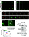
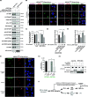
Photoactivation experiments reveals that knockout of ACBD3 results in acceleration of KDELR1 retrograde transport. A, B HeLa or HeLa-ACBD3-KO cells were co-transfected with KDELR1-FM4-SNAP and ST-RFP. After D/D solubilizer treatment, ER-Golgi antegrade transport of KDELR1-FM4-SNAP is monitored by live cell imaging acquired every 30 s for 10 min. Imaging sequences at the indicated time points are presented here. Scale bars = 10 μm. C To measure the amount of KDELR1 outflux from the Golgi, WT, or ACBD3-depleted HeLa cells were co-transfected with sialyltransferase-RFP (ST-RFP, a Golgi marker) and photoactivatable KDELR1-PA-GFP plasmids for 18 h. The KDELR1-PA-GFP in the Golgi were then activated by selecting an ROI of ST-RFP-positive region for intense 405-nm laser irradiation and the transport out of the Golgi are monitored by live cell imaging acquired every 5 s for 5 min. Imaging sequences prior to photoactivation (−10 s), immediately after photoactivation (0 s) and the indicated times following photoactivation are presented here. Magnified regions of interest (indicated by white boxes) from WT and ACBD3-KO cells at 100 s time point shows Golgi-derived tubules which are highlighted by white arrowheads. Scale bars = 10 μm. D The intensity of photoactivated GFP remaining in the Golgi area was expressed as a percentage of the intensity of photoactivated GFP at time point 0 and plotted as a function of time. The integrated fluorescence over the entire cell was normalized to the integrated fluorescence at time point 0 as a function of time (green and black lines), showing the photobleaching effect during live imaging, serving as a control for fluorescence (n = 22). (***p < 0.001) E Co-immunoprecipitation experiments indicate that ACBD3 depletion results in increased association of p24, ArfGAP1, coatomer, and Arf1 with KDELR1-mCherry.
Fig. 5
Fig. 5
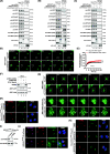
ACBD3 depletion alters KDELR association with Arf1, promoting Arf1-dependent tubular carrier formation at the Golgi. AC myc-ACBD3, ARFGAP1-myc, or ARFGAP3-myc can efficiently pull down KDELR1-mCherry, but myc-ACBD3 does not co-immunoprecipitate with the key components of the retrograde trafficking machinery. ARFGAP1-myc co-immunoprecipitates with γCOP, ARFGAP3, ARF1, and p24 proteins, while ARFGAP3-myc only co-immunoprecipitates with γCOP. HeLa cells were co-transfected KDELR1-mCherry with myc-ACBD3, ARFGAP1-myc, or ARFGAP3-myc, respectively, and the extracts were immunoprecipitated with a myc-trap agarose beads. The lysates and the immunoprecipitates were analyzed by western blotting, as indicated. D, E Kinetics of Arf1-EGFP binding to and dissociation from Golgi membranes in WT and ACBD3-KO HeLa cells. D Representative FRAP experiments in WT or ACBD3-KO HeLa cells transiently expressing Arf1-EGFP. The Golgi was selectively photobleached (white circle). The first frames show an initial prebleached image. After photobleaching, image frames were selected at times 10, 50, 100, and 300 s. Scale bar, 10 μm. E Quantification of Golgi intensity in Arf1-EGFP–expressing WT or ACBD3-KO HeLa cells. Statistical analysis was performed using two-tailed, paired t test (mean±SEM, N = 10, ****p < 0.0001). F HeLa WT, ACBD3-KD (stably knockdown) or ACBD3-KO cells were analyzed for levels of Arf1-GTP, as described in the methods. G Live-cell imaging of transiently expressed Arf1-EGFP in HeLa WT or ACBD3-KO cells. Zoomed Golgi areas were shown in the bottom panels. Examples of Golgi-derived ARF1-rich tubules are highlighted by arrowheads. H Confocal images of KDELR1endo-3xFlag-mCherry in HeLa cells showing that depletion of PI4KB does not result in re-distribution of KDELR1endo-3xFlag-mCherry to the ER in vivo. Scale bars = 10 μm. I Western blots showing depletion of PI4KB, by RNA interference. J Confocal images of KDELR1endo-3xFlag-mCherry in HeLa cells showing that overexpression of PI4KB does not result in its re-distribution to the ER in vivo. K Golgi localization of KDELR1endo-3xFlag-mCherry is unaffected by chemical inhibition of PI4K using PIK-93. Scale bars = 10 μm
Fig. 6
Fig. 6
Activation of PKA and increased Arf1-GTP level plays a crucial role in KDELR relocalization. A Effect of the co-expression of KDEL-cargo (hGH-GFP-myc-KDEL) on KDELR1-mCherry co-immunoprecipitation in HeLa cells. HeLa cells co-transfected KDELR1-mCherry with hGH-GFP-myc or hGH-GFP-myc-KDEL were firstly treated with DSP crosslinker in PBS at 37 °C for 30 min. Cells were lysed and the extracts were immunoprecipitated with anti-RFP agarose beads. B Effect of ACBD3-KO on KDELR1-mCherry co-immunoprecipitation in HeLa cells. HeLa-WT or ACBD3-KO cells transfected with KDELR1-mCherry (mCherry-SNX3 transfection as a control) were lysed and immunoprecipitated with anti-RFP agarose beads. C Both PKA-Cα-EGFP and PKA-RII-EGFP co-immunoprecipitate with ACBD3 in HeLa cells. D Effect of ACBD3-KD/KO or ACBD3 over-expression on the luciferase expression in HeLa cells, expressing the CRE-controlled luciferase. HeLa-WT, ACBD3 stable knockdown, ACBD3-KO, or EGFP-ACBD3 stably over-expressing cells seeded in 24-well plate were transfected with pCRE-luciferase plasmid. Cells were then lysed, and equal protein amounts of lysate per condition were assayed using a luciferase assay system. Statistical analysis was performed using one-way ANOVA with a Tukey’s post hoc test (mean±SD, *p < 0.05; **p < 0.01; ****p < 0.0001). E HeLa WT, ACBD3-KD, or ACBD3-KO cells were analyzed for phosphorylation levels of Src family. F PKA inhibition by H89 decreases ARF1-GTP level in ACBD3-KO HeLa cells. HeLa-WT or ACBD3-KO cells were firstly treated with H89 for 2 h and then analyzed for levels of Arf1-GTP. Equal aliquots of lysates were analyzed for Arf1-GTP by using ARF1 activation assay kit utilizes pull-down with GST-GGA3. G, H Confocal images of KDELR1endo-3xFlag-mCherry in HeLa cells showing that PKA inhibition by H89 does not result in re-distribution of KDELR1endo-3xFlag-mCherry to the ER, while PKA activation by 8-Bromo-cAMP results in re-distribution of KDELR1endo-3xFlag-mCherry to the ER in vivo without affecting ManII localization. I Confocal images of KDELR1endo-3xFlag-mCherry in HeLa cells showing that only over-expression of PKA-Cα-EGFP results in re-distribution of KDELR1endo-3xFlag-mCherry to the ER in vivo. Scale bars = 10 μm
Fig. 7
Fig. 7
ACBD3 functions as a negative regulator of PKA activity on KDELR. A Effect of ACBD3 overexpression on KDELR1-mCherry co-immunoprecipitation in HeLa cells, as shown here by immunprecipitation experiments. B, C ACBD3 overexpression inhibits hGH-GFP-FM4-KDEL stimulated KDELR retrograde traffic. KDELR1endo-3xFlag-mCherry cells transfected with hGH-GFP-FM4-KDEL alone or co-transfected with hGH-GFP-FM4-KDEL and SNAP-ACBD3 were subjected for D/D solubilizer treatment to induce ER to Golgi trafficking of hGH-GFP-FM4-KDEL. KDELR1endo-3xFlag-mCherry localization was analyzed at 10min or 30min after drug treatment by confocal imaging. Scale bars = 10 μm. Histogram summarizing the percentage of cells with Golgi localized KDELR1 after D/D solubilizer treatment at indicated time point. N=30–40 cells. Statistical analysis was performed using two-tailed, unpaired t test (mean±SD, ****p < 0.0001; ***p < 0.001). D ACBD3 overexpression inhibits the effect of ER-to-Golgi traffic wave on PKA activity. ER-to-Golgi traffic wave was induced and PKA activity was analyzed by CRE-luciferase assay. Statistical analysis was performed using two-way ANOVA with a Tukey’s post hoc test for multiple comparisons (mean±SD, **p < 0.01). E ACBD3 over-expression inhibits 8-Bromo-cAMP activated PKA. Cells were treated by 500μM 8-Bromo-cAMP for 6h and then PKA activity was analyzed by CRE-luciferase assay. Statistical analysis was performed using two-way ANOVA with a Tukey’s post-hoc test for multiple comparisons (mean±SD, *p < 0.05; **p < 0.01). F ACBD3 over-expression inhibits 8-Bromo-cAMP stimulated KDELR1 re-distribution to the ER. KDELR1endo-3xFlag-mCherry HeLa cells with or without SNAP-ACBD3 expression. G Histogram summarizing the percentage of cells with Golgi localized KDELR1 after 8-Bromo-cAMP treatment shown in F. Statistical analysis was performed using two-tailed, unpaired t test (mean±SD, **p < 0.01). H Effect of ACBD3 over-expression on KDELR1-mCherry interacting with Gαs in HeLa cells, as shown here by immunoprecipitation experiments. I Schematic diagram depicting the proposed role of ACBD3 in regulating PKA activity and KDELR trafficking. ACBD3 likely plays a pivotal role in Golgi localization of KDELR under low cargo trafficking condition by inhibiting basal PKA activity on KDELR

Similar articles

Cited by

References

    1. Pelham HR. Evidence that luminal ER proteins are sorted from secreted proteins in a post-ER compartment. EMBO J. 1988;7(4):913–918. doi: 10.1002/j.1460-2075.1988.tb02896.x. - DOI - PMC - PubMed
    1. Lewis MJ, Pelham HR. A human homologue of the yeast HDEL receptor. Nature. 1990;348(6297):162–163. doi: 10.1038/348162a0. - DOI - PubMed
    1. Capitani M, Sallese M. The KDEL receptor: new functions for an old protein. FEBS Lett. 2009;583(23):3863–3871. doi: 10.1016/j.febslet.2009.10.053. - DOI - PubMed
    1. Munro S, Pelham HR. A C-terminal signal prevents secretion of luminal ER proteins. Cell. 1987;48(5):899–907. doi: 10.1016/0092-8674(87)90086-9. - DOI - PubMed
    1. Yamamoto K, Fujii R, Toyofuku Y, Saito T, Koseki H, Hsu VW, Aoe T. The KDEL receptor mediates a retrieval mechanism that contributes to quality control at the endoplasmic reticulum. EMBO J. 2001;20(12):3082–3091. doi: 10.1093/emboj/20.12.3082. - DOI - PMC - PubMed

Publication types

Substances