. 2021 Nov;81(15):1235-1251.
doi: 10.1002/pros.24225. Epub 2021 Sep 7.

SLX4IP N-terminus dictates telomeric localization in ALT-like castration-resistant prostate cancer cell lines

Affiliations

SLX4IP N-terminus dictates telomeric localization in ALT-like castration-resistant prostate cancer cell lines

Tawna L Mangosh et al. Prostate. 2021 Nov.

Abstract

Background: To ensure replicative immortality in cancer, telomeres must be maintained through activation of telomere maintenance mechanisms (TMMs) that are dependent on telomerase or the alternative lengthening of telomeres (ALT) pathway. Although TMM pathways have traditionally been considered to be mutually exclusive, ALT hallmarks have been identified in cancers defined as being telomerase-positive, supporting TMM coexistence. In castration-resistant prostate cancer (CRPC), in vitro models were thought to be universally dependent on telomerase as the primary TMM; however, CRPC models with androgen receptor (AR) loss demonstrate ALT hallmarks with limited telomerase activity and require ALT-associated PML bodies (APBs) for sustained telomere maintenance. The TMM coexistence in AR-negative CRPC is reliant on the ALT regulator protein, SLX4IP.

Methods: To identify the regions of SLX4IP responsible for the induction of APBs and telomere preservation in CRPC models, five 3xFLAG-tagged SLX4IP constructs were designed and stably introduced into parental C4-2B, DU145, and PC-3 cells. Once generated, these cell lines were interrogated for APB abundance and SLX4IP construct localization via immunofluorescence-fluorescence in situ hybridization (IF-FISH) and coimmunoprecipitation experiments for telomeric localization. Similarly, PC-3 cells with endogenous SLX4IP knockdown and SLX4IP construct introduction were interrogated for APB abundance, telomere length preservation, and senescent rescue.

Results: Here, we define the N-terminus of SLX4IP as being responsible for the promotion of the ALT-like phenotype of AR-negative CRPC models. Specifically, the N-terminus of SLX4IP was sufficient for promoting APB formation to a similar degree as full-length SLX4IP across CRPC cell lines. Additionally, APB promotion by the N-terminus of SLX4IP rescued telomere shortening and senescent induction triggered by SLX4IP knockdown in AR-negative CRPC cells. Moreover, APB formation and telomere maintenance were dependent on the ability of the N-terminus to direct SLX4IP localization at telomeres and APBs.

Conclusions: These findings identify the role of the uncharacterized ALT regulator SLX4IP in the promotion of TMM coexistence to perpetuate replicative immortality in CRPC in vitro.

Keywords: ALT-associated PML body; PML; androgen receptor-negative prostate cancer; androgen-independent prostate cancer; telomere maintenance mechanism.


Figures

Figure 1:
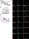
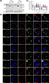
Figure 1:. N-terminus of SLX4IP promotes APB structures in C4–2B cells.
(a) Schematic representation of 3×FLAG-tagged SLX4IP truncation constructs. (b) Confirmation of stable overexpression of SLX4IP truncation constructs alongside full-length SLX4IP (1–408) and empty vector (EV) in C4–2B cells. (c) Representative IF-FISH images demonstrating the presence (arrowheads) or absence of PML (green) at telomeres (red) in C4–2B cells with SLX4IP overexpression. White outline represents nuclear boundary. Scale bar: 5 μm. (d) Quantification of (c) for percent positive cells with at least one APB. Data represented as mean±SD; n≥3; All constructs compared to 1–408 positive control; *p<0.05; **p<0.01; ***p<0.001; ****p<0.0001.
Figure 2:
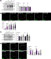
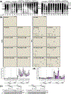
Figure 2:. N-terminus of SLX4IP supports additional APB-positive cells in DU145 and PC-3 cells.
(a) Confirmation of stable overexpression of SLX4IP truncation constructs alongside full-length (1–408) SLX4IP and empty vector (EV) in DU145 and PC-3 cells. (b) Representative IF-FISH images demonstrating the presence (arrowheads) or absence of PML (green) at telomeres (red) in DU145 and PC-3 cells with SLX4IP overexpression. White outline represents nuclear boundary. Scale bar: 5 μm. (c) Quantification of (b) for percent positive cells with at least one APB. Data represented as mean±SD; n≥3; All constructs compared to 1–408 positive control; *p<0.05; **p<0.01; ***p<0.001; ****p<0.0001.
Figure 3:
Figure 3:. SLX4IP expression directly correlates with endogenous PML expression and number of PML nuclear foci in CRPC in vitro models.
(a) PML and SLX4IP protein expression across CRPC cell lines. Quantification of PML and SLX4IP expression in (a) is shown to the right. (b) Relative PML expression plotted versus relative SLX4IP expression in CRPC cell lines. Simple linear regression model depicted by dotted line with R2 value reported. (c) Representative IF images demonstrating the presence of nuclear PML foci across CRPC cell lines. White outline represents nuclear boundary. Scale bar: 5 μm. (d) Quantification of (c) for number of nuclear PML foci per cell. Data represented as mean±SD; n≥3; *p<0.05; **p<0.01; ***p<0.001; ****p<0.0001.
Figure 4:
Figure 4:. Genetic manipulation of SLX4IP expression alters number of PML nuclear foci but not PML expression.
(a) PML expression in C4–2B, DU145 and PC-3 cells with full-length SLX4IP (1–408) overexpression versus empty vector (EV). Quantification of PML expression in (a) is shown to the right. (b) Representative IF images demonstrating the presence of nuclear PML foci in C4–2B, DU145 and PC-3 cells with full-length SLX4IP (1–408) overexpression versus empty vector (EV). White outline represents nuclear boundary. Scale bar: 5 μm. Quantification of (b) for number of nuclear PML foci per cell is shown below. (c) PML expression in DU145 and PC-3 cells with SLX4IP knockdown (KD.1, KD.2) versus non-targeting control (NS). Quantification of PML expression in (c) is shown to the right. (d) Representative IF images demonstrating the presence of nuclear PML foci in DU145 and PC-3 cells with SLX4IP knockdown (KD.1, KD.2) versus non-targeting control (NS). White outline represents nuclear boundary. Scale bar: 5 μm. Quantification of (d) for number of nuclear PML foci per cell is shown to the right. Data represented as mean±SD; n≥3; *p<0.05; **p<0.01; ***p<0.001; ****p<0.0001.
Figure 5:
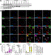
Figure 5:. Telomeric localization of SLX4IP at APBs occurs in DU145 and PC-3 cells.
(a) Representative immunoblots of CoIP and whole cell lysate (input) samples from CRPC cells. CoIP was completed with SLX4IP antibody and then complexes were probed for SLX4IP, TRF2, and GAPDH. (b) Representative IF-FISH images demonstrating the presence (arrowheads) or absence of PML (green) and SLX4IP (blue) at telomeres (red) in CRPC cell lines. White outline represents nuclear boundary. Scale bar: 5 μm. (c) Quantification of (b) for percent positive cells with at least one SLX4IP-positive telomere (magenta), percent positive cells with at least one SLX4IP-positive APB (white), percent APB-positive cells (yellow), and percent SLX4IP-positive APBs (white). Data represented as mean±SD; n≥3; *p<0.05; **p<0.01; ***p<0.001; ****p<0.0001.
Figure 6:
Figure 6:. N-terminus of SLX4IP coordinates telomeric localization at APBs.
(a) Representative immunoblots of CoIP and whole cell lysate (input) samples from C4–2B and PC-3 cells with stable overexpression of SLX4IP constructs. CoIP was completed with FLAG antibody and then complexes were probed for FLAG, TRF2, and GAPDH. (b) Representative IF-FISH images demonstrating the presence (arrowheads) or absence of PML (green) and FLAG (blue) at telomeres (red) in C4–2B and PC-3 cells with stable overexpression of SLX4IP constructs. White outline represents nuclear boundary. Scale bar: 5 μm. (c) Quantification of (b) for percent positive cells with at least one FLAG-positive telomere (magenta) and percent positive cells with at least one FLAG-positive APB. (white). Data represented as mean±SD; n≥3; All constructs compared to 1–408 positive control; *p<0.05; **p<0.01; ***p<0.001; ****p<0.0001.
Figure 7:
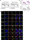
Figure 7:. Loss of APBs following SLX4IP knockdown is rescued with SLX4IP N-terminal region introduction.
(a) Confirmation of stable overexpression of SLX4IP truncation constructs alongside full-length (1–408) SLX4IP in PC-3 cells with endogenous SLX4IP knockdown (KD.1 and KD.2). PC-3.NS represents non-targeting control. (b) Representative IF-FISH images demonstrating the presence (arrowheads) or absence of PML (green) at telomeres (red) in PC-3 cells with SLX4IP construct overexpression on a background of SLX4IP knockdown. White outline represents nuclear boundary. Scale bar: 5 μm. (c) Quantification of (b) for percent APB-positive cells. All constructs compared to 1–408 positive control; Data represented as mean±SD; n≥3; *p<0.05; **p<0.01; ***p<0.001; ****p<0.0001.
Figure 8:
Figure 8:. SLX4IP N-terminal region aids in telomere length preservation and prevents accumulation of senescent markers.
(a) TRF analysis demonstrating telomere length changes over 50 population doublings (PD) in PC-3 cells with SLX4IP construct introduction and SLX4IP knockdown (KD.1 and KD.2). White dots within blot denote quantified average telomere length at each PD. (b) Representative bright field images demonstrating β-gal staining (arrowheads) for senescence in early and late-passage PC-3-derived cells. Scale bar: 20 μm. Quantification of percent β-gal positive cells in (b) is shown below. (c) p21 expression following knockdown of SLX4IP in early and late passage PC-3-derived cells. (d) Quantification of relative p21 expression in (c). All constructs compared to KD.1 or KD.2 control; Data represented as mean±SD; n≥3; *p<0.05; **p<0.01; ***p<0.001; ****p<0.0001.

Similar articles

References

    1. Blackburn EH. Structure and function of telomeres. Nature. 1991. doi:10.1038/350569a0 - DOI - PubMed
    1. Blackburn EH. Telomeres: No end in sight. Cell. 1994;77(5):621–623. doi:10.1016/0092-8674(94)90046-9 - DOI - PubMed
    1. Moyzis RK, Buckingham JM, Cram LS, et al.A highly conserved repetitive DNA sequence, (TTAGGG)(n), present at the telomeres of human chromosomes. Proc Natl Acad Sci U S A. 1988. doi:10.1073/pnas.85.18.6622 - DOI - PMC - PubMed
    1. De Lange T. Shelterin: The protein complex that shapes and safeguards human telomeres. Genes Dev. 2005. doi:10.1101/gad.1346005 - DOI - PubMed
    1. De Lange T. How telomeres solve the end-protection problem. Science (80- ). 2009. doi:10.1126/science.1170633 - DOI - PMC - PubMed

Publication types

MeSH terms