. 2021 Aug 30;17(8):e1009780.
doi: 10.1371/journal.pgen.1009780. eCollection 2021 Aug.

Cancer associated mutations in Sec61γ alter the permeability of the ER translocase

Affiliations

Cancer associated mutations in Sec61γ alter the permeability of the ER translocase

Christopher M Witham et al. PLoS Genet. .

Abstract

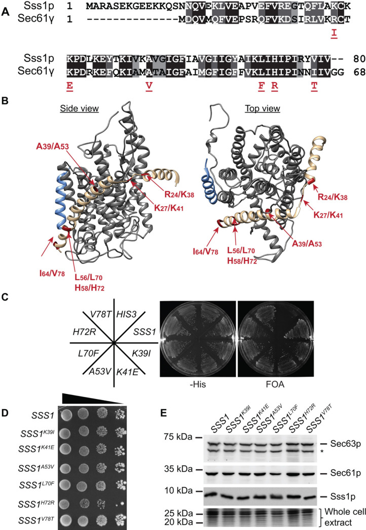
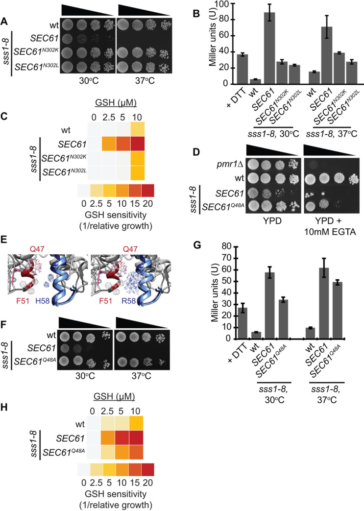
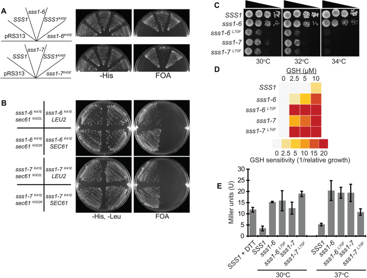
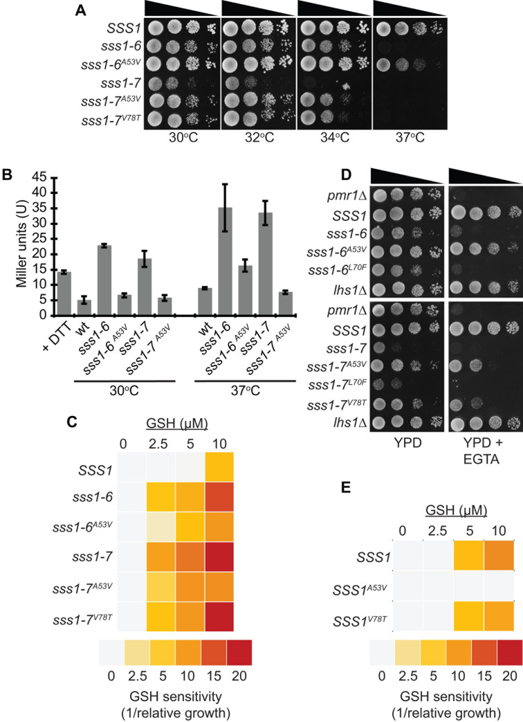
Translocation of secretory and integral membrane proteins across or into the ER membrane occurs via the Sec61 complex, a heterotrimeric protein complex possessing two essential sub-units, Sec61p/Sec61α and Sss1p/Sec61γ and the non-essential Sbh1p/Sec61β subunit. In addition to forming a protein conducting channel, the Sec61 complex maintains the ER permeability barrier, preventing flow of molecules and ions. Loss of Sec61 integrity is detrimental and implicated in the progression of disease. The Sss1p/Sec61γ C-terminus is juxtaposed to the key gating module of Sec61p/Sec61α and is important for gating the translocon. Inspection of the cancer genome database identifies six mutations in highly conserved amino acids of Sec61γ/Sss1p. We identify that five out of the six mutations identified affect gating of the ER translocon, albeit with varying strength. Together, we find that mutations in Sec61γ that arise in malignant cells result in altered translocon gating dynamics, this offers the potential for the translocon to represent a target in co-therapy for cancer treatment.


Conflict of interest statement

The authors have declared that no competing interests exist.

Figures

Fig 1
Fig 1. The Sss1p C-terminus is highly conserved.
(A) The sequence of Sec61γ and Sss1p are aligned using clustal omega sequence alignment software and the position of cancer associated mutations indicated. (B) Ribbon diagram of the Sec61 complex crystal structure (4CG7.pdb) (32) was composed using Chimera software. Sec61α, Sec61β and Sec61γ, are coloured grey, blue and sand respectively. Cancer associated mutations are indicated in red. (C) BWY530 yeast transformed with either YCp HIS3, YCp SSS1, YCp SSS1K39I, YCp SSS1K41E, YCp SSS1A53V, YCp SSS1L70F, YCp SSS1H72R or YCp SSS1V78T were streaked onto–His selective medium and medium containing FOA and incubated at 30°C for 2 days. (D) Wildtype or cells expressing either SSS1K39I, SSS1K41E, SSS1A53V, SSS1L70F, SSS1H72R or SSS1V78T as the sole source of SSS1 were spotted in a 10 fold dilution series and grown on YPD at 30°C for 3 days. (E) Cell extracts derived from wildtype cells or cells expressing either SSS1K39I, SSS1K41E, SSS1A53V, SSS1L70F, SSS1H72R or SSS1V78T were immunoblotted with anti-Sss1p, anti-Sec61p or anti-Sec63p antibodies. * identifies a proteolysed product of Sec63p.
Fig 2
Fig 2. The sss1H72R mutation disrupts ER homeostasis.
(A) Wildtype or sss1-8 yeast transformed with either YCp SEC61, YCp SEC61N302K or YCp SEC61N302L were spotted on YPD agar in a 10-fold dilution series and incubated at 30°C or 37°C for 2 days. (B) Wildtype or sss1-8 yeast transformed with either YCp SEC61, YCp SEC61N302K or YCp SEC61N302L and with pJT30 (UPRE-LacZ) were grown in–Ura selective medium and β-Galactosidase activity determined. As a positive control wildtype cells were treated with 5mM DTT for 2 hours. (C) Wildtype or sss1-8 yeast transformed with either YCp SEC61, YCp SEC61N302K or YCp SEC61N302L and with YEp HGT1 were grown in–Ura selective medium with increasing concentrations of GSH. The relative growth of each strain determined and the GSH sensitivity (1/relative growth) presented. (D) Wildtype or sss1-8 (sss1H72R) yeast transformed with either YCp SEC61 or YCp SEC61Q48A were spotted on YPD agar or YPD agar containing 10mM EGTA in a 10-fold dilution series and incubated at 30°C for 3 days. (E) Ribbon diagram of the open (4CG5/7.pdb) and closed (4CG5/7.pdb) Sec61 complex crystal structure (34) was composed and overlayed using Chimera software. The position of Q47 and F51 in Sec61α relative to H58 in Sec61γ are indicated. (F) Wildtype or sss1-8 (sss1H72R) yeast transformed with either YCp SEC61 or YCp SEC61Q48A were spotted on YPD agar in a 10-fold dilution series and incubated at 30°C or 37°C for 2 days. (G) Wildtype or sss1-8 (sss1H72R) yeast transformed with either YCp SEC61 or YCp SEC61Q48A and with pJT30 (UPRE-LacZ) were grown in–Ura selective medium and β-Galactosidase activity determined. (H) Wildtype or sss1-8 (sss1H72R) yeast transformed with either YCp SEC61 or YCp SEC61Q48A and with YEp HGT1 were grown in–Ura selective medium with increasing concentrations of GSH. The relative growth of each strain determined and the GSH sensitivity (1/relative growth) presented.
Fig 3
Fig 3. The K27E and L56F mutations destabilise the closed conformation of the translocon.
(A) BWY530 yeast transformed with either YCp HIS3, YCp SSS1, YCp SSS1P74A,I75A, YCp SSS1K41E, P74A,I75A, YCp SSS1H72K, YCp SSS1K41E,H72K were streaked onto–His selective medium and medium containing FOA and incubated at 30°C for 2 days. (B) BWY530 yeast transformed with YCp SSS1K41E, P74A,I75A and YCp LEU2, YCp SEC61, YCp SEC61N302K or YCp SEC61N302L or YCp SSS1K41E, H72KA and YCp LEU2, YCp SEC61, YCp SEC61N302K or YCp SEC61N302L were streaked onto–His selective medium and medium containing FOA and incubated at 30°C for 2 days. (C) Wildtype or cells expressing either SSS1P74A,I75A, SSS1L70F, P74A,I75A, SSS1H72K or SSS1L70F,H72K as the sole source of SSS1 were spotted on YPD agar in a 10-fold dilution series and incubated at 30°C, 32°C or 34°C for 2 days. (D) Wildtype or cells expressing either SSS1P74A,I75A, SSS1L70F, P74A,I75A, SSS1H72K or SSS1L70F,H72K as the sole source of SSS1 transformed with YEp HGT1 were grown in–Ura selective medium with increasing concentrations of GSH. The relative growth of each strain determined and the GSH sensitivity (1/relative growth) presented. (E) Wildtype or cells expressing either SSS1P74A,I75A, SSS1L70F, P74A,I75A, SSS1H72K or SSS1L70F,H72K as the sole source of SSS1 transformed with pJT30 (UPRE-LacZ) were grown in–Ura selective medium and β-Galactosidase activity determined. As a positive control wildtype cells were treated with 5mM DTT for 2 hours.
Fig 4
Fig 4. The A39V and I64T mutations destabilise the open conformation of the translocon.
(A) Wildtype or cells expressing either SSS1P74A,I75A, SSS1A53V, P74A,I75A, SSS1H72K, SSS1A53V, H72K or SSS1H72K, V78T as the sole source of SSS1 were spotted on YPD agar in a 10-fold dilution series and incubated at 30°C, 32°C, 34°C or 37°C for 2 days. (B) Wildtype or cells expressing either SSS1P74A,I75A, SSS1A53V, P74A,I75A, SSS1H72K, SSS1A53V, H72K or SSS1H72K, V78T as the sole source of SSS1 transformed with pJT30 (UPRE-LacZ) were grown in–Ura selective medium and β-Galactosidase activity determined. As a positive control wildtype cells were treated with 5mM DTT for 2 hours. (C) Wildtype or cells expressing either SSS1P74A,I75A, SSS1A53V, P74A,I75A, SSS1H72K, SSS1A53V, H72K or SSS1H72K, V78T as the sole source of SSS1 transformed with YEp HGT1 were grown in–Ura selective medium with increasing concentrations of GSH. The relative growth of each strain determined and the GSH sensitivity (1/relative growth) presented. (D) Wildtype or cells expressing either SSS1P74A,I75A, SSS1A53V, P74A,I75A, SSS1H72K, SSS1A53V, H72K or SSS1H72K, V78T as the sole source of SSS1 were spotted on YPD agar or YPD agar containing 5 mM (sss1-7 derivatives) or 10 mM (sss1-6 derivatives) EGTA in a 10-fold dilution series and incubated at 30°C for 3 days. (E) Wildtype or cells expressing either SSS1A53V or SSS1 V78T as the sole source of SSS1 transformed with YEp HGT1 were grown in–Ura selective medium with increasing concentrations of GSH. The relative growth of each strain determined and the GSH sensitivity (1/relative growth) presented.

Similar articles

Cited by

References

    1. Schwarz DS, Blower MD. The endoplasmic reticulum: structure, function and response to cellular signaling. Cell. Mol. Life Sci. 2016; 73(1):79–94. doi: 10.1007/s00018-015-2052-6 - DOI - PMC - PubMed
    1. English AR, Voeltz GK. Endoplasmic reticulum structure and interconnections with other organelles. Cold Spring Harb. Perspect. Biol. 2013; 5(4):a013227. doi: 10.1101/cshperspect.a013227 - DOI - PMC - PubMed
    1. Görlich D, Prehn S, Hartmann E, Kalies K-U, Rapoport TA. A mammalian homolog of SEC61p and SECYp is associated with ribosomes and nascent polypeptides during translocation. Cell. 1992; 71(3):489–503. doi: 10.1016/0092-8674(92)90517-g - DOI - PubMed
    1. Blobel G, Dobberstein B. Transfer of proteins across membranes. I. Presence of proteolytically processed and unprocessed nascent immunoglobulin light chains on membrane-bound ribosomes of murine myeloma. J. Cell Biol. 1975; 67(3):835–51. doi: 10.1083/jcb.67.3.835 - DOI - PMC - PubMed
    1. von Heijne G, Abrahmsen L. Species-specific variation in signal peptide design. Implications for protein secretion in foreign hosts. FEBS Lett. 1989; 244(2):439–46. doi: 10.1016/0014-5793(89)80579-4 - DOI - PubMed

Publication types

MeSH terms

Substances

Grants and funding

C.M.W received a Research Training Program Scholarship from the Graduate Research School at Curtin University. C.M.W. and C.J.M. received funding from the Curtin Medical School to undertake work completed in this project. The funders had no role in study design, data collection and analysis, decision to publish, or preparation of the manuscript.

LinkOut - more resources