. 2021 Aug 12;13(8):1595.
doi: 10.3390/v13081595.

Plant-Produced Glycosylated and In Vivo Deglycosylated Receptor Binding Domain Proteins of SARS-CoV-2 Induce Potent Neutralizing Responses in Mice

Affiliations

Plant-Produced Glycosylated and In Vivo Deglycosylated Receptor Binding Domain Proteins of SARS-CoV-2 Induce Potent Neutralizing Responses in Mice

Tarlan Mamedov et al. Viruses. .

Abstract

The COVID-19 pandemic, caused by SARS-CoV-2, has rapidly spread to more than 222 countries and has put global public health at high risk. The world urgently needs cost-effective and safe SARS-CoV-2 vaccines, antiviral, and therapeutic drugs to control it. In this study, we engineered the receptor binding domain (RBD) of the SARS-CoV-2 spike (S) protein and produced it in the plant Nicotiana benthamiana in a glycosylated and deglycosylated form. Expression levels of both glycosylated (gRBD) and deglycosylated (dRBD) RBD were greater than 45 mg/kg fresh weight. The purification yields were 22 mg of pure protein/kg of plant biomass for gRBD and 20 mg for dRBD, which would be sufficient for commercialization of these vaccine candidates. The purified plant-produced RBD protein was recognized by an S protein-specific monoclonal antibody, demonstrating specific reactivity of the antibody to the plant-produced RBD proteins. The SARS-CoV-2 RBD showed specific binding to angiotensin converting enzyme 2 (ACE2), the SARS-CoV-2 receptor. In mice, the plant-produced RBD antigens elicited high titers of antibodies with a potent virus-neutralizing activity. To our knowledge, this is the first report demonstrating that mice immunized with plant-produced deglycosylated RBD form elicited high titer of RBD-specific antibodies with potent neutralizing activity against SARS-CoV-2 infection. Thus, obtained data support that plant-produced glycosylated and in vivo deglycosylated RBD antigens, developed in this study, are promising vaccine candidates for the prevention of COVID-19.

Keywords: COVID-19; SARS-CoV-2; plant; receptor binding domain (RBD) vaccine; spike protein; transient expression system.


Conflict of interest statement

T.M. is named as the inventor on the patent applications covering plant-produced COVID-19 vaccine development.

Figures
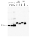
Figure 1
Figure 1
Western blot analysis of the expression of RBD (receptor binding domain) and co-expression of RBD with bacterial Endo H in N. benthamiana. N. benthamiana plants were infiltrated with pEAQ-RBD or with pEAQ-RBD/pGreenII-Endo H (non-tagged) constructs, to produce glycosylated RBD (gRBD) or in vivo deglycosylated RBD (dRBD) of SARS-CoV-2, respectively. Leaf samples were collected at 4 dpi and 5 dpi (days post-infiltration) and were homogenized in three volumes of extraction buffer. RBD protein bands were probed using the anti-4×His tag mAb. Purified plant-produced FLAG-tagged Endo H protein (~30 kDa, 25, 50, and 100 ng) was used as a standard protein. C—crude extract prepared from a control, non-infiltrated plant. gRBD—glycosylated RBD; dRBD—deglycosylated RBD; pp-plant produced.
Figure 2
Figure 2
SDS-PAGE analysis of purified plant-produced gRBD and dRBD proteins. RBD protein variants were purified from N. benthamiana using anti-FLAG antibody resin. (A) gRBD—3 or 6 μg plant-produced glycosylated RBD; dRBD—3 or 6 μg plant-produced deglycosylated RBD; BSA standards—1.0, 2.5 and 5.0 μg; M—color prestained protein standard (NEB). (B) Western blot analysis of gRBD and dRBD using anti-Flag antibody: pp Endo H—plant-produced and purified Endo H protein with molecular mass of ~30 kDa [37]. pp PNGase F—plant-produced and purified PNGase F protein with a molecular mass of ~35 kDa [36]. (C) Western blot analysis of gRBD and dRBD using commercially available, purified anti-SARS-CoV-2 S protein S1 mAb (cat. no. 945102, BioLegend). (D) Western blot analysis of gRBD and dRBD using commercially available anti-RBD antibody (cat. no. MBS2563840, MyBiosource, San Diego, CA, USA); commercial spike protein—COVID 19 spike protein (RBD) active protein, sequence positions Arg319–Phe541 (MBS2563882, MyBiosource); pp—plant-produced; R—reducing; NR—non-reducing condition.
Figure 3
Figure 3
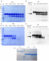
Gel filtration chromatography and glycan detection of plant-produced gRBD or dRBD proteins. Profiles of plant-produced gRBD (A) and dRBD (B) proteins, eluted from Sephacryl® S-200 HR column. The column was equilibrated with 50 mM phosphate buffer (with 150 mM NaCl, pH 7.4). The plant-produced dRBD and gRBD proteins (0.25 mg), purified using FLAG affinity chromatography, were loaded onto column. Gel filtration was performed with ÄKTA start using 60 cm × 16 mm column (cat. no. 19-5003-01, GEHealthcare, Chicago, IL, USA), packed with Sephacryl® S-200 HR (cat. no. 17-0584-10, GE Healthcare). (C) SDS-PAGE analysis of plant-produced gRBD and dRBD proteins eluted from Sephacryl® S-200 HR column. (D) Glycan detection in gRBD and dRBD proteins. Protein (250 ng) from each sample (eluted from Sephacryl® S-200 HR column) was separated on a 10% SDS-PAGE gel followed by in-gel glycan detection using the Pro-Q Emerald 300 glycoprotein staining kit. Stained proteins were visualized by UV illumination. M—CandyCane glycoprotein molecular weight standards (Molecular Probes), 250 ng of each protein per lane; gRBD—glycosylated RBD; dRBD—deglycosylated RBD; gPA83—glycosylated PA83; dPA83—deglycosylated PA83. (E) Western blot analysis of the same sample using the anti-FLAG antibody. Purified Endo H protein (25, 50, and 100 ng) and gRBD or dRBD proteins (50 ng) were loaded into wells.
Figure 4
Figure 4
Binding affinity of gRBD, dRBD, and commercial RBDs of SARS-CoV-2 to ACE2, the receptor of SARS-CoV-2. The samples of 100 ng of plant-produced gRBD, dRBD, and commercially available (A) recombinant SARS-CoV-2 S protein RBD (amino acids Arg319–Phe541, with a C-terminal 8×His tag, expressed in 293E cells, cat. no. 793606, BioLegend, USA), or (B) SARS-CoV-2 spike protein (RBD) coronavirus active protein (amino acids Arg319–Phe541, produced in baculovirus–insect cells, cat. no. MBS2563882, MyBioSource, San Diego, CA, USA) were incubated on plates coated with ACE2. After incubation, anti-SARS-CoV-2 spike RBD polyclonal antibodies were added into each well and detected with rabbit IgG conjugated with HRP. Each point on the graph was derived from three replica for each dilution. Data are shown as mean ± standard error of the mean (SEM) of triplicates in each sample dilution. Statistical significance (p < 0.05) was calculated using the one-way ANOVA test with Tukey’s multiple comparisons. p value for each group is shown in parentheses. ** p < 0.01, *** p < 0.001; OD—optical density; Kd—dissociation constant.
Figure 5
Figure 5
Stability of glycosylated and deglycosylated RBD proteins. Plant-produced, FLAG-antibody affinity column-purified glycosylated (gRBD, A,B) or in vivo Endo H deglycosylated (dRBD, C,D) proteins were incubated at 37 °C for 24, 48, 72, and 96 h and analyzed in SDS-PAGE (A,C) and Western blotting (B,D). Lanes were loaded with ~4.0 μg of gRBD (A) or dRBD (C) and ~200 ng of gRBD (B) or dRBD (D). M—color prestained protein standard. Proteins on the blot were probed with anti-SARS-CoV-2 spike RBD polyclonal antibodies. (E) Proteins were stored at −80 °C for 3 and 6 months as indicated and then analyzed using SDS-PAGE: I—initial sample, after purification of gRBD or dRBD proteins, SDS sample buffer was added to it and it was stored at −80 °C. The image was taken using the highly sensitive GeneGnomeXRQ Chemiluminescence imaging system. Protein bands were calculated and quantified based on SDS-PAGE and WB analyses using the highly sensitive Gene Tools software (Syngene Bioimaging, Cambridge, UK) and ImageJ software, as described previously [38].
Figure 6
Figure 6
Immunogenicity of plant-produced RBD variants in mice. Mice (6–7-week-old Balb/c male) were immunized on days 0 and 21 IM with 5 μg (with alum) of plant-produced gRBD and dRBD using Alhydrogel as an adjuvant. IgG titers were determined by ELISA in sera collected on days 21 or 42 post-vaccination from mice immunized with plant-produced gRBD and dRBD. For immunogenicity analysis, 102 to 108 dilutions of sera were assessed by ELISA. Endpoint titer refers to reciprocal serum dilutions that give a mean OD value four times greater than the pre-immune control samples. Control group received PBS with Alhydrogel. (A) Detection of the SARS-CoV-2 RBD-specific IgG in different dilutions of sera from day 21 or 42. (B) Detection of IgG titers in the murine serum with different dilutions specific to commercially available S protein, gRBD, dRBD at day 21 or 42. Each point on the graph was derived from three replicas for each dilution. Data are shown as mean ± standard error of the mean (SEM) of triplicates in each sample dilution. Statistical significance (p < 0.05) was calculated using the one-way ANOVA test with Tukey’s multiple comparisons. *** p < 0.001 (n = 6 mice/group).
Figure 7
Figure 7
In vitro microneutralization assay of mouse sera against live SARS-CoV-2 in the Vero E6 cell line immunized with plant-produced gRBD or dRBD. In the assay, 32 to 1024 dilutions of the sera were used. Statistical significance (p < 0.05) was calculated using the one-way ANOVA test. ** p < 0.01; *** p < 0.001; n = 6 mice/group.

Similar articles

Cited by

References

    1. Gralinski L.E., Menachery V.D. Return of the Coronavirus: 2019-nCoV. Viruses. 2020;12:135. doi: 10.3390/v12020135. - DOI - PMC - PubMed
    1. Chen J., Wang R., Wang M., Wei G.W. Mutations Strengthened SARS-CoV-2 Infectivity. J. Mol. Biol. 2020;432:5212–5226. doi: 10.1016/j.jmb.2020.07.009. - DOI - PMC - PubMed
    1. Duchene S., Featherstone L., Haritopoulou-Sinanidou M., Rambaut A., Philippe L., Baele G. Temporal signal and the phylodynamic threshold of SARS-CoV-2. Virus Evol. 2020;6:2. doi: 10.1093/ve/veaa061. - DOI - PMC - PubMed
    1. Worobey M., Pekar J., Larsen B.B., Nelson M.I., Hill V., Joy J.B., Rambaut A., Suchard M.A., Wertheim J.O., Lemey P. The emergence of SARS-CoV-2 in Europe and North America. Science. 2020;370:564–570. doi: 10.1126/science.abc8169. - DOI - PMC - PubMed
    1. Hou Y.J., Chiba S., Halfmann P., Ehre C., Kuroda M., Dinnon K.H., Leist S.R., Schafer A., Nakajima N., Takahashi K., et al. SARS-CoV-2 D614G variant exhibits efficient replication ex vivo and transmission in vivo. Science. 2020;370:1464–1468. doi: 10.1126/science.abe8499. - DOI - PMC - PubMed

Publication types

MeSH terms

LinkOut - more resources