. 2021 Aug 27;17(8):e1009771.
doi: 10.1371/journal.pgen.1009771. eCollection 2021 Aug.

Allele-specific mitochondrial stress induced by Multiple Mitochondrial Dysfunctions Syndrome 1 pathogenic mutations modeled in Caenorhabditis elegans

Affiliations

Allele-specific mitochondrial stress induced by Multiple Mitochondrial Dysfunctions Syndrome 1 pathogenic mutations modeled in Caenorhabditis elegans

Peter A Kropp et al. PLoS Genet. .

Abstract

Multiple Mitochondrial Dysfunctions Syndrome 1 (MMDS1) is a rare, autosomal recessive disorder caused by mutations in the NFU1 gene. NFU1 is responsible for delivery of iron-sulfur clusters (ISCs) to recipient proteins which require these metallic cofactors for their function. Pathogenic variants of NFU1 lead to dysfunction of its target proteins within mitochondria. To date, 20 NFU1 variants have been reported and the unique contributions of each variant to MMDS1 pathogenesis is unknown. Given that over half of MMDS1 individuals are compound heterozygous for different NFU1 variants, it is valuable to investigate individual variants in an isogenic background. In order to understand the shared and unique phenotypes of NFU1 variants, we used CRISPR/Cas9 gene editing to recreate exact patient variants of NFU1 in the orthologous gene, nfu-1 (formerly lpd-8), in C. elegans. Five mutant C. elegans alleles focused on the presumptive iron-sulfur cluster interaction domain were generated and analyzed for mitochondrial phenotypes including respiratory dysfunction and oxidative stress. Phenotypes were variable between the mutant nfu-1 alleles and generally presented as an allelic series indicating that not all variants have lost complete function. Furthermore, reactive iron within mitochondria was evident in some, but not all, nfu-1 mutants indicating that iron dyshomeostasis may contribute to disease pathogenesis in some MMDS1 individuals.


Conflict of interest statement

The authors have declared that no competing interests exist.

Figures

Fig 1
Fig 1. C. elegans nfu-1/lpd-8 is orthologous to human and yeast NFU1.
(A) Partial Clustal Omega alignment of yeast Nfu1, C. elegans NFU-1/LPD-8, mouse Nfu1, and human NFU1 highlighting five residues mutated in MMDS1 and the conserved CXXC ISC-interaction motif. (B) Predicted structure of the C. elegans NFU-1 C-terminal domain with mutated residues annotated in red and labeled with corresponding mutations. (C) Representative complementation assay with yeast nfu1, C. elegans nfu-1/lpd-8, and human NFU1 on complete (SC) and acetate (OAc) medium. Cell density indicated above images. (D) Known targets of NFU1/NFU-1 and downstream pathways/complexes affected. NFU1/NFU-1 predominantly functions as a homodimer, but other binding partners are possible (indicated by dotted line and question mark) (E) Representative western blot analysis of lipoylated proteins from L4 WT and nfu-1 mutants. Lipoylated proteins are above and tubulin (loading control) is below. DLAT-1 (E2 subunit of PDH; 53kD) and DLST-1 (E2 subunit of KGDH; 49.8kD) indicated to the right. The lower band is consistent with LIAS-1 (39.8kD) but could not be verified.
Fig 2
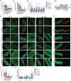
Fig 2. Mutations to nfu-1 affect lifespan and fecundity.
(A-A’) Kaplan-Meier survival curve and analysis of nfu-1 mutants (n = 26–30). (B) Total brood size (n = 11–15 broods), (C) embryonic viability (n = 11–15 broods), and (D) L4 germline nuclei of nfu-1 mutants (n = 12–22). A’: Log-rank (Mantle-Cox) analysis with Bonferroni correction for multiple comparisons. B-D: *: p≤0.05; ****: p≤0.0001 difference between WT and mutant by One-way ANOVA with Dunnett correction for multiple comparisons. NS: Not significant.
Fig 3
Fig 3. Mitochondrial physiology and function are impaired by nfu-1 mutations.
(A-A’) OCR as measured via Seahorse XFe96 (n = 11–14). (A) Basal OCR. (A’) Maximal OCR. (B) Gene expression of ETC components and accessory factors (n = 3–5). Genes assessed in order presented: gas-1 (CI), mev-1 (CII), clk-1 (Coenzyme Q), isp-1 (CIII), cox-4 (CIV), and asg-2 (CV). (B’) ETC schematic with the factor measured in (B) indicated for each complex or accessory factor. (C) Representative COX-4::GFP expression in mitochondria of WT and nfu-1 mutants in the gonad, body wall muscle, and hypodermis. Images captured at 63X or 63X + 3X digital zoom. White boxes indicate regions enhanced at right. Green: COX-4::GFP. Scale bar: 10μm. (D) TMRE labeling of hypodermal mitochondria in WT and nfu-1 mutants. Green: COX-4::GFP; Red: TMRE. Scale bar: 20μm. (E) Quantification of (D). Ratiometric measure of pixel intensity (561nm/488nm) (n = 11–32). (F) Gene expression mitochondrial fission (fzo-1), fusion (drp-1) and mitophagy (dct-1) positive regulators (n = 3–5). A-B; E-F: *: p≤0.05; **:p≤0.01; ***:p≤0.001; ****:p≤0.0001 difference between control and nfu-1 mutant by One-way ANOVA with Dunnett correction for multiple comparisons. OCR: oxygen consumption rate; ETC: electron transport chain; TMRE: Tetramethylrhodamine, Ethyl Ester.
Fig 4
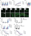
Fig 4. Activation of multiple stress response pathways in nfu-1 mutants.
(A) Gene expression of atfs-1 and direct targets in the UPRmt (n = 3). (B-B’) H2DCFDA fluorescent signal for ROS (n = 7). (B) Fluorescent signal over time. (B’) End point fluorescent signal. (C) Representative images of the Pgst-4::gst-4::GFP transgene in control (transgene only) and nfu-1 mutants. Scale bar: 100μm in 10X; 20μm in 60X images. Insets at 10X show DIC image of animals in the field of view. (D-D’) Gene expression of superoxide dismutase genes (n = 3–4). (E-E’) Gene expression of catalase genes (n = 3–4). (F, G) Kaplan-Meier survival curves with exposure to exogenous oxidants. (F) H2O2 exposure (n = 134–175). (G) Paraquat exposure (n = 102–251). Data in (F-G) represented as percentages ± standard error. Statistical analysis in Table 2. *: p≤0.05; **:p≤0.01; ***:p≤0.001; ****:p≤0.0001 difference between (A-B,D,E) WT and nfu-1 mutants by One-way ANOVA with Dunnett correction for multiple comparisons; (D’,E’) all groups by One-way ANOVA with Tukey correction for multiple comparisons. NS: Not significant. RFU: Relative fluorescence units.
Fig 5
Fig 5. DAF-16-mediated iron response activation in nfu-1 mutants.
(A) Schematic of the DAF-16-mediated iron response pathway. DAF-16 is normally inhibited (indirectly) by DAF-2. Upon disinhibition, DAF-16 translocates to the nucleus where it can activate its transcriptional program including the C. elegans iron response. (B-B”) Gene expression of ftn-1 and smf-3 (n = 3–6). (B) WT and nfu-1 mutants; (B’) WT and daf-2(e1370) at 15°C or 25°C; (B”) Veh. (H2O) or FAC treatment. (C): Schematic of 2–2’-BP treatment. (D-D’) Gene expression of ftn-1 and smf-3 following treatment with Veh. (EtOH) or 2–2’-BP (n = 3–5). (D) Expression of ftn-1; (D’) Expression of smf-3. (E-E’) Fecundity assays following treatment with Veh. (DMSO) or fer-1 (n = 9–10). (E) Total brood size. (E’) Embryonic viability. B-B”: *: p≤0.05; **:p≤0.01; ***:p≤0.001; ****:p≤0.0001 difference between (B) WT and nfu-1 mutants by One-way ANOVA with Dunnett correction for multiple comparisons; (B’) all groups by One-way ANOVA with Tukey correction for multiple comparisons; or (B”) Vehicle and FAC by parallel two-tailed Student’s T-test with Holm-Sidak correction for multiple comparisons. D-D’ black bars: *: p≤0.05; **:p≤0.01; ***:p≤0.001 between Veh. and 2–2’-BP by parallel two-tailed Student’s T-test with Holm-Sidak correction for multiple comparisons. D-D’ gray bars: *: p≤0.05; **:p≤0.01; ****:p≤0.0001 between fold change of WT and fold change of nfu-1 mutants as post-hoc One-way ANOVA with Dunnet correction for multiple comparisons. ROS: reactive oxygen species;: Veh.: Vehicle; FAC: ferric ammonium citrate; 2–2’-BP: 2–2’-bipyridyl; ND: Not detected; NS: Not significant.
Fig 6
Fig 6. Antioxidant treatment partially suppresses the nfu-1 phenotype.
(A) Kaplan-Meier survival curve of nfu-1 mutants with either Veh. (H2O) or NAC treatment (n = 58–60). Detailed statistical analysis presented in Table 3. (B-B”) Fecundity assays following treatment with Veh. (H2O) or NAC (n = 9–10). (B) Total brood size. (B’) Embryonic viability. No statistical differences between treated and untreated. B-B’: Analysis by parallel two-tailed Student’s T-test with Holm-Sidak correction for multiple comparisons. Veh.: Vehicle; NAC: N-acetyl-L-cysteine.
Fig 7
Fig 7. Proposed model for ISC handling and phenotypes of nfu-1 mutants.
Proposed model for how nfu-1 mutants affect ISC handling and ISC delivery. Model assumes NFU-1 protein functioning as a homodimer. Gly147Arg variant is biased toward monomeric state impairing ISC delivery to ISPs and potentially exposing ISC to reactive environment and producing ROS. Gly166Cys variant is biased towards the dimeric state impairing ISC release and delivery to ISPs. Increased dimerization indicated by NFU-1 proteins more closely surrounding the ISC. Gly148Arg and Cys168Phe are nonfunctional and do not bind ISCs eliminating ISC delivery to ISPs. Gly148Arg, Gly166Cys and Cys168Phe accumulate mitochondrial iron which is reactive via Fenton Chemistry to produce ROS. ISP dysfunction results because of impaired ISC delivery, which leads to elevated ROS and decreased metabolism which in turn activate mitochondrial stress responses. ISP: Iron-Sulfur protein; ISC: Iron-Sulfur cluster; ROS: reactive oxygen species; Fe: labile iron; UPRmt: mitochondrial unfolded protein response.

Similar articles

Cited by

References

    1. Andreini C, Banci L, Rosato A. Exploiting Bacterial Operons To Illuminate Human Iron-Sulfur Proteins. Journal of proteome research. 2016;15: 1308–1322. doi: 10.1021/acs.jproteome.6b00045 - DOI - PubMed
    1. Rouault TA, Maio N. Biogenesis and functions of mammalian iron-sulfur proteins in the regulation of iron homeostasis and pivotal metabolic pathways. The Journal of biological chemistry. 2017;292: 12744–12753. doi: 10.1074/jbc.R117.789537 - DOI - PMC - PubMed
    1. Braymer JJ, Lill R. Iron-sulfur cluster biogenesis and trafficking in mitochondria. The Journal of biological chemistry. 2017;292: 12754–12763. doi: 10.1074/jbc.R117.787101 - DOI - PMC - PubMed
    1. Kim KS, Maio N, Singh A, Rouault TA. Cytosolic HSC20 integrates de novo iron–sulfur cluster biogenesis with the CIAO1-mediated transfer to recipients. Hum Mol Genet. 2018;27: 837–852. doi: 10.1093/hmg/ddy004 - DOI - PMC - PubMed
    1. Cai K, Liu G, Frederick RO, Xiao R, Montelione GT, Markley JL. Structural/Functional Properties of Human NFU1, an Intermediate [4Fe-4S] Carrier in Human Mitochondrial Iron-Sulfur Cluster Biogenesis. Structure (London, England: 1993). 2016;24: 2080–2091. doi: 10.1016/j.str.2016.08.020 - DOI - PMC - PubMed

Publication types

MeSH terms

Supplementary concepts

Grants and funding

This work was supported, in part, by the Intramural Research Program of the National Institutes of Health, National Institute of Diabetes and Digestive and Kidney Diseases (A.G.) and National Heart, Lung, and Blood Institute (M.N.S.). The funders had no role in the study design, data collection and analysis, decision to publish, or preparation of the manuscript.