. 2021 Aug 27;17(8):e1009757.
doi: 10.1371/journal.pgen.1009757. eCollection 2021 Aug.

AKTIP interacts with ESCRT I and is needed for the recruitment of ESCRT III subunits to the midbody

Affiliations

AKTIP interacts with ESCRT I and is needed for the recruitment of ESCRT III subunits to the midbody

Chiara Merigliano et al. PLoS Genet. .

Abstract

To complete mitosis, the bridge that links the two daughter cells needs to be cleaved. This step is carried out by the endosomal sorting complex required for transport (ESCRT) machinery. AKTIP, a protein discovered to be associated with telomeres and the nuclear membrane in interphase cells, shares sequence similarities with the ESCRT I component TSG101. Here we present evidence that during mitosis AKTIP is part of the ESCRT machinery at the midbody. AKTIP interacts with the ESCRT I subunit VPS28 and forms a circular supra-structure at the midbody, in close proximity with TSG101 and VPS28 and adjacent to the members of the ESCRT III module CHMP2A, CHMP4B and IST1. Mechanistically, the recruitment of AKTIP is dependent on MKLP1 and independent of CEP55. AKTIP and TSG101 are needed together for the recruitment of the ESCRT III subunit CHMP4B and in parallel for the recruitment of IST1. Alone, the reduction of AKTIP impinges on IST1 and causes multinucleation. Our data altogether reveal that AKTIP is a component of the ESCRT I module and functions in the recruitment of ESCRT III components required for abscission.


Conflict of interest statement

The authors have declared that no competing interests exist.

Figures
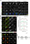
Fig 1
Fig 1. AKTIP localizes as a ring at the dark zone of the midbody.
(A) Confocal immunofluorescence images showing the distribution of AKTIP during mitosis and cytokinesis. HeLa cells were stained with anti-AKTIP (red), anti-α-tubulin (green) and DAPI to visualize DNA (blue). Scale bar, 5μm. (B) Representative 3D-SIM images of cells observed for AKTIP at the midbody in early, mid, late and cut stages. HeLa cells were stained with α-tubulin (green) and AKTIP (red) antibodies. Scale bar, 5μm. (C) Representative fluorescence intensity profile plotted for AKTIP and α-tubulin along the midbody at different stages; early/forming (n = 6), mid (n = 7), late (n = 8), cut (n = 8). (D-E) Representative 3D-SIM images of AKTIP ring (D) and relative measurements (E). Size of AKTIP structure measured in mid (n = 8), late (n = 7) and cut (n = 7) midbodies. Scale bar, 2.5μm.
Fig 2
Fig 2. AKTIP is associated with the ESCRT I subunits VPS28 and TSG101.
(A) AKTIP fused to the Gal4 DNA binding domain was tested for interactions with the human components of ESCRT I, II, III, and ESCRT associated proteins fused to the VP16 activation domain by yeast two-hybrid assay. Error bars indicate the SEM from the mean of triplicate measurements. (B) Western blotting showing that AKTIP interacts with GST-VPS28 but not with GST alone. Cells were transfected with plasmids encoding the indicated fusion proteins. Purified VPS28-GST or GST alone were used to pull down interacting proteins; cell lysates and glutathione-bound fractions were then analyzed with MYC antisera. GST-pull down was repeated three times. (C) Spinning disk microscopy images of AKTIP (green) and VPS28 (red). Scale bar, 5μm. (D) 3D rendering of spinning disk imaging as in (C) showing that AKTIP and VPS28 are in proximity at midbody. VPS28 in late midbodies displays also an asymmetric protruding element. Scale bar, 0.5 μm. (E) Western blotting showing that HA-AKTIP, GFP-TSG101 and GST-VPS28 are captured together in GST pull down experiment. Cells were co-transfected with a fixed amount of plasmid encoding GFP-TSG101 (500 ng) and GST-VPS28 (1000 ng) and increasing amounts of HA-AKTIP (0, 50, 100, 500 or 1000 ng) encoding plasmid. Cell lysates and GST pull down fractions were then analyzed with GST, GFP, HA antisera. GST-pull down was repeated three times. (F) Spinning disk microscopy images of AKTIP (green) and TSG101 (red). Scale bar, 5μm. (G) 3D rendering of spinning disk imaging as in (E) showing that AKTIP and TSG101 are near each other. Scale bar, 0.5 μm. (H) Quantification of the percentage of midbodies positive both for AKTIP and TSG101 showing that the two proteins are concomitantly present at the midbody. For results in (H) at least 100 midbodies per condition were counted.
Fig 3
Fig 3. The central region of AKTIP interacts with the N-terminus of VPS28.
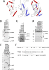
(A) Superimposing of AKTIP model on TSG101 X-ray solved structure highlights similarities in the central region and two main different elements outside of it. Namely, the AKTIP central UEV domain presents two C-terminal helices (H5 and H6), absent in TSG101; TSG101 contains two N-terminal helices (H1 and H2), while AKTIP only one (H2). (B-C) Western blotting showing that central UEV region of AKTIP interacts with GST-VPS28 (B) and that AKTIP interacts with N-terminal domain of VPS28 (C). Cells were transfected with plasmids encoding the indicated fusion proteins and the AKTIP protein N-terminal (1–76 aa, NT), central domain (78–220 aa), C-terminal (221–292 aa) fragments. Purified VPS28-GST, or N-terminal (1–120 aa, NT) VPS28, or C-terminal (121-221aa, CT) or GST alone were used to pull down interacting proteins; cell lysates and glutathione-bound fractions were then analyzed with GST, GFP or MYC antisera. GST-pull downs were repeated three times. (D) Western blotting showing that AKTIP does not interact with VPS37 A to D isotypes and confirming its interaction with VPS28. Cells were transfected with plasmids encoding the indicated fusion proteins. Purified VPS28-GST or VPS37(A-D)-GST or GST alone were used to pull down interacting proteins; cell lysates and glutathione-bound fractions were then analyzed with MYC antisera. GST-pull downs were repeated two times. (E) Western blotting showing that AKTIP does not interact with SEPT9 (A, B and E) and confirming its interaction with VPS28. Cells were transfected with the indicated fusion proteins. Purified SEPT9 (isoforms A, B and E)-GFP, VPS28-GFP or GFP alone were used to trap interacting proteins; then cell lysates and GFP-trapped fractions were analyzed with MYC antisera. GFP-TRAP were repeated three times. (F) Schematic representation of the interacting regions of AKTIP and TSG101 with VPS28. UEV (Ubiquitin E2 variant domain); TSG101 PRR (Proline Rich Region).
Fig 4
Fig 4. The supra-molecular structure of AKTIP is flanked by the rings formed by the ESCRT III components.
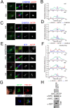
(A, C, E) 3D-SIM images of ESCRT III components CHMP4B (A), CHMP2A (C), IST1 (E) and AKTIP in HeLa cells. Staining with antibodies against ESCRT III (blue), AKTIP (red) and α-tubulin (green). (B, D, F) Representative fluorescence intensity profile plotted for AKTIP, ESCRT III subunits and α-tubulin along the midbodies at different stages. (B) mid, n = 6; late, n = 4. (D) mid, n = 6; late, n = 3. (F) early/forming, n = 6; mid, n = 7; late, n = 8; cut, n = 6. (G) Spinning disk microscopy images of IST1 (blue), AKTIP (green) and TSG101 (red). Scale bars, 2.5μm. (H) Western blotting showing that AKTIP interacts with GST-VPS28, but not with GST-IST1 or GST alone. Cells were transfected with plasmids encoding the indicated fusion proteins. Purified GST-VPS28 or GST-IST1 or GST alone were used to pull down interacting proteins; cell lysates and glutathione-bound fractions were then analyzed with MYC and GST antisera. GST-pull downs were repeated three times.
Fig 5
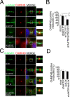
Fig 5. AKTIP recruitment to the midbody is CEP55 independent.
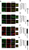
(A-B) Representative images and quantification of sictr and siCEP55 HeLa cells stained for ALIX (red) and α-tubulin (green). (C-D) Representative images and quantification of sictr and siCEP55 HeLa cells stained for TSG101 (red) and α-tubulin (green). (E-F) Representative images and relative quantification of sictr and siCEP55 HeLa cells stained for AKTIP (green) and α-tubulin (red). (G-H) Representative images and relative quantification of shctr and shAKTIP HeLa cells stained for CEP55 (red) and α-tubulin (green). For results in (B, D, F and H) at least 100 midbodies per condition were counted. Scale bar, 5μm.
Fig 6
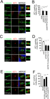
Fig 6. AKTIP, TSG101 and VPS28 are independently recruited to the midbody.
(A-B) Representative images and relative quantification of shctr and shAKTIP HeLa cells stained for TSG101 (red) and α-tubulin (green) showing that TSG101 is present at the midbody in shAKTIP cells. (C-D) Representative images and relative quantification of sictr and siTSG101 HeLa cells stained for AKTIP (red) and α-tubulin (green) showing that AKTIP is present at the midbody in siTSG101 cells (E-F) Representative images and relative quantification of shctr and shAKTIP HeLa cells stained for VPS28 (red) and α-tubulin (green) showing that VPS28 is present at the midbody in shAKTIP cells. (G-H) Representative images and relative quantification of sictr and siVPS28 HeLa cells stained for AKTIP (red) and α-tubulin (green) showing that AKTIP is present at the midbody in siVPS28 cells. For results in (B, D, F and H) at least 100 midbodies per conditions were counted. Scale bars, 5μm.
Fig 7
Fig 7. AKTIP recruitment to the midbody is MKLP1 dependent.
(A) Spinning disk microscopy images of MKLP1 (blue), AKTIP (red) and α-tubulin (green). (B) Representative fluorescence intensity profile plotted for AKTIP, MKLP1 and α-tubulin along the midbodies at different stages; mid, n = 3; late, n = 3. (C) MSIM images of MKLP1 (blue), AKTIP (red) and α-tubulin (green). (D) Representative fluorescence intensity profile plotted for AKTIP, MKLP1 and α-tubulin along mid midbodies from (C); n = 3. (E) Representative images of sictr and siMKLP1 HeLa cells stained for AKTIP (red), MKLP1 (blue) and α-tubulin (green) showing that AKTIP recruitment is MKLP1 dependent. (F) Quantification of AKTIP positive midbodies in sictr and siMKLP1 cells. For results in (F) at least 25 midbodies were counted for each sample. Scale bars, 5μm.
Fig 8
Fig 8. AKTIP cooperates with TSG101 but not with ALIX for CHMP4B recruitment.
(A-B) Representative images of shctr, shAKTIP, siTSG101 and shAKTIP/siTSG101 HeLa cells stained for CHMP4B (red) and α-tubulin (green) and relative quantification. (C-D) Representative images of shctr, shAKTIP, siALIX and shAKTIP/siALIX HeLa cells stained for CHMP4B (red) and α-tubulin (green) and relative quantification. For results in (B and D) at least 100 midbodies per condition were counted. Scale bars, 5μm.
Fig 9
Fig 9. AKTIP and TSG101 act in a common pathway to promote IST1 recruitment.
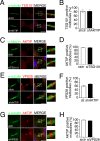
(A-B) Representative images of shctr and shAKTIP, siTSG101 and shAKTIP/siTSG101 HeLa cells stained for IST1 (red) and α-tubulin (green) and relative quantification. (C-D) Representative images of sictr, siTSG101 and AKTIP-FLAG/siTSG101 HeLa cells stained for IST1 (red) and α-tubulin (green) and relative quantification. (E-F) Representative images of shctr and 3’UTR shAKTIP, and AKTIP-FLAG/3’UTR shAKTIP HeLa cells stained for IST1 (red) and α-tubulin (green) and relative quantification. For results in (B, D and F) at least 100 midbodies per condition were counted. Scale bars, 5μm.
Fig 10
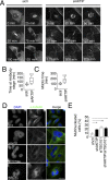
Fig 10. The reduction of AKTIP is associated with cytokinesis defects.
(A) Selected frames from time-lapse microscopy of ctr or AKTIP depleted (siAKTIP) HeLa cells stably expressing mCherry-tubulin. The arrow points to an example of two cells that remain connected. Elapsed times are provided in each panel. (B-C) Quantitative analysis of time-lapse microscopy showing the time from prometaphase to abscission and from telophase to abscission. (D-E) Representative images and quantification of the percentage of multinucleated cells compared to normal interphase nuclei in shctr, shAKTIP, siTSG101, shAKTIP/siTSG101 HeLa cells. For results in (E) at least 200 nuclei per condition were counted. Scale bars, 5μm.
Fig 11
Fig 11. Schematic representation of AKTIP structural and functional association with the ESCRT machinery.
(A) AKTIP localizes at the midbody in proximity with the annular structure formed by MKLP1 and with that formed by the ESCRT I subunit TSG101 and VPS28. AKTIP super-structure is flanked by the rings formed by the ESCRT III subunits CHMP4B, CHMP2A and IST1. (B) AKTIP is recruited to the midbody in a MKLP1 dependent, CEP55 independent way. AKTIP and TSG101 cooperate in the recruitment of CHMP4B to the midbody through independent routes, likely involving the ESCRT I subunit VPS28, that interacts with both TSG101 and AKTIP. AKTIP and TSG101 are needed for the recruitment of IST1 to the midbody, and act in a common pathway leading to abscission.

Similar articles

Cited by

References

    1. Carlton J. The ESCRT machinery: a cellular apparatus for sorting and scission. Biochem Soc Trans. 2010;38(6):1397–412. Epub 2010/12/02. doi: 10.1042/BST0381397 . - DOI - PubMed
    1. Schoneberg J, Lee IH, Iwasa JH, Hurley JH. Reverse-topology membrane scission by the ESCRT proteins. Nat Rev Mol Cell Biol. 2017;18(1):5–17. Epub 2016/11/04. doi: 10.1038/nrm.2016.121 ; PubMed Central PMCID: PMC5198518. - DOI - PMC - PubMed
    1. Morita E, Sandrin V, Chung HY, Morham SG, Gygi SP, Rodesch CK, et al.. Human ESCRT and ALIX proteins interact with proteins of the midbody and function in cytokinesis. EMBO J. 2007;26(19):4215–27. doi: 10.1038/sj.emboj.7601850 ; PubMed Central PMCID: PMC2230844. - DOI - PMC - PubMed
    1. Carlton JG, Martin-Serrano J. The ESCRT machinery: new functions in viral and cellular biology. Biochem Soc Trans. 2009;37(Pt 1):195–9. Epub 2009/01/16. doi: 10.1042/BST0370195 . - DOI - PubMed
    1. Carlton JG, Martin-Serrano J. Parallels between cytokinesis and retroviral budding: a role for the ESCRT machinery. Science. 2007;316(5833):1908–12. Epub 2007/06/09. doi: 10.1126/science.1143422 . - DOI - PubMed

Publication types

MeSH terms

LinkOut - more resources