

Neuroglobin: A New Possible Marker of Estrogen-Responsive Breast Cancer
- PMID: 34440755
- PMCID: PMC8393432
- DOI: 10.3390/cells10081986
Neuroglobin: A New Possible Marker of Estrogen-Responsive Breast Cancer
Abstract
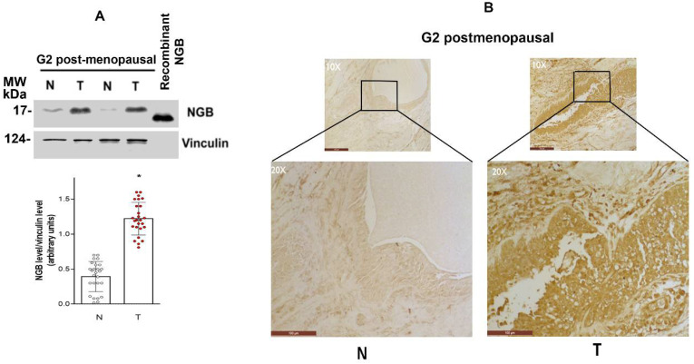
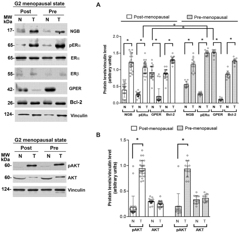
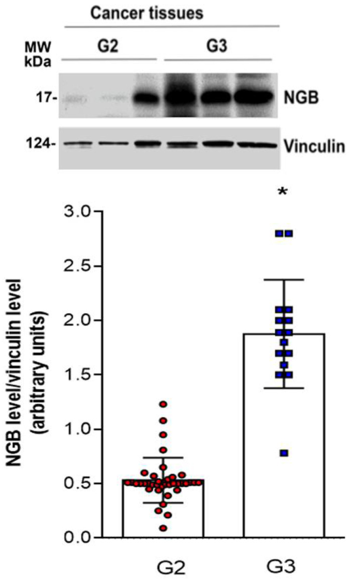
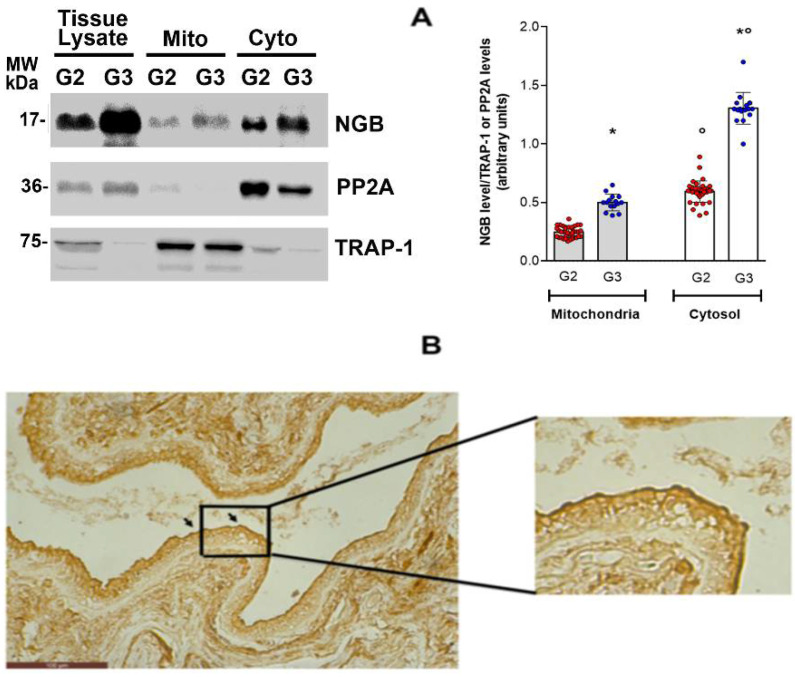
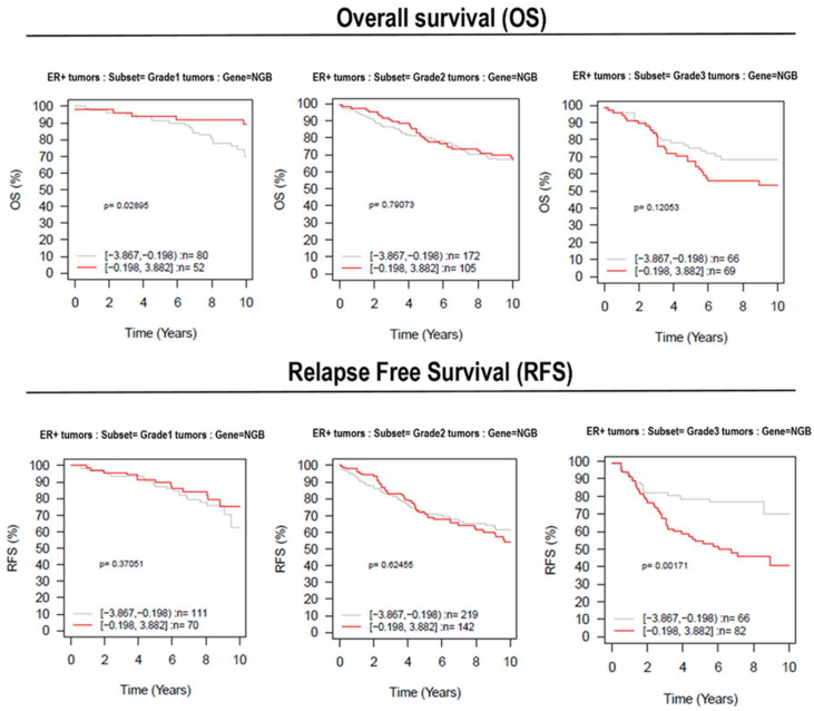
The expression of the α-subtype of Estrogen Receptor (ERα) characterizes most breast cancers (more than 75%), for which endocrine therapy is the mainstay for their treatment. However, a high percentage of ERα+ breast cancers are de novo or acquired resistance to endocrine therapy, and the definition of new targets for improving therapeutic interventions and the prediction of treatment response is demanding. Our previous data identified the ERα/AKT/neuroglobin (NGB) pathway as a common pro-survival process activated in different ERα breast cancer cell lines. However, no in vivo association between the globin and the malignity of breast cancer has yet been done. Here, we evaluated the levels and localization of NGB in ERα+ breast ductal carcinoma tissue of different grades derived from pre-and post-menopausal patients. The results indicate a strong association between NGB accumulation, ERα, AKT activation, and the G3 grade, while no association with the menopausal state has been evidenced. Analyses of the data set (e.g., GOBO) strengthen the idea that NGB accumulation could be linked to tumor cell aggressiveness (high grade) and resistance to treatment. These data support the view that NGB accumulation, mainly related to ER expression and tumor grade, represents a compensatory process, which allows cancer cells to survive in an unfavorable environment.
Keywords: AKT; breast cancer survival; ductal infiltrating adenocarcinoma; estrogen receptor α; neuroglobin; tumor microenvironment.
Conflict of interest statement
The authors declare that they have no conflict of interest.
Figures
Similar articles
-
Dissecting the 17β-estradiol pathways necessary for neuroglobin anti-apoptotic activity in breast cancer.J Cell Physiol. 2018 Jul;233(7):5087-5103. doi: 10.1002/jcp.26378. Epub 2018 Jan 19. J Cell Physiol. 2018. PMID: 29219195
-
Potentiation of paclitaxel effect by resveratrol in human breast cancer cells by counteracting the 17β-estradiol/estrogen receptor α/neuroglobin pathway.J Cell Physiol. 2019 Apr;234(4):3147-3157. doi: 10.1002/jcp.27309. Epub 2018 Nov 13. J Cell Physiol. 2019. PMID: 30421506
-
Neuroglobin overexpression induced by the 17β-Estradiol-Estrogen receptor-α Pathway reduces the sensitivity of MCF-7 Breast cancer cell to paclitaxel.IUBMB Life. 2016 Aug;68(8):645-51. doi: 10.1002/iub.1522. Epub 2016 Jun 16. IUBMB Life. 2016. PMID: 27312786
-
Gli1 is a potential cancer stem cell marker and predicts poor prognosis in ductal breast carcinoma.Hum Pathol. 2017 Nov;69:38-45. doi: 10.1016/j.humpath.2017.08.038. Epub 2017 Sep 28. Hum Pathol. 2017. PMID: 28965964
-
HER2 as a prognostic factor in breast cancer.Oncology. 2001;61 Suppl 2:67-72. doi: 10.1159/000055404. Oncology. 2001. PMID: 11694790 Review.
Cited by
-
Structural and (Pseudo-)Enzymatic Properties of Neuroglobin: Its Possible Role in Neuroprotection.Cells. 2021 Nov 30;10(12):3366. doi: 10.3390/cells10123366. Cells. 2021. PMID: 34943874 Free PMC article. Review.
-
Editorial for Special Issue: Neuroglobin from Brain Protection to Cancer Progression.Cells. 2022 Jul 13;11(14):2181. doi: 10.3390/cells11142181. Cells. 2022. PMID: 35883624 Free PMC article.
-
A functional genetic screen for metabolic proteins unveils GART and the de novo purine biosynthetic pathway as novel targets for the treatment of luminal A ERα expressing primary and metastatic invasive ductal carcinoma.Front Endocrinol (Lausanne). 2023 Apr 18;14:1129162. doi: 10.3389/fendo.2023.1129162. eCollection 2023. Front Endocrinol (Lausanne). 2023. PMID: 37143728 Free PMC article.
References
-
- Skandalis S.S., Afratis N., Smirlaki G., Nikitovic D., Theocharis A.D., Tzanakakis G.N., Karamanos N.K. Cross-Talk between Estradiol Receptor and EGFR/IGF-IR Signaling Pathways in Estrogen-Responsive Breast Cancers: Focus on the Role and Impact of Proteoglycans. Matrix Biol. 2014;35:182–193. doi: 10.1016/j.matbio.2013.09.002. - DOI - PubMed
Publication types
MeSH terms
Substances
Grants and funding
LinkOut - more resources
Full Text Sources
Medical
Molecular Biology Databases
Miscellaneous
