Review
. 2021 Aug 12;13(8):565.
doi: 10.3390/toxins13080565.

In Vivo Models and In Vitro Assays for the Assessment of Pertussis Toxin Activity

Affiliations
Review

In Vivo Models and In Vitro Assays for the Assessment of Pertussis Toxin Activity

Marieke Esther Hoonakker. Toxins (Basel). .

Abstract

One of the main virulence factors produced by Bordetella pertussis is pertussis toxin (PTx) which, in its inactivated form, is the major component of all marketed acellular pertussis vaccines. PTx ADP ribosylates Gαi proteins, thereby affecting the inhibition of adenylate cyclases and resulting in the accumulation of cAMP. Apart from this classical model, PTx also activates some receptors and can affect various ADP ribosylation- and adenylate cyclase-independent signalling pathways. Due to its potent ADP-ribosylation properties, PTx has been used in many research areas. Initially the research primarily focussed on the in vivo effects of the toxin, including histamine sensitization, insulin secretion and leukocytosis. Nowadays, PTx is also used in toxicology research, cell signalling, research involving the blood-brain barrier, and testing of neutralizing antibodies. However, the most important area of use is testing of acellular pertussis vaccines for the presence of residual PTx. In vivo models and in vitro assays for PTx often reflect one of the toxin's properties or details of its mechanism. Here, the established and novel in vivo and in vitro methods used to evaluate PTx are reviewed, their mechanisms, characteristics and limitations are described, and their application for regulatory and research purposes are considered.

Keywords: acellular pertussis vaccines; in vitro assays; in vivo models; pertussis toxin.


Conflict of interest statement

The author declares no conflict of interest. The funders had no role in the design of the study; in the collection, analyses, or interpretation of data; in the writing of the manuscript, or in the decision to publish the results.

Figures

Figure 1
Figure 1
The classical models for PTx binding, internalisation, ADP ribosylation, and its effect on cell signalling. The B oligomer of PTx binds to glycoconjugate proteins on the cell surface, upon which the holotoxin enters the cell by endocytosis, followed by retrograde transport through the endosome, Golgi and the endoplasmatic reticulum. Subsequently, the S1 subunit is released into the cytosol. Within the cytosol, the S1 subunit catalyses the transfer of ADP-ribose from NAD+ to the α-subunit of Gαi/o/t proteins, thereby preventing interaction of these proteins with their cognate receptors. ADP ribosylation fixes the α-subunit of the G-proteins in their inactive (GDP-bound) form, thereby rendering it unable to inhibit its target enzymes; ACs. ACs catalyse the conversion of ATP into cAMP. This second messenger binds to and activates protein kinase A (PKA), which is involved in a range of pathways, one of which is the phosphorylation of the cAMP response element-binding protein (CREB). CREB binds to the DNA sequence cAMP response elements (CRE) and thereby increases the transcription of CRE responsive genes. PTx-induced cAMP might also directly bind to “exchange protein directly activated by cAMP” (EPAC), which are guanine nucleotide exchange factors for Rap molecules.
Figure 2
Figure 2
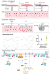
The classical in vivo methods for PTx and models for the mechanisms. (A). The histamine sensitization test is based on the sensitizing effects of PTx in mice to histamine doses which would normally cause no effect. Under “normal” conditions, vascular permeability and blood pressure is maintained by the vascular endothelial cells and vascular smooth muscle cells. Administration of histamine results in the reorganization of actin filament structure and adherence junctions, increased endothelial permeability and vascular leakiness. Contraction of vascular smooth muscle cells can (partly) compensate for the blood volume loss, but this compensatory mechanism is inhibited by PTx, causing a significantly reduction in blood pressure. (B). In the leukocytosis promotion test, the effect of PTx on leukocyte numbers is measured. PTx inhibits lymphocyte extravasation and restores lymphocyte egress from lymph nodes to the lymph, resulting in a rise in circulating leukocyte number. Migration involves rolling mediated by attachment to selectins and arrest mediated by integrins. The binding of integrins and endothelial adhesion molecules triggers the opening of endothelial junctions, allowing leukocytes to transmigrate. PTx does not affect selectins and rolling, but does inhibit integrin mediated arrest. Integrins can only bind to adhesion molecules on the high endothelial venule cells upon activation by Gαi-coupled chemokine receptors. These Gαi-coupled receptors might be the target of PTx, although additional research will be necessary to confirm whether these receptors are responsible for the leukocytosis. (C). The mouse weight gain test is based upon the negative effect of PTx on the weight gain of mice throughout a period of seven days. Although considered a general toxicity test, its mechanism is unknown. (D). In the islet activation protein test, PTx enhances the glucose-induced release of insulin. In the test, PTx is responsible for ADP ribosylation of Gαi/o proteins in β-cell, resulting in accumulation of cAMP. Although not studied directly in relation to PTx, enhanced cAMP levels can activate PKA and EPAC and stimulate the glucose pathway that induces the release of insulin. The other pathway shown to be involved in the IAP test is mediated by ghrelin. Ghrelin is an endogenous ligand for the Gαi-coupled growth hormone secretagogue receptor, which can suppress K+ exflux. As a result of the ADP ribosylation of this Gαi-coupled receptor, K+ exflux is inhibited. The resulting depolarization causes Ca2+ influx and enhanced levels of Ca2+ are essential for the release of insulin.
Figure 3
Figure 3
The cellular in vitro assays for PTx and their mechanisms. (A) The CHO cell clustering assay is based on the clustered growth pattern of CHO cells in response to PTx and requires an active S1 subunit. The morphological changes require rearrangement the cytoskeletal structures, but the underlying responsible mechanisms have not been fully elucidated. The first proposed mechanism (A1) involves the RhoA pathway, which normally results in polymerization of actin filaments. cAMP-dependent activation of PKA might result in an inactive state of RhoA, thereby diminishing or preventing actin polymerisation. Alternatively (A2), PTx-induced uncoupling of αi proteins might directly affect the functioning of Rap1 and thereby reduce polymerization of cytoskeletal structures. In the third proposed mechanism (A3), uncoupling of Gαi/o protein causes cGMP accumulation which also suppresses actin polymerization, affecting both cell shape and motility. Additional studies will be required to determine the involvement these three mechanisms in the CHO cell clustering assay. Clustering can be analysed manually and quantified by electrical impedance, continuous phase-contrast imaging or by measuring the distance between nearest neighbouring nuclei. (B) The ATP and cAMP PTx assays are based on the effects of PTx on the conversion of ATP to cAMP by the ACs. In the ATP-PTx assay (B1), the effect of PTx on ATP levels in peripheral blood mononuclear cells is measured using an ATP-luminescence assay. The cAMP-PTx assay is based on the rise in cAMP upon exposure of cells to PTx. Initially, A10 cells (B2) were used to monitor for PTx in combination with isoprenaline as an activator of AC using and a commercially available ELISA kits to measure cAMP levels. Subsequently, a CHO cell line stably expressing a CRE controlled NanoLuc construct was developed (B3). Upon stimulation with forskolin, PTx enhances the production of cAMP, leading to the activation of PKA, resulting in enhanced transcription of NanoLuc. The iGIST assay (C) is based upon HEK293 cells that co-express the Gαi-coupled SSTR2 receptor and a luminescent cAMP probe. In combination with octreotide and forskolin, these cells allow for real time assessment of cellular cAMP levels. PTx ADP ribosylates the αi subunit of SSTR2, diminishing the effect of octreotide and results in enhanced levels of cAMP. cAMP binding to the probe causes a conformational change and increases the luminescence signal. In the MoDC assay (D), the effect of PTx on the production of the cytokines IL-2 and IFN-γ by MoDC is assessed. As PTx is described to stimulate the TLR4 receptor, this pathway may be involved in MoDC activation.

Similar articles

Cited by

References

    1. Yeung K.H.T., Duclos P., Nelson E.A.S., Hutubessy R.C.W. An update of the global burden of pertussis in children younger than 5 years: A modelling study. Lancet Infect. Dis. 2017;17:974–980. doi: 10.1016/S1473-3099(17)30390-0. - DOI - PubMed
    1. Libster R., Edwards K.M. Re-emergence of pertussis: What are the solutions? Expert Rev. Vaccines. 2012;11:1331–1346. doi: 10.1586/erv.12.118. - DOI - PubMed
    1. Sato H., Ito A., Chiba J., Sato Y. Monoclonal antibody against pertussis toxin: Effect on toxin activity and pertussis infections. Infect. Immun. 1984;46:422–428. doi: 10.1128/iai.46.2.422-428.1984. - DOI - PMC - PubMed
    1. Black W.J., Munoz J.J., Peacock M.G., Schad P.A., Cowell J.L., Burchall J.J., Lim M., Kent A., Steinman L., Falkow S. ADP-ribosyltransferase activity of pertussis toxin and immunomodulation by Bordetella pertussis. Science. 1988;240:656–659. doi: 10.1126/science.2896387. - DOI - PubMed
    1. Markey K., Asokanathan C., Feavers I. Assays for Determining Pertussis Toxin Activity in Acellular Pertussis Vaccines. Toxins. 2019;11:417. doi: 10.3390/toxins11070417. - DOI - PMC - PubMed

Publication types

Substances

LinkOut - more resources