124I-Iodo-DPA-713 Positron Emission Tomography in a Hamster Model of SARS-CoV-2 Infection
- PMID: 34424479
- PMCID: PMC8381721
- DOI: 10.1007/s11307-021-01638-5
124I-Iodo-DPA-713 Positron Emission Tomography in a Hamster Model of SARS-CoV-2 Infection
Abstract
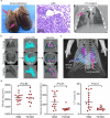
Purpose: Molecular imaging has provided unparalleled opportunities to monitor disease processes, although tools for evaluating infection remain limited. Coronavirus disease (COVID-19) caused by severe acute respiratory syndrome coronavirus 2 (SARS-CoV-2) is mediated by lung injury that we sought to model. Activated macrophages/phagocytes have an important role in lung injury, which is responsible for subsequent respiratory failure and death. We performed pulmonary PET/CT with 124I-iodo-DPA-713, a low-molecular-weight pyrazolopyrimidine ligand selectively trapped by activated macrophages cells, to evaluate the local immune response in a hamster model of SARS-CoV-2 infection.
Procedures: Pulmonary 124I-iodo-DPA-713 PET/CT was performed in SARS-CoV-2-infected golden Syrian hamsters. CT images were quantified using a custom-built lung segmentation tool. Studies with DPA-713-IRDye680LT and a fluorescent analog of DPA-713 as well as histopathology and flow cytometry were performed on post-mortem tissues.
Results: Infected hamsters were imaged at the peak of inflammatory lung disease (7 days post-infection). Quantitative CT analysis was successful for all scans and demonstrated worse pulmonary disease in male versus female animals (P < 0.01). Increased 124I-iodo-DPA-713 PET activity co-localized with the pneumonic lesions. Additionally, higher pulmonary 124I-iodo-DPA-713 PET activity was noted in male versus female hamsters (P = 0.02). DPA-713-IRDye680LT also localized to the pneumonic lesions. Flow cytometry demonstrated a higher percentage of myeloid and CD11b + cells (macrophages, phagocytes) in male versus female lung tissues (P = 0.02).
Conclusion: 124I-Iodo-DPA-713 accumulates within pneumonic lesions in a hamster model of SARS-CoV-2 infection. As a novel molecular imaging tool, 124I-Iodo-DPA-713 PET could serve as a noninvasive, clinically translatable approach to monitor SARS-CoV-2-associated pulmonary inflammation and expedite the development of novel therapeutics for COVID-19.
Keywords: COVID-19; Immune response; Macrophage; Molecular imaging; PET/CT; SARS-CoV-2; Sex difference.
© 2021. World Molecular Imaging Society.
Conflict of interest statement
Ali Ghayoor works at Invicro, Boston, MA, USA. A. A. O. receives consulting fees from Cubresa Inc. S. K. J. is a Senior Editor for Molecular Imaging and Biology.
Figures


Similar articles
-
Imaging Pulmonary Foreign Body Reaction Using [125I]iodo-DPA-713 SPECT/CT in Mice.Mol Imaging Biol. 2019 Apr;21(2):228-231. doi: 10.1007/s11307-018-1249-0. Mol Imaging Biol. 2019. PMID: 29987615 Free PMC article.
-
Noninvasive molecular imaging of tuberculosis-associated inflammation with radioiodinated DPA-713.J Infect Dis. 2013 Dec 15;208(12):2067-74. doi: 10.1093/infdis/jit331. Epub 2013 Jul 30. J Infect Dis. 2013. PMID: 23901092 Free PMC article.
-
Longitudinal analyses using 18F-Fluorodeoxyglucose positron emission tomography with computed tomography as a measure of COVID-19 severity in the aged, young, and humanized ACE2 SARS-CoV-2 hamster models.Antiviral Res. 2023 Jun;214:105605. doi: 10.1016/j.antiviral.2023.105605. Epub 2023 Apr 15. Antiviral Res. 2023. PMID: 37068595 Free PMC article.
-
The Role of Imaging in the Detection and Management of COVID-19: A Review.IEEE Rev Biomed Eng. 2021;14:16-29. doi: 10.1109/RBME.2020.2990959. Epub 2021 Jan 22. IEEE Rev Biomed Eng. 2021. PMID: 32356760 Review.
-
SARS-CoV replication and pathogenesis in an in vitro model of the human conducting airway epithelium.Virus Res. 2008 Apr;133(1):33-44. doi: 10.1016/j.virusres.2007.03.013. Epub 2007 Apr 23. Virus Res. 2008. PMID: 17451829 Free PMC article. Review.
Cited by
-
Dynamic single-cell RNA sequencing reveals BCG vaccination curtails SARS-CoV-2 induced disease severity and lung inflammation.bioRxiv [Preprint]. 2022 Mar 15:2022.03.15.484018. doi: 10.1101/2022.03.15.484018. bioRxiv. 2022. PMID: 35313583 Free PMC article. Preprint.
-
Intramuscular [18F]F-FDG Administration for Successful PET Imaging of Golden Hamsters in a Maximum Containment Laboratory Setting.Viruses. 2022 Nov 11;14(11):2492. doi: 10.3390/v14112492. Viruses. 2022. PMID: 36423101 Free PMC article.
-
The SARS-CoV-2 spike protein binds and modulates estrogen receptors.Sci Adv. 2022 Dec 2;8(48):eadd4150. doi: 10.1126/sciadv.add4150. Epub 2022 Nov 30. Sci Adv. 2022. PMID: 36449624 Free PMC article.
-
Medical imaging of pulmonary disease in SARS-CoV-2-exposed non-human primates.Trends Mol Med. 2022 Feb;28(2):123-142. doi: 10.1016/j.molmed.2021.12.001. Epub 2021 Dec 7. Trends Mol Med. 2022. PMID: 34955425 Free PMC article. Review.
-
Intravenous BCG vaccination reduces SARS-CoV-2 severity and promotes extensive reprogramming of lung immune cells.iScience. 2023 Aug 24;26(10):107733. doi: 10.1016/j.isci.2023.107733. eCollection 2023 Oct 20. iScience. 2023. PMID: 37674985 Free PMC article.
References
-
- GlobalHealth5050 The sex, gender and COVID-19 Project. https://globalhealth5050.org/the-sex-gender-and-covid-19-project/ (accessed Dec 28, 2020)
Publication types
MeSH terms
Substances
Grants and funding
LinkOut - more resources
Full Text Sources
Medical
Research Materials
Miscellaneous

