. 2021 Sep 1;134(17):jcs247171.
doi: 10.1242/jcs.247171. Epub 2021 Sep 9.

SUMOylation of RepoMan during late telophase regulates dephosphorylation of lamin A

Affiliations

SUMOylation of RepoMan during late telophase regulates dephosphorylation of lamin A

Takanobu Moriuchi et al. J Cell Sci. .

Abstract

Dephosphorylation of lamin A, which triggers nuclear lamina reconstitution, is crucial for the completion of mitosis. However, the specific phosphatase and regulatory mechanism that allow timely lamin A dephosphorylation remain unclear. Here, we report that RepoMan (also known as CDCA2), a regulatory subunit of protein phosphatase 1γ (PP1γ) is transiently modified with SUMO-2 at K762 during late telophase. SUMOylation of RepoMan markedly enhanced its binding affinity with lamin A. Moreover, SUMOylated RepoMan contributes to lamin A recruitment to telophase chromosomes and dephosphorylation of the mitotic lamin A phosphorylation. Expression of a SUMO-2 mutant that has a defective interaction with the SUMO-interacting motif (SIM) resulted in failure of the lamin A and RepoMan association, along with abrogation of lamin A dephosphorylation and subsequent nuclear lamina formation. These findings strongly suggest that RepoMan recruits lamin A through SUMO-SIM interaction. Thus, transient SUMOylation of RepoMan plays an important role in the spatiotemporal regulation of lamin A dephosphorylation and the subsequent nuclear lamina formation at the end of mitosis.

Keywords: Lamin; Mitosis; Nuclear lamina; Protein phosphatase; SIM; SUMO-interacting motif; Sumoylation.


Conflict of interest statement

Competing interests The authors declare no competing or financial interests.

Figures

Fig. 1.
Fig. 1.
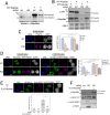
SUMOylation of RepoMan at K762. (A) HeLa cells were transfected with the indicated combinations of expression plasmids encoding CFP–SUMO-2 [WT (W) or GA mutant], CFP–Ubc9 and FLAG–RepoMan and directly lysed in boiled Laemmli's sample buffer. Polypeptides in whole-cell lysates were separated on a 5–20% gradient SDS polyacrylamide gel and subjected to western blot analysis using anti-RepoMan, anti-FLAG and anti-GFP antibodies. The asterisks and arrowhead indicate the CFP–SUMO-2-modified FLAG–RepoMan and endogenous RepoMan, respectively. (B) 293FT cells were transfected with plasmids expressing FLAG–RepoMan (WT) and CFP–SUMO-2 plasmid as indicated. At 24 h after DNA transfection, cell lysates were prepared and subjected to immunoprecipitation (IP) using an anti-FLAG antibody, then proteins were separated by SDS-PAGE and detected by western blots using anti-FLAG and anti-GFP antibodies. The asterisks indicate the CFP–SUMO-2-modified FLAG–RepoMan. (C) Conjugation of endogenous RepoMan with CFP–SUMO isoforms in HeLa cells were analyzed by western blot analysis. HeLa cells transfected with plasmids expressing CFP, CFP–SUMO-1, CFP–SUMO-2 or CFP–SUMO-3 were lysed in boiled Laemmli's sample buffer and proteins were separated on a 5–20% gradient gel by SDS-PAGE and subjected to western blot analysis using anti-RepoMan and anti-GFP antibodies. The arrowhead indicates the endogenous RepoMan conjugated to CFP-SUMO isoforms. (D) HeLa cells were lysed and subjected to immunoprecipitation using normal rabbit IgG or rabbit anti-RepoMan antibody, then proteins were separated on a 5–20% gradient gel by SDS-PAGE and detected by western blotting using anti-RepoMan, anti-SUMO-1 and anti-SUMO-2/3 antibodies. The arrowhead indicates the 125 kDa band specifically recognized by anti-SUMO-2/3. (E) 293FT cells were transfected with plasmids expressing FLAG–RepoMan(WT) or FLAG–RepoMan(K762R) with CFP or CFP–SUMO-2 and treated as in B. The asterisk indicates the FLAG–RepoMan modified with CFP-SUMO-2. Blots shown are representative of triplicate experiments.
Fig. 2.
Fig. 2.
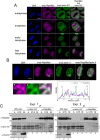
Colocalizaion and interaction between lamin A and RepoMan during mitosis. (A) HeLa cells were fixed with 4% formaldehyde in PBS, permeabilized with 0.3% Triton X-100 in PBS and immunofluorescently stained with anti-RepoMan (magenta) and anti-lamin A/C (green) antibodies. DNA was stained with Hoechst 33258 dye (blue). The confocal images depict cells at each stage of mitosis. Scale bar: 5 µm. The images are representative cells examined in two independent experiments (n>5 for each mitosis stage). (B) HeLa cells were incubated for 20 h in medium containing 1.5 mM hydroxyurea. At 8 h after release, cells were arrested at the prometaphase by adding monastrol to the medium at the finial concentration of 150 nM for 6 h. Cells were collected by a shaking off method, suspended in 0.5 ml of the medium at 37°C for a further 90 min, and immobilized on a glass slide using cytospinning. After fixation with 4% formaldehyde, telophase cells were immunostained with anti-RepoMan and anti-lamin A/C antibodies. DNA was also stained with Hoechst 33258 dye (blue). The images are representative cells examined in three independent experiments (n=6). The colocalizations of two proteins are represented by merged images of green (lamin A) and magenta (RepoMan) immunofluorescence signals. Enlarged images within the white squares (a, b, c) are shown in the lower left panel. The white line (d–e) in the merge image corresponds to the line profile plot shown in the lower right panel. Arrows (NE) indicate the position corresponding to the newly formed nuclear envelope. Scale bar: 5 µm. (C) HeLa cells were arrested at G1/S boundary or M phase by incubating cells in medium containing 1.5 mM hydroxyurea or 1 µg/ml nocodazole for 16 h. The cell lysates were subjected to immunoprecipitation (IP) using an anti-lamin A/C or anti-RepoMan antibody, then proteins were separated on a 5–20% gradient gel by SDS-PAGE and subjected to western blot analysis using anti-lamin A/C and anti-RepoMan antibodies. Synchronization at mitosis with nocodazole treatment was monitored by western blot analysis using antibody specific for anti-mitotic phosphorylation of histone H3 (H3pS10). A, asynchronous; G1/S, hydroxyurea-treated cells; M, nocodazole-treated cells. Blots shown are representative of triplicate experiments.
Fig. 3.
Fig. 3.
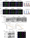
Knockdown of RepoMan disturbs lamin A reassembly and delays dephosphorylation of lamin A/C. (A) HeLa cells were transfected with plasmid expressing RFP–RepoMan and plasmid encoding ECFP and scramble (scr) shRNA or shRNA against RepoMan (KD) mRNA. Cells at 24 h after transfection were subjected to western blotting analysis using anti-RepoMan antibody. The transfection efficiency was ∼70% based on numbers of ECFP-positive cells. (B) HeLa cells were transfected with plasmid expressing WT RFP–RepoMan or shRNA-resistant RFP–RepoMan (RFP–RepoManr) and plasmid encoding ECFP and scramble (scr) shRNA or shRNA against RepoMan (KD) mRNA. Cells at 24 h after transfection were subjected to western blotting analysis using anti-RFP antibody. (C) HeLa cells were transfected with shRNA plasmid (scr or KD), then synchronized by thymidine-hydroxyurea-nocodazole block. At 90 min after the release, cells were immunofluorescently stained using anti-H3 pT3 antibody (magenta). The images are representative late telophase cells examined in two independent experiments (scr, n=5; KD, n=6). Scale bar: 5 µm. The fluorescence signals of H3 pT3 in each ECFP-positive cell at metaphase, anaphase and telophase were quantified and average intensities of signals relative to that of metaphase cells were summarized in the right panel. Data are shown as means±s.e.m. *P<0.05 (Student's t-test). (D) HeLa cells were transfected with shRNA plasmid (scr or KD), HA–lamin A plasmid and ECFP plasmid, then synchronized by thymidine-hydroxyurea-nocodazole block. At 90 min after the release, cells were immunofluorescently stained using anti-HA (green) and anti-lamin ApS22 (magenta) antibodies. The images are representative telophase cells (scr, n=5; KD, n=6) and cells during cytokinesis (scr, n=6; KD, n=6) examined in two independent experiments. Scale bars: 5 µm. The fluorescence signals of lamin ApS22 in each ECFP-positive cell at metaphase, anaphase and telophase were quantified and expressed relative to the signals of metaphase cells expressing scr shRNA. Data are shown as means±s.e.m. *P<0.05; **P<0.01 (Student's t-test). (E) HeLa cells were co-transfected with KD shRNA plasmid, HA–lamin A plasmid and plasmid carrying shRNA-resistant cDNA for FLAG–RepoMan. Cells were synchronized by thymidine-hydroxyurea-nocodazole block. At 90 min after the release, cells were immunofluorescently stained using anti-HA (green), anti-lamin A pS22 (magenta) and anti-FLAG (white) antibodies. The ECFP signal, indicating cells with shRNA plasmid, is also shown. DNA was stained with Hoechst 33258 (blue). Representative images of cell are shown in two independent experiments (n=6). Scale bar: 5 µm. The fluorescence signals of lamin A pS22 in each ECFP-positive cell at late telophase were quantified and expressed relative to those of cells transfected with scr shRNA plasmid. Data are shown as means±s.e.m. *P<0.05 (Student's t-test). (F) HeLa cells were transfected with scr or KD shRNA plasmid with or without plasmid carrying shRNA-resistant cDNA for FLAG-RepoMan. Cells at 24 h after transfection were subjected to western blotting analysis using anti-RepoMan, anti-lamin A/C, anti-lamin ApS22 and anti-H3 pT3 antibodies. Blots shown are representative of triplicate experiments.
Fig. 4.
Fig. 4.
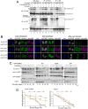
The SUMOylated RepoMan–PP1 holoenzyme is likely responsible for dephosphorylation of lamin A. HeLa cells were transfected with shRNA plasmid for RepoMan knockdown (KD), FLAG–RepoMan plasmid encoding shRNA-resistant RepoMan mRNA (WT, K762R mutant or RATA mutant) and HA–lamin A plasmid as indicated and synchronized by the hydroxyurea-nocodazole method. (A,B) Cells were fixed with 4% formaldehyde in PBS at 60, 75 and 90 min after release from the nocodazole blockage and immunostained with anti-FLAG (white), anti-HA (green), anti-lamin ApS22 (magenta) antibodies (A), or immunostained with anti-FLAG (white), anti-HA (green), and anti-H3 pT3 (magenta) antibodies (B). DNA was stained with the Hoechst 33258 (blue). Representative images of cells (of more than four cells for each mitosis stage) are shown from two independent experiments. Scale bars: 5 µm. The fluorescence intensities of lamin A pS22 or H3 pT3 signals of each cell at metaphase (meta), anaphase (ana) and telophase (telo) were quantified and expressed relative to those of metaphase cells. Data are shown as means±s.e.m. **P<0.01 (Student's t-test). (C) HeLa cells transiently expressing FLAG–RepoMan (WT, K762R mutant or RATA mutant) and HA–lamin A were collected at the indicated time point after release from the hydroxyurea-nocodazole blockage and directly lysed in Laemmli's sample buffer. The samples were then subjected to SDS-PAGE and proteins were detected by western blot analysis using anti-FLAG, anti-HA, anti-lamin ApS22 and anti-H3 pT3 antibodies. (D) Quantification of lamin ApS22 and H3 pT3 signals after the release from the nocodazole blockage. Data are mean±s.e.m. and represent three independent experiments. *P<0.05; **P<0.01; ***P<0.001 versus WT RepoMan transfection (Student's t-test). (E) 293FT cells were simultaneously transfected with plasmids expressing FLAG–RepoMan (WT, K762R, or RATA) and Myc–PP1γ as indicated. The cell lysates were subjected to immunoprecipitation (IP) using an anti-FLAG antibody, then proteins were separated by SDS-PAGE and detected by western blotting using anti-FLAG and anti-Myc antibodies. Input represents 25% of lysate. Blot shown is representative of three experiments. (F) HeLa cells were simultaneously transfected with plasmids expressing FLAG–RepoMan (WT, K762R or RATA) and Myc–PP1γ. At 24 h after transfection, cells were fixed and stained using anti-FLAG (green) and anti-Myc (magenta) antibodies. DNA was stained with Hoechst 33258 (blue). Scale bar: 5 µm. Representative images of anaphase cells are shown from two independent experiments (WT, n=4; K762R, n=3; RATA, n=3).
Fig. 5.
Fig. 5.
SUMOylation of RepoMan enhances the interaction between RepoMan and lamin A. (A) 293FT cells were transfected with plasmids expressing HA–lamin A(WT) (W) or HA–lamin A[SIM3(AA)] (SIM), FLAG–RepoMan(WT)–Ubc9 (W) or FLAG-RepoMan(K762R)-Ubc9 (KR), and CFP–SUMO-2(R59E) as indicated and subjected to immunoprecipitation (IP) using an anti-FLAG antibody, anti-HA antibody or normal mouse IgG. FLAG–RepoMan with or without SUMOylation and HA–lamin A in input and immunoprecipitated samples were detected using anti-FLAG and anti-HA antibodies, respectively. The asterisk indicates the CFP–SUMO-2 modified FLAG–RepoMan–Ubc9. Relative amounts of SUMOylated RepoMan and non-SUMOylated RepoMan in each sample were determined by quantifying the signals of immunoblots (IB) using ImageJ. The amounts of HA–lamin A precipitated using anti-FLAG antibody were quantified from signals of triplicate experiments. (B) Quantification of RepoMan with or without SUMOylation in immunoprecipitation samples. 293FT cells were transfected with the indicated combinations of expression plasmids and subjected to immunoprecipitation using anti-HA antibody. HA–lamin A and FLAG–RepoMan in input and immunoprecipitated samples were detected using anti-HA and anti-FLAG antibodies, respectively. The asterisk indicates the CFP–SUMO-2 modified FLAG–RepoMan–Ubc9. Relative amounts of SUMOylated RepoMan and non-SUMOylated RepoMan in each sample were determined by quantifying the signals of immunoblots using ImageJ. Blots shown are representative of triplicate experiments.
Fig. 6.
Fig. 6.
SUMO–SIM interaction is required for interaction between lamin A and RepoMan and also dephosphorylation of lamin A. (A) 293FT cells were transfected with expression plasmids for CFP–SUMO-2 (WT, R59E, G93A or QFI), FLAG–RepoMan–Ubc9 and HA–lamin A as indicated. At 24 h after DNA transfection, cells were lysed and subjected to immunoprecipitation using an anti-FLAG or anti-HA antibody. Proteins were detected by western blot analysis using anti-FLAG, anti-HA and anti-GFP antibodies. The asterisks indicate the CFP–SUMO-2-modified FLAG–RepoMan–Ubc9. The values represent relative amount of SUMOylated and non-SUMOylated FLAG–RepoMan–Ubc9, which were derived from signals of triplicate immuoblots. (B) HeLa cells were simultaneously transfected with Myc–SUMO-2 (WT or QFI) and HA–lamin A [WT or SIM3(EE)] expression plasmids. At 36 h after DNA transfection, cells were fixed with 4% formaldehyde in PBS and immunofluorescently stained with anti-myc (white), anti-HA (green), and anti-lamin Ap22 (magenta) antibodies. DNA was stained with Hoechst 33258 dye (blue). The confocal images depict cells at each stage of mitosis. Representative images of cells (more than four cells for each mitosis stage) are shown from two independent experiments. Scale bar: 5 µm. (C) HeLa cells transiently expressing HA–lamin A and CFP–SUMO-2 (WT, G93A or QFI) were synchronized by the hydroxyurea-nocodazole method. Cells were collected at the indicated time point after release from the nocodazole blockage and directly lysed in Laemmli's sample buffer. The samples were then subjected to SDS-PAGE and proteins were detected by western blot analysis using anti-HA, anti-lamin A pS22, anti-H3 pT3, and anti-GFP antibodies. (D) Quantification of lamin ApS22 and H3 pT3 signals after the release from the nocodazole blockage. Data are mean±s.e.m.; n=3 independent experiments. *P<0.05; **P<0.01; ***P<0.001 versus WT SUMO-2 expression (Student's t-test).
Fig. 7.
Fig. 7.
RepoMan SUMOylation peaks during telophase. (A) HeLa cells transiently expressing FLAG–RepoMan and CFP–SUMO-2(WT) were synchronized by the thymidine-hydroxyurea method. Cells were collected at the indicated time point after release from the thymidine blockage and directly lysed in Laemmli's sample buffer. The samples were then subjected to SDS-PAGE and proteins were detected by western blot analysis using anti-FLAG, anti-lamin A/C pS22, and anti-H3 pS10 antibodies. The asterisk, A and C indicate the CFP–SUMO-2-modified FLAG–RepoMan, lamin A and lamin C, respectively. The data is representative of three independent experiments with similar results. Amounts of FLAG-RepoMan with or without SUMOylation were determined by quantifying the signals of immunoblots using ImageJ and summarized in the graph. Data are mean±s.e.m. and represent three independent experiments. (B) HeLa cells transiently expressing CFP–SUMO-2(WT) were synchronized by the hydroxyurea-monastrol method. Prometaphase cells were collected by shake-off, seeded and cultured in 12-well plates for the indicated time. Cells were then lysed in Laemmli's sample buffer. The samples were then subjected to SDS-PAGE and proteins were detected by western blot analysis using anti-FLAG, anti-lamin A/C pS22, anti-H3 pS10, anti-cyclin B and anti-GFP antibodies. The asterisk indicates the CFP–SUMO-2-modified FLAG–RepoMan. The data is representative of two independent experiments with similar results. (C) HeLa cells transiently expressing CFP–SUMO-2(WT) were synchronized and processed as in B. The arrowhead indicates endogenous RepoMan modified with CFP–SUMO-2. The data is representative of two independent experiments with similar results.

Similar articles

Cited by

References

    1. Andrés, V. and González, J. M. (2009). Role of A-type lamins in signaling, transcription, and chromatin organization. J. Cell Biol. 187, 945-957. 10.1083/jcb.200904124 - DOI - PMC - PubMed
    1. Combes, G., Alharbi, I., Braga, L. G. and Elowe, S. (2017). Playing polo during mitosis: PLK1 takes the lead. Oncogene 36, 4819-4827. 10.1038/onc.2017.113 - DOI - PubMed
    1. Dawlaty, M. M., Malureanu, L., Jeganathan, K. B., Kao, E., Sustmann, C., Tahk, S., Shuai, K., Grosschedl, R. and van Deursen, J. M. (2008). Resolution of sister centromeres requires RanBP2-mediated SUMOylation of topoisomerase IIα. Cell 133, 103-115. 10.1016/j.cell.2008.01.045 - DOI - PMC - PubMed
    1. de Castro, I. J., Budzak, J., Di Giacinto, M. L., Ligammari, L., Gokhan, E., Spanos, C., Moralli, D., Richardson, C., de Las Heras, J. I., Salatino, S.et al. (2017). Repo-Man/PP1 regulates heterochromatin formation in interphase. Nat. Commun. 8, 14048. 10.1038/ncomms14048 - DOI - PMC - PubMed
    1. Dechat, T., Pfleghaar, K., Sengupta, K., Shimi, T., Shumaker, D. K., Solimando, L. and Goldman, R. D. (2008). Nuclear lamins: major factors in the structural organization and function of the nucleus and chromatin. Genes Dev. 22, 832-853. 10.1101/gad.1652708 - DOI - PMC - PubMed

Publication types