

Molecular Regulation of Paused Pluripotency in Early Mammalian Embryos and Stem Cells
- PMID: 34386497
- PMCID: PMC8353277
- DOI: 10.3389/fcell.2021.708318
Molecular Regulation of Paused Pluripotency in Early Mammalian Embryos and Stem Cells
Abstract
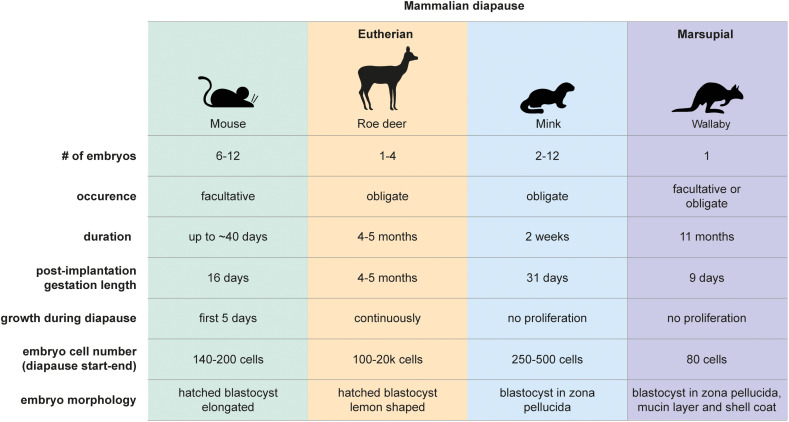
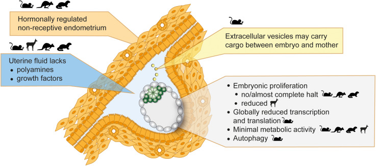
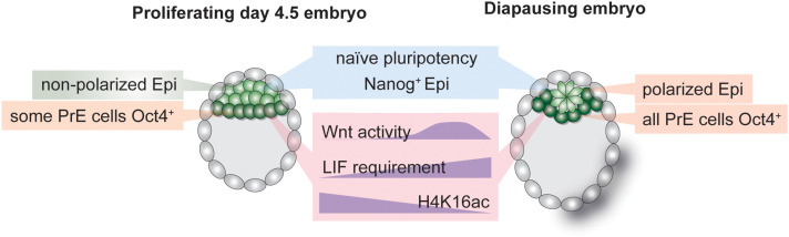
The energetically costly mammalian investment in gestation and lactation requires plentiful nutritional sources and thus links the environmental conditions to reproductive success. Flexibility in adjusting developmental timing enhances chances of survival in adverse conditions. Over 130 mammalian species can reversibly pause early embryonic development by switching to a near dormant state that can be sustained for months, a phenomenon called embryonic diapause. Lineage-specific cells are retained during diapause, and they proliferate and differentiate upon activation. Studying diapause thus reveals principles of pluripotency and dormancy and is not only relevant for development, but also for regeneration and cancer. In this review, we focus on the molecular regulation of diapause in early mammalian embryos and relate it to maintenance of potency in stem cells in vitro. Diapause is established and maintained by active rewiring of the embryonic metabolome, epigenome, and gene expression in communication with maternal tissues. Herein, we particularly discuss factors required at distinct stages of diapause to induce, maintain, and terminate dormancy.
Keywords: dormancy; embryonic diapause; metabolism; miRNA; pluripotency; signaling pathways; stem cells; transcription.
Copyright © 2021 van der Weijden and Bulut-Karslioglu.
Conflict of interest statement
The authors declare that the research was conducted in the absence of any commercial or financial relationships that could be construed as a potential conflict of interest.
Figures
Similar articles
-
Embryonic diapause in mammals and dormancy in embryonic stem cells with the European roe deer as experimental model.Reprod Fertil Dev. 2021 Jan;33(2):76-81. doi: 10.1071/RD20256. Reprod Fertil Dev. 2021. PMID: 38769673
-
FOXO1-mediated lipid metabolism maintains mammalian embryos in dormancy.Nat Cell Biol. 2024 Feb;26(2):181-193. doi: 10.1038/s41556-023-01325-3. Epub 2024 Jan 4. Nat Cell Biol. 2024. PMID: 38177284 Free PMC article.
-
Distinct dormancy progression depending on embryonic regions during mouse embryonic diapause†.Biol Reprod. 2019 May 1;100(5):1204-1214. doi: 10.1093/biolre/ioz017. Biol Reprod. 2019. PMID: 30715198
-
New insights into how to induce and maintain embryonic diapause in the blastocyst.Curr Opin Genet Dev. 2024 Jun;86:102192. doi: 10.1016/j.gde.2024.102192. Epub 2024 Apr 11. Curr Opin Genet Dev. 2024. PMID: 38604005 Review.
-
The mTOR Pathway in Pluripotent Stem Cells: Lessons for Understanding Cancer Cell Dormancy.Membranes (Basel). 2021 Nov 7;11(11):858. doi: 10.3390/membranes11110858. Membranes (Basel). 2021. PMID: 34832087 Free PMC article. Review.
Cited by
-
Beyond energy and growth: the role of metabolism in developmental signaling, cell behavior and diapause.Development. 2023 Oct 15;150(20):dev201610. doi: 10.1242/dev.201610. Epub 2023 Oct 26. Development. 2023. PMID: 37883062 Free PMC article.
-
Perspective: Might Maternal Dietary Monosodium Glutamate (MSG) Consumption Impact Pre- and Peri-Implantation Embryos and Their Subsequent Development?Int J Environ Res Public Health. 2022 Oct 20;19(20):13611. doi: 10.3390/ijerph192013611. Int J Environ Res Public Health. 2022. PMID: 36294193 Free PMC article.
-
Metabolic regulation of the hallmarks of stem cell biology.Cell Stem Cell. 2024 Feb 1;31(2):161-180. doi: 10.1016/j.stem.2024.01.003. Cell Stem Cell. 2024. PMID: 38306993 Free PMC article. Review.
-
The stem cell zoo for comparative studies of developmental tempo.Curr Opin Genet Dev. 2024 Feb;84:102149. doi: 10.1016/j.gde.2023.102149. Epub 2024 Jan 9. Curr Opin Genet Dev. 2024. PMID: 38199063 Free PMC article. Review.
-
m 6 A RNA methylation orchestrates transcriptional dormancy during developmental pausing.bioRxiv [Preprint]. 2023 Feb 1:2023.01.30.526234. doi: 10.1101/2023.01.30.526234. bioRxiv. 2023. Update in: Nat Cell Biol. 2023 Sep;25(9):1279-1289. doi: 10.1038/s41556-023-01212-x. PMID: 36778216 Free PMC article. Updated. Preprint.
References
Publication types
LinkOut - more resources
Full Text Sources