. 2021 Aug 12;11(1):16394.
doi: 10.1038/s41598-021-95225-x.

Predicted limited redistribution of T cells to secondary lymphoid tissue correlates with increased risk of haematological malignancies in asplenic patients

Affiliations

Predicted limited redistribution of T cells to secondary lymphoid tissue correlates with increased risk of haematological malignancies in asplenic patients

Aleksandra E Kmieciak et al. Sci Rep. .

Abstract

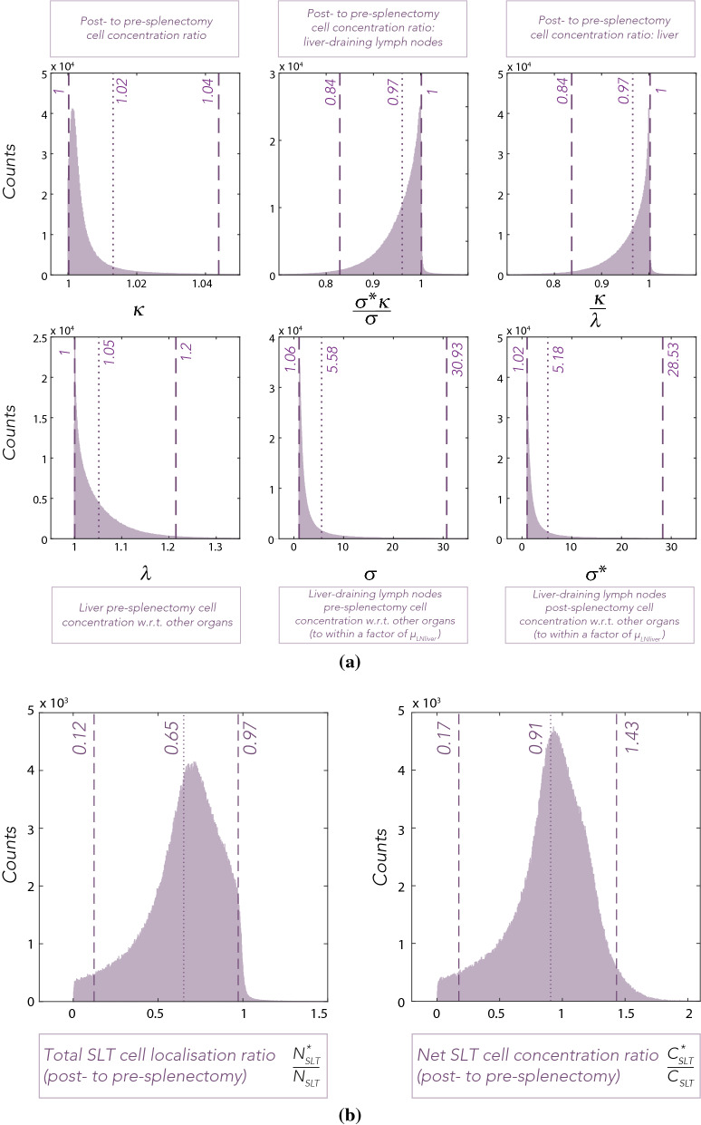
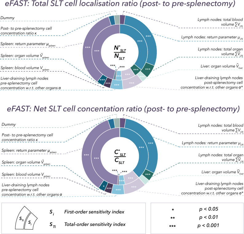
The spleen, a secondary lymphoid tissue (SLT), has an important role in generation of adaptive immune responses. Although splenectomy remains a common procedure, recent studies reported poor prognosis and increased risk of haematological malignancies in asplenic patients. The high baseline trafficking of T lymphocytes to splenic tissue suggests splenectomy may lead to loss of blood-borne malignant immunosurveillance that is not compensated for by the remaining SLT. To date, no quantitative analysis of the impact of splenectomy on the human T cell trafficking dynamics and tissue localisation has been reported. We developed a quantitative computational model that describes organ distribution and trafficking of human lymphocytes to explore the likely impact of splenectomy on immune cell distributions. In silico splenectomy resulted in an average reduction of T cell numbers in SLT by 35% (95%CI 0.12-0.97) and a comparatively lower, 9% (95%CI 0.17-1.43), mean decrease of T cell concentration in SLT. These results suggest that the surveillance capacity of the remaining SLT insufficiently compensates for the absence of the spleen. This may, in part, explain haematological malignancy risk in asplenic patients and raises the question of whether splenectomy has a clinically meaningful impact on patient responses to immunotherapy.


Conflict of interest statement

L.V.B, M.C.C. and E.A.G. declare no competing interests. At the time the work presented in this manuscript was completed, A.P. and J.W. were employees of and shareholders in F. Hoffmann-La Roche AG. F. Hoffmann-La Roche AG have contributed to the costs of the doctoral student, A.E.K., under the supervision of E.A.G. and M.C.C.

Figures

Figure 1
Figure 1
Standardised Incidence Ratio (SIR) and Standardised Mortality Ratio (SMR) due to selected malignancies following splenectomy as reported by Kristinsson et al.. Reported malignancies were plotted on one graph (a) to highlight the visible distinction between solid tumours (b) and haematological malignancies (c). Cross-lines represent the reported 95% confidence intervals (95%CI). Note the difference in scale.
Figure 2
Figure 2
Model schematic of the ODE system (right) compared to the simplified schematic of the blood and lymph circulation (left). As a PBPK framework, the T cell trafficking model comprises of anatomical compartments corresponding directly and faithfully to the organs and tissues of the body, connected by the cardiovascular and lymphatic system. Such a description of anatomy is used to predict a systemic and tissue exposure to the circulating T cells. Cells flow from the left atrium and left ventricle of the heart to each organ (red lines), via lymphatics (blue lines) into organ-draining lymph nodes and return back to the right atrium and right ventricle of the heart. Each box represents a vasculature and interstitium compartment. Chambers of the heart: RA—right atrium, RV—right ventricle, LA—left atrium, LV—left ventricle. Other organs compartments include: adrenals, bladder, brain, adipose tissue, gonads, kidneys, skeletal muscles, skeleton, skin, thyroid. Note that the skeleton compartment in the model includes the bone marrow and bones and is parameterised accordingly (see Table 2).
Figure 3
Figure 3
Organ, o, and draining lymph nodes for organ, o, are represented in the model as separate vascular and interstitial compartments. T cells flow from the cardiac output compartment (previously defined as left atrium, left ventricle, aorta and large arteries) to each organ, o, with a blood flow Bo, from which a proportion, eoBo, enters the interstitial space per unit time. A proportion of T cells, eoμoBo, flows via lymphatics from the interstitial sub-compartment of organ, o, into the draining lymph nodes per unit time. T cells also enter organ, o, -draining lymph nodes through the lymph node blood supply, denoted BLNo. From the lymph node vasculature compartment, a proportion of T cells extravasates into the lymph node interstitial compartment (at a rate eLNoBLNo). T cells drained to organ, o, -draining lymph nodes are returned to the systemic circulation (at a rate eLNoμLNoBLNo+μLNoeoμoBo). T cells that have not extravasated to the interstitium of organ, o, and organ, o, -draining lymph nodes are, similarly, returned to the systemic circulation (at rates (1-eo)Bo and (1-eLNo)BLNo, respectively). Different suffixes, such as LA&LV (combined left atrium, left ventricle, and large arteries), RA&RV (combined right atrium, right ventricle, and large veins), PC (pulmonary circuit), med (mediastinal lymph nodes) are also used.
Figure 4
Figure 4
Example results from the ODE system, subject to the initial distribution of T cells administered intravenously (Eq. 4), with baseline parameter values (see Table S.1, Supplementary Material). Each curve represents the sum of the number of T cells in vascular (No) and interstitial localisations (N~o) expressed as fraction of the total number of administered T cells (Relative localisation). For all organs (left) and all organ-draining groups of lymph nodes (right), extravasation, eo, and return parameters, μo, were assumed to be equal to 0.25 and 0.05, respectively. Note that the contents of heart chambers (RA&RV and LA&LV) and the blood supply to the heart (Heart blood supply) are plotted separately. Similarly, the lung curve does not include the pulmonary circuit.
Figure 5
Figure 5
Distributions of (a) post- to pre-splenectomy cell concentration ratios: κ, σκ/σ and κ/λ, as well as λ, σ and σ, and (b) the post- to pre-splenectomy total T cell localisation ratio and the net T cell concentration ratio in the SLT. Dotted and dashed lines correspond to means and 95% confidence intervals of the plotted distributions. See “Parameterisation” and “In silico splenectomy—simulation” sections for details on parameters.
Figure 6
Figure 6
Results of the global sensitivity analysis eFAST on the post- to pre-splenectomy total T cell localisation to SLT ratio (upper) and the post- to pre-splenectomy net T cell concentration in SLT ratio (lower). The inner (outer) ring indicates the proportional size of individual sensitivity indices (total sensitivity indices). A dummy parameter is used as a baseline for statistical comparison and a p-value was assigned to each sensitivity index using a two-sample t-test. Parameters with statistically significant sensitivity indices were labelled according to the assigned p-value (,,). The eFAST sensitivity analysis considered with a sampling of the lumped parameters κ, σ, σ from their distributions in Fig. 5a, cut off at the 95% confidence interval, together with the other parameters to be sampled uniformly, as detailed in “Global sensitivity analysis” section, with further details on parameter distributions in “Parameterisation” and “In silico splenectomy—simulation” sections.

Similar articles

Cited by

References

    1. Aide N, et al. FDG PET/CT for assessing tumour response to immunotherapy. Eur. J. Nucl. Med. Mol. Imaging. 2019;46:238–250. doi: 10.1007/s00259-018-4171-4. - DOI - PMC - PubMed
    1. Im A, Pavletic S. Immunotherapy in hematologic malignancies: Past, present, and future. J. Hematol. Oncol. 2017;10:94. doi: 10.1186/s13045-017-0453-8. - DOI - PMC - PubMed
    1. Miyasaka M, Hata E, Tohya K, Hayasaka H. Lymphocyte recirculation. Encycl. Immunobiol. 2016 doi: 10.1016/B978-0-12-374279-7.07013-2. - DOI
    1. Butcher E, Picker L. Lymphocyte homing and homeostasis. Science. 1996;272:60–66. doi: 10.1126/science.272.5258.60. - DOI - PubMed
    1. Weninger W, Crowley MA, Manjunath N, von Andrian UH. Migratory properties of naive, effector, and memory CD8(+) T cells. J. Exp. Med. 2001;194:953–966. doi: 10.1084/jem.194.7.953. - DOI - PMC - PubMed

Publication types