Human umbilical cord mesenchymal stem cells reduce oxidative damage and apoptosis in diabetic nephropathy by activating Nrf2
- PMID: 34380544
- PMCID: PMC8356418
- DOI: 10.1186/s13287-021-02447-x
Human umbilical cord mesenchymal stem cells reduce oxidative damage and apoptosis in diabetic nephropathy by activating Nrf2
Abstract
Background: Mesenchymal stem cells (MSCs) have a therapeutic effect on diabetic nephropathy (DN) but the underlying mechanism remains unclear. This study was conducted to investigate whether human umbilical cord-MSCs (hUCMSCs) can induce oxidative damage and apoptosis by activating Nrf2.
Methods: We used a type 2 diabetic rat model and a high-glucose and fat-stimulated human glomerular mesangial cell (hGMC) model. Western blotting, RT-qPCR, and TUNEL staining were performed on animal tissues and cultured cells. Nuclear expression of Nrf2 was detected in the renal tissue. Furthermore, Nrf2 siRNA was used to examine the effects of hUCMSCs on hGMCs. Finally, the effect of hUCMSCs on the Nrf2 upstream signalling pathway was investigated.
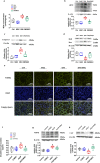
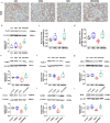
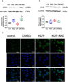
Results: After treatment with hUCMSCs, Nrf2 showed increased expression and nuclear translocation. After Nrf2-specific knockout in hGMCs, the protective effect of hUCMSCs on apoptosis induced by high-glucose and fat conditions was reduced. Activation of the PI3K signalling pathway may be helpful for ameliorating DN using hUCMSCs.
Conclusions: hUCMSCs attenuated renal oxidative damage and apoptosis in type 2 diabetes mellitus and Nrf2 activation is one of the important mechanisms of this effect. hUCMSCs show potential as drug targets for DN.
Keywords: Apoptosis; Diabetic nephropathy; Mesenchymal stem cell; Nrf2; Oxidative damage.
© 2021. The Author(s).
Conflict of interest statement
The authors declare that they have no competing interests.
Figures


Similar articles
-
Intravenous injection of human umbilical cord-derived mesenchymal stem cells ameliorates not only blood glucose but also nephrotic complication of diabetic rats through autophagy-mediated anti-senescent mechanism.Stem Cell Res Ther. 2023 May 29;14(1):146. doi: 10.1186/s13287-023-03354-z. Stem Cell Res Ther. 2023. PMID: 37248536 Free PMC article.
-
Tetramethylpyrazine enhanced the therapeutic effects of human umbilical cord mesenchymal stem cells in experimental autoimmune encephalomyelitis mice through Nrf2/HO-1 signaling pathway.Stem Cell Res Ther. 2020 May 19;11(1):186. doi: 10.1186/s13287-020-01700-z. Stem Cell Res Ther. 2020. PMID: 32430010 Free PMC article.
-
Identifying key antioxidative stress factors regulating Nrf2 in the genioglossus with human umbilical cord mesenchymal stem-cell therapy.Sci Rep. 2024 Mar 10;14(1):5838. doi: 10.1038/s41598-024-55103-8. Sci Rep. 2024. PMID: 38462642 Free PMC article.
-
Human umbilical cord-derived mesenchymal stem cells not only ameliorate blood glucose but also protect vascular endothelium from diabetic damage through a paracrine mechanism mediated by MAPK/ERK signaling.Stem Cell Res Ther. 2022 Jun 17;13(1):258. doi: 10.1186/s13287-022-02927-8. Stem Cell Res Ther. 2022. PMID: 35715841 Free PMC article.
-
A systematic review and meta-analysis of cell-based interventions in experimental diabetic kidney disease.Stem Cells Transl Med. 2021 Sep;10(9):1304-1319. doi: 10.1002/sctm.19-0419. Epub 2021 Jun 9. Stem Cells Transl Med. 2021. PMID: 34106528 Free PMC article. Review.
Cited by
-
Therapeutic potential of conditioned medium obtained from deferoxamine preconditioned umbilical cord mesenchymal stem cells on diabetic nephropathy model.Stem Cell Res Ther. 2022 Sep 2;13(1):438. doi: 10.1186/s13287-022-03121-6. Stem Cell Res Ther. 2022. PMID: 36056427 Free PMC article.
-
Human Umbilical Cord Mesenchymal Stem Cells Improve Premature Ovarian Failure through Cell Apoptosis of miR-100-5p/NOX4/NLRP3.Biomed Res Int. 2022 Jul 7;2022:3862122. doi: 10.1155/2022/3862122. eCollection 2022. Biomed Res Int. 2022. Retraction in: Biomed Res Int. 2023 Jun 21;2023:9848903. doi: 10.1155/2023/9848903 PMID: 35845923 Free PMC article. Retracted.
-
Research progress and prospects of benefit-risk assessment methods for umbilical cord mesenchymal stem cell transplantation in the clinical treatment of spinal cord injury.Stem Cell Res Ther. 2024 Jul 2;15(1):196. doi: 10.1186/s13287-024-03797-y. Stem Cell Res Ther. 2024. PMID: 38956734 Free PMC article. Review.
-
Human umbilical cord mesenchymal stem cells attenuate diabetic nephropathy through the IGF1R-CHK2-p53 signalling axis in male rats with type 2 diabetes mellitus.J Zhejiang Univ Sci B. 2024 Jul 10;25(7):568-580. doi: 10.1631/jzus.B2300182. J Zhejiang Univ Sci B. 2024. PMID: 39011677 Free PMC article.
-
Human umbilical cord mesenchymal stem cells in diabetes mellitus and its complications: applications and research advances.Int J Med Sci. 2023 Sep 11;20(11):1492-1507. doi: 10.7150/ijms.87472. eCollection 2023. Int J Med Sci. 2023. PMID: 37790847 Free PMC article. Review.
References
-
- Navarro-González JF, Sánchez-Niño MD, Donate-Correa J, Martín-Núñez E, Ferri C, Pérez-Delgado N, Górriz JL, Martínez-Castelao A, Ortiz A, Mora-Fernández C. Effects of pentoxifylline on soluble klotho concentrations and renal tubular cell expression in diabetic kidney disease. Diabetes Care. 2018;41(8):1817–1820. doi: 10.2337/dc18-0078. - DOI - PubMed
-
- Lv S, Cheng J, Sun A, Li J, Wang W, Guan G, Liu G, Su M. Mesenchymal stem cells transplantation ameliorates glomerular injury in streptozotocin-induced diabetic nephropathy in rats via inhibiting oxidative stress. Diabetes Res Clin Pract. 2014;104(1):143–154. doi: 10.1016/j.diabres.2014.01.011. - DOI - PubMed
Publication types
MeSH terms
Substances
LinkOut - more resources
Full Text Sources
Medical

