. 2021 Oct 13;95(21):e0089721.
doi: 10.1128/JVI.00897-21. Epub 2021 Aug 11.

Enterovirus A71 Induces Neurological Diseases and Dynamic Variants in Oral Infection of Human SCARB2-Transgenic Weaned Mice

Affiliations

Enterovirus A71 Induces Neurological Diseases and Dynamic Variants in Oral Infection of Human SCARB2-Transgenic Weaned Mice

Jing-Yi Lin et al. J Virol. .

Abstract

Enterovirus A71 (EV-A71) and many members of the Picornaviridae family are neurotropic pathogens of global concern. These viruses are primarily transmitted through the fecal-oral route, and thus suitable animal models of oral infection are needed to investigate viral pathogenesis. An animal model of oral infection was developed using transgenic mice expressing human SCARB2 (hSCARB2 Tg), murine-adapted EV-A71/MP4 virus, and EV-A71/MP4 virus with an engineered nanoluciferase gene that allows imaging of viral replication and spread in infected mice. Next-generation sequencing of EV-A71 genomes in the tissues and organs of infected mice was also performed. Oral inoculation of EV-A71/MP4 or nanoluciferase-carrying MP4 virus stably induced neurological symptoms and death in infected 21-day-old weaned mice. In vivo bioluminescence imaging of infected mice and tissue immunostaining of viral antigens indicated that orally inoculated virus can spread to the central nervous system (CNS) and other tissues. Next-generating sequencing further identified diverse mutations in viral genomes that can potentially contribute to viral pathogenesis. This study presents an EV-A71 oral infection murine model that efficiently infects weaned mice and allows tracking of viral spread, features that can facilitate research into viral pathogenesis and neuroinvasion via the natural route of infection. IMPORTANCE Enterovirus A71 (EV-A71), a positive-strand RNA virus of the Picornaviridae, poses a persistent global public health problem. EV-A71 is primarily transmitted through the fecal-oral route, and thus suitable animal models of oral infection are needed to investigate viral pathogenesis. We present an animal model of EV-A71 infection that enables the natural route of oral infection in weaned and nonimmunocompromised 21-day-old hSCARB2 transgenic mice. Our results demonstrate that severe disease and death could be stably induced, and viral invasion of the CNS could be replicated in this model, similar to severe real-world EV-A71 infections. We also developed a nanoluciferase-containing EV-A71 virus that can be used with this animal model to track viral spread after oral infection in real time. Such a model offers several advantages over existing animal models and can facilitate future research into viral spread, tissue tropism, and viral pathogenesis, all pressing issues that remain unaddressed for EV-A71 infections.

Keywords: enterovirus A71 (EV-A71); hSCARB2 transgenic mouse; in vivo imaging system (IVIS); quasispecies.


Figures
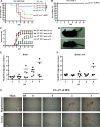
FIG 1
FIG 1
Pathogenesis of EV-A71 in mice. Survival curves of 21-day-old hSCARB2 Tg10 mice (A) or control wild-type C57BL/6 mice (B) orally inoculated with murine-adapted EV-A71/MP4 virus or its EV-A71/4643 parental strain. Mice were intragastrically (i.g.) inoculated with a specified number of PFU of each virus strain and then monitored daily for 14 days postinfection (dpi). (C) Neurological diseases in 21-day-old hSCARB2 transgenic mice inoculated i.g. with the indicated PFU of EV-A71/MP4. Diseases were monitored daily until 14 dpi and scored as follows: 0 = healthy, 1 = weakness of a limb, 2 = paralysis of a single limb, 3 = paralysis of two or more limbs, 4 = death. (D) Limb paralysis (white arrowheads) in hSCARB2 Tg mice infected with 105 PFU (top panel) or 106 PFU (bottom panel) of EV-A71/MP4 at 9 and 7 dpi, respectively. Viral genomic RNA levels in the brains (E) and spinal cords (F) of EV-A71/MP4-infected hSCARB2 Tg10 mice, which were harvested on the specified dpi. (G) EV-A71 capsid protein (VP1) expression in the hypothalamus and cortex of mock or orally infected (106 PFU of EV-A71/MP4) hSCARB2 mice at 6 dpi. Disease scores (DS) of observed neurological diseases in infected mice are indicated. Scale bar = 200 μm. CA1, hippocampal CA1; opt, optic nerve.
FIG 2
FIG 2
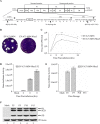
Construction and characterization of the EV-A71-MP4-Nluc5 reporter virus. (A) An infectious cDNA clone, pCR-XL-TOPO-EV-A71-MP4, was used as the backbone, and the nanoluciferase reporter gene was inserted between the 5′ UTR and VP4 sequences. The 2A protease cleavage site (ITTLG) is indicated by an arrow. (B) Plaque morphology of EV-A71/MP4 and EV-A71-MP4-Nluc5 in RD cell cultures. (C) Viral growth of EV-A71/MP4 and EV-A71-MP4-Nluc5. RD cells were infected with a multiplicity of infection (MOI) of 10 for the EV-A71/MP4 or EV-A71-MP4-Nluc5 virus, and virus titers were detected by plaque assay. (D) Nanoluciferase activity of EV-A71-MP4-Nluc5 virus. RD cells were infected with a multiplicity of infection of 10 for the EV-A71-MP4-Nluc5 virus, and cell lysates were collected at the stated times for assessment of nanoluciferase activity. (E) Summary of nanoluciferase activity at different EV-A71-MP4-Nluc5 passages. RD cells were infected with an MOI of 10 for the EV-A71-MP4-Nluc5 virus of the indicated passages, and cell lysates were collected at the stated times for assessment of nanoluciferase activity. (F) Summary of viral protein (P3, 3CD, and 3D) expression levels at different EV-A71-MP4-Nluc5 virus passages. RD cells were infected with an MOI of 10 for the EV-A71-MP4-Nluc5 virus of the indicated passages, and cell lysates were collected to assess viral protein expression levels by Western blotting.
FIG 3
FIG 3
EV-A71-MP4-Nluc5 virus infection in 21-day-old hSCARB2 Tg10 mice. (A) Body weight change in 21-day-old hSCARB2 Tg10 mice orally inoculated with the specified PFU of EV-A71-MP4-Nluc5 virus and then monitored daily for 12 dpi. (B) Neurological symptoms in 21-day-old hSCARB2 Tg10 mice orally inoculated with the indicated PFU of EV-A71-MP4-Nluc5 virus. Disease score was monitored daily until 12 dpi as follows: 0 = healthy, 1 = weakness of a limb, 2 = paralysis of a single limb, 3 = paralysis of two or more limbs, and 4 = death. (C) Survival rates of 21-day-old hSCARB2 Tg10 mice orally inoculated with the indicated PFU of EV-A71-MP4-Nluc5 virus.
FIG 4
FIG 4
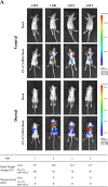
Real-time in vivo imaging of EV-A71-MP4-Nluc5 infection. (A) Ventral and dorsal views of hSCARB2 Tg10 mice after infection with EV-A71-MP4-Nluc5. Viral spread was monitored in real-time after 5.6 × 108 PFU of the virus was intragastrically inoculated into 21-day-old hSCARB2 Tg10 mice. Bioluminescence was visualized in the brain of infected hSCARB2 Tg10 mice at 2 days postinfection. Luminescence was undetected in hSCARB2 Tg10 mice (mock), which served as a negative control. The body weight of EV-A71-MP4-Nluc5-infected mice decreased, even without visible disease symptoms at 1 to 4 dpi. Histopathological changes and viral antigen (VP1) expression in different organs of 21-day-old hSCARB2 Tg10 mice i.g. inoculated with 5.6 × 108 PFU of EV-A71-MP4-Nluc5 at 4 dpi, with the brain, spinal cord, stomach, intestine, liver, spleen, heart, lung, muscle (forelimb), and muscle (hindlimb) shown in panel B. HE, hematoxylin and eosin stain; IHC, immunohistochemistry staining.
FIG 4
FIG 4
Real-time in vivo imaging of EV-A71-MP4-Nluc5 infection. (A) Ventral and dorsal views of hSCARB2 Tg10 mice after infection with EV-A71-MP4-Nluc5. Viral spread was monitored in real-time after 5.6 × 108 PFU of the virus was intragastrically inoculated into 21-day-old hSCARB2 Tg10 mice. Bioluminescence was visualized in the brain of infected hSCARB2 Tg10 mice at 2 days postinfection. Luminescence was undetected in hSCARB2 Tg10 mice (mock), which served as a negative control. The body weight of EV-A71-MP4-Nluc5-infected mice decreased, even without visible disease symptoms at 1 to 4 dpi. Histopathological changes and viral antigen (VP1) expression in different organs of 21-day-old hSCARB2 Tg10 mice i.g. inoculated with 5.6 × 108 PFU of EV-A71-MP4-Nluc5 at 4 dpi, with the brain, spinal cord, stomach, intestine, liver, spleen, heart, lung, muscle (forelimb), and muscle (hindlimb) shown in panel B. HE, hematoxylin and eosin stain; IHC, immunohistochemistry staining.
FIG 5
FIG 5
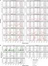
Genetic diversity of EV-A71-MP4-Nluc5 derived from different organs of infected mice. Following intragastric inoculation of 21-day-old hSCARB2 Tg10 mice with 5.6 × 108 PFU of EV-A71-MP4-Nluc5 virus, the brain, spinal cord, heart, lung, stomach, intestine, forelimb muscle, hindlimb muscle, spleen, liver, kidney, and blood were harvested from the infected mouse at 3 dpi when neurological symptoms presented (disease score = 1). Total RNAs from these organs were extracted and analyzed by next-generation sequencing (NGS). (A) NGS depths of the EV-A71 genome from different organs of infected mice. (B) Single-nucleotide variants detected in EV-A71-MP4-Nluc5 harvested from different organs in infected mice. Single-nucleotide variants of the EV-A71 genome untranslated and protein-coding regions in different organs were plotted by nucleotide position.

Similar articles

Cited by

References

    1. Huang PN, Shih SR. 2014. Update on enterovirus 71 infection. Curr Opin Virol 5:98–104. 10.1016/j.coviro.2014.03.007. - DOI - PubMed
    1. Ho M. 2000. Enterovirus 71: the virus, its infections and outbreaks. J Microbiol Immunol Infect 33:205–216. - PubMed
    1. Lum LC, Wong KT, Lam SK, Chua KB, Goh AY, Lim WL, Ong BB, Paul G, AbuBakar S, Lambert M. 1998. Fatal enterovirus 71 encephalomyelitis. J Pediatr 133:795–798. 10.1016/s0022-3476(98)70155-6. - DOI - PubMed
    1. Weng KF, Chen LL, Huang PN, Shih SR. 2010. Neural pathogenesis of enterovirus 71 infection. Microbes Infect 12:505–510. 10.1016/j.micinf.2010.03.006. - DOI - PubMed
    1. Wang YF, Chou CT, Lei HY, Liu CC, Wang SM, Yan JJ, Su IJ, Wang JR, Yeh TM, Chen SH, Yu CK. 2004. A mouse-adapted enterovirus 71 strain causes neurological disease in mice after oral infection. J Virol 78:7916–7924. 10.1128/JVI.78.15.7916-7924.2004. - DOI - PMC - PubMed

MeSH terms

Substances