. 2021 Aug 9;17(8):e1009863.
doi: 10.1371/journal.ppat.1009863. eCollection 2021 Aug.

Cytomegalovirus immediate-early 1 proteins form a structurally distinct protein class with adaptations determining cross-species barriers

Affiliations

Cytomegalovirus immediate-early 1 proteins form a structurally distinct protein class with adaptations determining cross-species barriers

Johannes Schweininger et al. PLoS Pathog. .

Abstract

Restriction factors are potent antiviral proteins that constitute a first line of intracellular defense by blocking viral replication and spread. During co-evolution, however, viruses have developed antagonistic proteins to modulate or degrade the restriction factors of their host. To ensure the success of lytic replication, the herpesvirus human cytomegalovirus (HCMV) expresses the immediate-early protein IE1, which acts as an antagonist of antiviral, subnuclear structures termed PML nuclear bodies (PML-NBs). IE1 interacts directly with PML, the key protein of PML-NBs, through its core domain and disrupts the dot-like multiprotein complexes thereby abrogating the antiviral effects. Here we present the crystal structures of the human and rat cytomegalovirus core domain (IE1CORE). We found that IE1CORE domains, also including the previously characterized IE1CORE of rhesus CMV, form a distinct class of proteins that are characterized by a highly similar and unique tertiary fold and quaternary assembly. This contrasts to a marked amino acid sequence diversity suggesting that strong positive selection evolved a conserved fold, while immune selection pressure may have fostered sequence divergence of IE1. At the same time, we detected specific differences in the helix arrangements of primate versus rodent IE1CORE structures. Functional characterization revealed a conserved mechanism of PML-NB disruption, however, primate and rodent IE1 proteins were only effective in cells of the natural host species but not during cross-species infection. Remarkably, we observed that expression of HCMV IE1 allows rat cytomegalovirus replication in human cells. We conclude that cytomegaloviruses have evolved a distinct protein tertiary structure of IE1 to effectively bind and inactivate an important cellular restriction factor. Furthermore, our data show that the IE1 fold has been adapted to maximize the efficacy of PML targeting in a species-specific manner and support the concept that the PML-NBs-based intrinsic defense constitutes a barrier to cross-species transmission of HCMV.


Conflict of interest statement

The authors have declared that no competing interests exist.

Figures

Fig 1
Fig 1. Analysis of the domain organization of ratIE1.
(A) In silico disorder prediction analysis of human (hum), rhesus (rhes) and rat (rat) cytomegalovirus IE1 sequences using IUPred2A [69]. The disorder score for all three proteins suggest a globular domain with disordered N- and C-termini (scores ≥ 0.5 indicate disorder). (B) Limited proteolysis of recombinant ratIE1. Purified ratIE1 was incubated with subtilisin (1 mU protease per mg ratIE1) for different times, and samples were analyzed by SDS-PAGE and Coomassie blue staining. (C) CD spectroscopy of humIE1 14–382, ratIE1 1–392 and ratIE1 30–392. The spectra were normalized at 207 nm as suggested by Raussens and coworkers [57].
Fig 2
Fig 2. RatIE1CORE, humIE1CORE and previously characterized rhesIE1CORE share a common and unique fold.
Ribbon representation of ratIE1 30–392 (A), humIE1 14–382 (B) and rhesIE1 36–395 (C) (PDB: 4WID, chain B). The helices are colored from blue to pink for ratIE1 and from blue to red for humIE1 and rhesIE1. N- and C-terminal residues as well as residues flanking chain breaks are labeled.
Fig 3
Fig 3. Shared dimerization mode in ratIE1CORE, humIE1CORE and rhesIE1CORE.
(A) Dimers of IE1CORE proteins are depicted viewing along or perpendicular to the dimerization axis as well as with cylinders placed through all atoms of the respective molecule. (A) ratIE1CORE, (B) humIE1CORE and (C) superposition of ratIE1CORE, humIE1CORE and rhesIE1CORE (the latter is taken from PDB entry 4WID). The dimeric assembly is characterized by a two-fold rotation axis that interrelates the monomers in the dimer (highlighted in panel C). The cylinder representations show that the two monomer axes of least inertia form an angle of about 23° in the dimers. Panel C shows that this angle is highly similar in ratIE1CORE (24.4°), humIE1CORE (22.4°) and rhesIE1CORE (21.2°).
Fig 4
Fig 4. Occurrence and distribution of left- and right-handed coiled-coils in rodent and primate IE1 proteins.
Ribbon representation of ratIE1CORE (A), humIE1CORE (B) and rhesIE1CORE (C) colored according to the handedness of helix-pairings. Yellow: left-handed coiled-coils. Cyan: right-handed coiled-coils. Magenta: three-residue insertion.
Fig 5
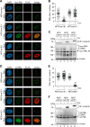
Fig 5. Interaction of ratIE1 with ratPML followed by ratPML deSUMOylation and dispersion.
(A) Schematic overview of full-length ratPML and deletion mutants. (B) Efficient interaction of ratIE1CORE with ratPML in co-immunoprecipitation analysis. HEK293T cells were co-transfected with expression plasmids encoding FLAG-tagged ratIE1 or ratIE1core (residues 1–392) and Myc-tagged ratPML variants. After cell lysis, immunoprecipitation was performed with an anti-FLAG antibody. Co-precipitated ratPML proteins (IP), precipitated ratIE1 proteins, and proteins within the cell lysate (input) were analyzed by Western blotting as indicated. (C) Binding of ratIE1CORE to ratPML requires both the coiled-coil and the RING domain. HEK293T cells were co-transfected with expression plasmids encoding FLAG-tagged ratPML variants and Myc-tagged ratIE1CORE (residues 1–382) as indicated. Upper two panels: Western blot detection of ratIE1 and ratPML after immunoprecipitation using an anti-FLAG antibody. Lower two panels: detection of ratIE1 and ratPML in cell lysates before precipitation (input). (D) Inhibition of ratPML SUMOylation by ratIE1 expression. HEK293T cells were transfected with expression plasmids encoding Myc-ratPML, HA-SUMO2 and FLAG-ratIE1 as indicated. After cell harvest, ratPML and SUMOylated ratPML were visualized by Western blotting using anti-Myc and anti-HA antibodies, respectively. Expression of IE1 was analyzed with an anti-FLAG antibody and β-actin was included as internal control. (E) Impact of RCMV infection on ratPML SUMOylation. Rat embryonic fibroblast (REF) cells were infected with RCMV at an MOI of 1.5 or mock infected, and were harvested at indicated times for Western Blot analysis of ratPML (upper panel), ratIE1 (middle panel), and β-actin (lower panel) as loading control. (F) Impact of RCMV infection on ratPML-NB integrity. REF cells were infected with RCMV at an MOI of 0.7 or mock infected, and were harvested at indicated times for immunofluorescence analysis of ratIE1 (left panel) or ratPML (right panel). Cell nuclei were stained with DAPI. F, FLAG; M, Myc; R, RING domain; B, B-boxes; CC, coiled-coil domain.
Fig 6
Fig 6. Species-specific disruption of PML-NBs during CMV infection.
(A) Analysis of PML-NB integrity in rat fibroblasts after HCMV infection. REF cells were infected with HCMV strain AD169 (MOI = 0.5) or mock infected. Cells were harvested at indicated times after infection to analyze the subcellular localization of ratPML (left panel) and humIE1 (right panel). Cell nuclei were stained with DAPI. (B) Species-specific binding of IE1 proteins to ratPML in co-immunoprecipitation analysis. HEK293T cells were co-transfected with expression plasmids coding for the TRIM motif of ratPML fused to a myc-tag (ratPML RBCC) and either FLAG-ratIE1CORE (residues 1–392), FLAG-humIE1CORE (residues 1–382) or an empty plasmid (pcDNA3). Afterwards, immunoprecipitation was performed with an anti-FLAG antibody. Left panels: Western blot detection of precipitated IE1 proteins and co-precipitated ratPML RBCC (IP). Right panels: detection of IE1 proteins and ratPML RBCC in cell lysates before precipitation (input). (C) Analysis of PML-NB integrity in human fibroblasts after RCMV infection. HFF cells were infected with RCMV-E (MOI = 0.5) or mock infected. Cells were fixed at indicated times for immunofluorescence analysis of humPML and ratIE1. Cell nuclei were visualized by DAPI staining. (D) Species-specific binding of IE1 proteins to humPML in co-immunoprecipitation analysis. HEK293T cells were co-transfected with expression plasmids encoding myc-tagged humPML and either FLAG-ratIE1CORE (residues 1–392), FLAG-humIE1CORE (residues 1–382) or an empty plasmid (pcDNA3). After immunoprecipitation of IE1 with an anti-FLAG antibody, co-precipitated humPML (left panels) as well as proteins in the lysate before precipitation (right panels) were detected by Western blotting.
Fig 7
Fig 7. Species-specific disruption of PML-NBs in cells stably expressing IE1.
(A, B) Effect of humIE1 and ratIE1 on the integrity of PML foci in human fibroblasts. Human fibroblasts with doxycycline-inducible expression of FLAG-tagged humIE1 (HFF/humIE1), FLAG-tagged ratIE1 (HFF/ratIE1) or control cells (HFF/control) were either left untreated (- Dox) or were treated with doxycycline (+ Dox) for 24 h. The cells were fixed for immunofluorescence staining of endogenous humPML and of IE1 proteins using an anti-FLAG antibody (A), followed by quantitation of humPML foci numbers in 50 cell nuclei per sample (B). (C) Impact of humIE1 and ratIE1 on the SUMOylation state of humPML. HFF/humIE1, HFF/ratIE1 or control cells were either left untreated (- Dox) or were treated with doxycycline (+ Dox). 24 h later, cells were harvested for Western Blot detection of IE1 proteins using an anti-FLAG antibody (upper panel), humPML (middle panel), and β-actin as loading control (lower panel). (D, E) Effect of humIE1 and ratIE1 on the integrity of PML foci in rat fibroblasts. Rat fibroblasts with doxycycline-inducible expression of FLAG-tagged humIE1 (REF/humIE1), FLAG-tagged rIE1 (REF/ratIE1) or control cells (REF/control) were either mock treated (- Dox) or were treated with doxycycline (+ Dox) for 24 h. The cells were fixed for immunofluorescence staining of endogenous ratPML and for IE1 proteins using an anti-FLAG antibody (D), followed by quantitation of ratPML foci numbers in 50 cell nuclei per sample (E). (F) Impact of humIE1 and ratIE1 on the SUMOylation state of ratPML. REF/humIE1, REF/ratIE1 or control REF were either left untreated (- Dox) or were treated with doxycycline (+ Dox). 24 h later, cells were harvested for Western Blot detection of IE1 proteins using an anti-FLAG antibody (upper panel), ratPML (middle panel), and β-actin as loading control (lower panel).
Fig 8
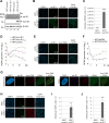
Fig 8. RCMV replication in human fibroblasts expressing humIE1.
(A, B) Increased initiation of RCMV gene expression in humIE1-expressing HFF. HFF with doxycycline-inducible expression of FLAG-tagged humIE1 (HFF/humIE1) or control cells (HFF/control) were treated with doxycycline (+ Dox) or mock treated (- Dox) for 24 h and subsequently infected with RCMV-E (MOI = 0.1). At 8 h post-infection (hpi), cells were harvested for Western Blot analysis of ratIE1 as well as humIE1 with an anti-FLAG antibody and β-actin as loading control (A) or for immunofluorescence detection of ratIE1, humIE1 (FLAG), and cell nuclei by DAPI staining (B). The percentage of rat IE1-positive cells was determined from triplicate samples. (C) Release of infectious RCMV particles from humIE1-expressing HFF. HFF/control and HFF/humIE1 were infected with RCMV-E at an MOI of 0.01 after 24 h of doxycycline treatment. Supernatants were harvested at 6 d post infection and titrated on REF cells. Values are derived from triplicate samples and represent mean values ± SD. P-values were calculated using two-tailed Student’s t-test. ***, p ≤ 0.001. (D) Multistep growth curve analysis of RCMV in humIE1-expressing HFF. HFF/control, HFF/humIE1 and HFF/humIE1CORE, which express residues 1–382 of humIE1, were treated with doxycycline for 24 h and subsequently infected with RCMV-E at an MOI of 0.01. Supernatants were harvested at indicated times after infection and analyzed for genome equivalents by RCMV gB-specific quantitative real-time PCR. (E) Increased initiation of RCMV gene expression in PML-depleted human fibroblasts. HFF expressing a control shRNA (HFF/shControl) or a shRNA directed against PML (HFF/shPML) were infected with RCMV-E (MOI = 0.1). At 8 hpi, cells were fixed for immunofluorescence detection of ratIE1 and humPML. Cell nuclei were visualized by DAPI staining. The percentage of ratIE1-positive cells was quantified from triplicate samples. (F) Multistep growth curve analysis of RCMV in PML-knockdown HFF. HFF/shControl and HFF/shPML infected with RCMV-E at an MOI of 0.01. Supernatants were harvested at indicated times after infection and analyzed for genome equivalents by RCMV gB-specific quantitative real-time PCR. (G) Colocalization of RCMV genomes with PML-NBs in human fibroblasts. HFF cells were infected with RCMV-EdC at an MOI of 0.05 or were mock infected. At 8 hpi, cells were fixed for click labeling to visualize RCMV genomes (vDNA) in combination with immunofluorescence detection of ratIE1 and humPML. DAPI staining was performed to visualize cell nuclei. Arrows in the merged PML-vDNA image indicate RCMV genomes colocalizing with PML-NBs. Dashed lines indicate the position of the cell nuclei. (H) Increased initiation of HCMV gene expression in ratIE1-expressing REF. REF/control and REF/ratIE1 were treated with doxycycline for 24 h and subsequently infected with HCMV strain AD169 (MOI = 0.1). At 24 hpi, cells were harvested for immunofluorescence analysis of humIE1, followed by quantification of humIE1-positive cells from triplicate samples. RatIE1 expression was confirmed by staining with an anti-FLAG antibody and cell nuclei were detected with DAPI. (I, J) Release of infectious HCMV particles from ratIE1-expressing REF. REF/control and REF/ratIE1 were treated with doxycycline for 24h and subsequently infected with HCMV strain AD169 (I) or TB40/E (J) at an MOI of 0.1. Supernatants were harvested at 6 d post infection and directly subjected to titration on HFF cells. Values are derived from triplicate samples and represent mean values ± SD. P-values were calculated using two-tailed Student’s t-test. **, p ≤ 0.01.

Similar articles

Cited by

References

    1. Bieniasz PD. Intrinsic immunity: a front-line defense against viral attack. Nature immunology. 2004;5(11):1109–15. Epub 2004/10/22. doi: 10.1038/ni1125 . - DOI - PubMed
    1. Chemudupati M, Kenney AD, Bonifati S, Zani A, McMichael TM, Wu L, et al.. From APOBEC to ZAP: Diverse mechanisms used by cellular restriction factors to inhibit virus infections. Biochimica et biophysica acta Molecular cell research. 2019;1866(3):382–94. Epub 2018/10/06. doi: 10.1016/j.bbamcr.2018.09.012 . - DOI - PMC - PubMed
    1. Gaba A, Flath B, Chelico L. Examination of the APOBEC3 Barrier to Cross Species Transmission of Primate Lentiviruses. Viruses. 2021;13(6). Epub 2021/07/03. doi: 10.3390/v13061084. - DOI - PMC - PubMed
    1. Rivera-Molina YA, Martínez FP, Tang Q. Nuclear domain 10 of the viral aspect. World journal of virology. 2013;2(3):110–22. Epub 2013/11/21. doi: 10.5501/wjv.v2.i3.110 . - DOI - PMC - PubMed
    1. Bernardi R, Pandolfi PP. Structure, dynamics and functions of promyelocytic leukaemia nuclear bodies. Nature reviews Molecular cell biology. 2007;8(12):1006–16. Epub 2007/10/12. doi: 10.1038/nrm2277 . - DOI - PubMed

Publication types

Substances

Grants and funding

This work was supported by the Deutsche Forschungsgemeinschaft [grant number SFB796, A03 to YAM; grant number Mu 1477/10-1 to YAM; grant number SFB796, B03 to TS; grant number STA357/7-1 to TS]. The funders had no role in study design, data collection and analysis, decision to publish, or preparation of the manuscript.