. 2021 Jul 22:12:672192.
doi: 10.3389/fmicb.2021.672192. eCollection 2021.

Subcellular Localization of Epstein-Barr Virus BLLF2 and Its Underlying Mechanisms

Affiliations

Subcellular Localization of Epstein-Barr Virus BLLF2 and Its Underlying Mechanisms

Jingjing Li et al. Front Microbiol. .

Abstract

Epstein-Barr virus (EBV), the pathogen of several human malignancies, encodes many proteins required to be transported into the nucleus for viral DNA reproduction and nucleocapsids assembly in the lytic replication cycle. Here, fluorescence microscope, mutation analysis, interspecies heterokaryon assays, co-immunoprecipitation assay, RNA interference, and Western blot were performed to explore the nuclear import mechanism of EBV encoded BLLF2 protein. BLLF2 was shown to be a nucleocytoplasmic shuttling protein neither by a chromosomal region maintenance 1 (CRM1)- nor by a transporter associated with antigen processing (TAP)-dependent pathway. Yet, BLLF2's two functional nuclear localization signals (NLSs), NLS1 (16KRQALETVPHPQNRGR31) and NLS2 (44RRPRPPVAKRRRFPR58), were identified, whereas the predicted NES was nonfunctional. Finally, BLLF2 was proven to transport into the nucleus via a Ran-dependent and importin β1-dependent pathway. This mechanism may contribute to a more extensive insight into the assembly and synthesis of EBV virions in the nucleus, thus affording a new direction for the treatment of viruses.

Keywords: CRM1; EBV BLLF2; NES; NLS; TAP; importin.


Conflict of interest statement

The authors declare that the research was conducted in the absence of any commercial or financial relationships that could be construed as a potential conflict of interest.

Figures
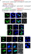
FIGURE 1
FIGURE 1
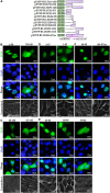
Subcellular localization and nucleocytoplasmic shuttling of BLLF2. (A) Potential NESs and NLSs of BLLF2 were predicted by the bioinformatics software NetNES 1.1 and PSORT II, respectively. Proteins assigned for trafficking into the nucleus encompass aa targeting sequences termed NLSs (Lange et al., 2007), and proteins bound for delivery out of the nucleus consisting of aa targeting sequences named NESs (La Cour et al., 2004). The basic aas are arginine (R), histidine (H), and lysine (K). PSORT II adopts the following two standards to dissect target protein: four-residue pattern (termed “pat4”) formed by four basic aa (K or R), or formed by three basic aa (K or R) and either H or P; the other (termed “pat7”) is a pattern beginning with P and followed within three residues by a basic segment including three K/R residues out of four. The identified NLSs 16KRQALETVPHPQNRGR31 (NLS1) and 44RRPRPPVAKRRRFPR58 (NLS2) were also indicated. (B) Subcellular distributions of EYFP-BLLF2 and EYFP vector in COS-7 cells. (C) Subcellular distributions of BLLF2-EYFP and BLLF2-Myc in COS-7 cells. (D) Co-expression of EYFP-BLLF2 and pNucleolin-EGFP was observed in COS-7 cells. (E) COS-7 and NIH3T3 cells were stained with DAPI. NIH3T3 cells were identified by their speckled nuclei. (F) Nucleocytoplasmic shuttling of BLLF2 was demonstrated by interspecies heterokaryon assays. EYFP-BFLF2 was used as nucleocytoplasmic shuttling positive control. COS-7 and NIH3T3 cells were discriminated against by nuclear staining with DAPI. NIH3T3 cells were identified by their speckled nuclei (red arrowhead). All scale bars indicate 20 μm.
FIGURE 2
FIGURE 2
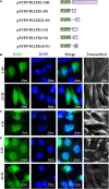
Identification of the predicted NES and functional NLS in BLLF2. (A) Schematic diagram of constructs encoding EYFP-tagged wild-type BLLF2 and its deletion mutants aa1–43, 1–47, 1–58, 44–58, 48–58, 48–82, 59–82, 59–148, 83–148, 83–110, and full-length mutant BLLF2(44–47)m. (B) Intracellular localization of deletion mutants BLLF2(1–58) and BLLF2(59–148) in COS-7 cells. (C) Intracellular localization of deletion mutants BLLF2(83–143) and BLLF2(83–110) in COS-7 cells. (D) Intracellular localization of deletion mutants BLLF2(1–47) and BLLF2(1–43) in COS-7 cells. (E) Intracellular localization of deletion mutants BLLF2(48–82), BLLF2(48–58), and BLLF2(59–82) in COS-7 cells. (F) Intracellular localization of deletion mutant BLLF2(44–58) and full-length mutant BLLF2(44–47)m in COS-7 cells. All scale bars indicate 20 μm.
FIGURE 3
FIGURE 3
aa16–31 is another functional NLS of BLLF2. (A) Schematic diagram of constructs encoding EYFP-tagged wild-type BLLF2 and its deletion mutants aa1–20, 21–43, 1–31, 6–31, and 16–31. (B) Intracellular localization of deletion mutants BLLF2(1–20) and BLLF2(21–43) in COS-7 cells. (C) Intracellular localization of deletion mutant BLLF2(1–31) in COS-7 cells. (D) Intracellular localization of deletion mutants BLLF2(6–31) and BLLF2(16–31) in COS-7 cells. All scale bars indicate 20 μm.
FIGURE 4
FIGURE 4
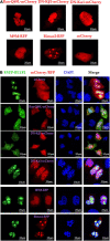
Nuclear import mechanism of BLLF2. (A) Individual subcellular localization of Ran-Q69L-mCherry, DN kα1-mCherry, DN kβ1-mCherry, M9M-RFP, Bimax2-RFP, or mCherry vector in COS-7 cells. (B) Co-expression of Ran-Q69L-mCherry/EYFP-BLLF2, DN kα1-mCherry/EYFP-BLLF2, DN kβ1-mCherry/EYFP-BLLF2, M9M-RFP/EYFP-BLLF2, Bimax2-RFP/EYFP-BLLF2, or mCherry/EYFP-BLLF2 in COS-7 cells. All scale bars indicate 20 μm.
FIGURE 5
FIGURE 5
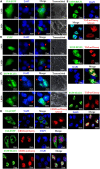
Nuclear export mechanism of BLLF2. Interspecies heterokaryon assays were performed to analyze the nuclear export of BLLF2. Mouse NIH3T3 cells were plated onto the CRM1-dependent positive control UL4 (A), CRM1-independent negative control EYFP vector (B), or pEYFP-BLLF2 (C) transfected COS-7 cells, with or without LMB treatment, as described in Section “Materials and Methods.” Cells were then stained with DAPI and imaged by fluorescence microscopy. NIH3T3 cells were identified by their speckled nuclei (red arrowhead). (D) COS-7 cells were individually transfected with UL4-EYFP or co-transfected with expression plasmids CRM1-mCherry/UL4-EYFP or CRM1-mCherry/EYFP-BLLF2 and then examined by confocal microscopy. (E) COS-7 cells were individually transfected with the TAP-dependent positive control EYFP-BFLF2 or co-transfected with expression plasmids TAP-mCherry/EYFP-BFLF2 or TAP-mCherry/EYFP-BLLF2, and then examined by confocal microscopy. All scale bars indicate 20 μm.
FIGURE 6
FIGURE 6
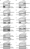
BLLF2 binds to importin β1. (A–G,L) Co-IP analysis of BLLF2 with importin β1 (A,G), importin α5 (kα1) (B), importin α1 (kα2) (C), importin α3 (kα4) (D), importin α7 (kα6) (E), importin β2 (F), or Flag vector (L). HEK293T cells were co-transected with expression plasmids combination of 3 × Flag-importin β1/EYFP-BLLF2 (A,G), Flag-kα1/EYFP-BLLF2 (B), Flag-kα2/EYFP-BLLF2 (C), Flag-kα4/EYFP-BLLF2 (D), Flag-kα6/EYFP-BLLF2 (E), Flag-importin β2/EYFP-BLLF2 (F), or Flag vector/EYFP-BLLF2 (L) for 24 h; cells were subsequently lysed and Co-IPed with anti-Flag mAb (A–F,L) or anti-YFP pAb (G) or control IgG, and then WB analysis was carried out with the indicated Abs. (H,I) Co-IP analysis of importin β1 with BFLF2 (H) or PRV UL31 (I). HEK293T cells were co-transected with expression plasmids combination of 3 × Flag-importin β1/EYFP-BFLF2 (H) or 3 × Flag-importin β1/PRV UL31-EYFP (I) for 24 h, and cells were then lysed and Co-IPed with anti-Flag mAb or control IgG, and then WB analysis was carried out with the indicated Abs. (J,K) Co-IP analysis of EYFP vector with Flag vector (J) or importin β1 (K). HEK293T cells were co-transected with expression plasmids combination of Flag vector/EYFP vector (J) or 3 × Flag-importin β1/EYFP vector (K) for 24 h; cells were then lysed and Co-IPed with anti-Flag mAb (J) or anti-YFP (K) or control IgG, and then WB analysis was carried out with the indicated Abs.
FIGURE 7
FIGURE 7
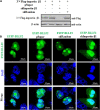
Subcellular localization of BLLF2 when importin β1 was knocked down. (A) Validation of knockdown efficiency of the constructed shImportin-β1 expression plasmid. HEK293T cells were transfected with 3 × Flag-importin β1 expression plasmid or co-transfected with the expression plasmids combination of 3 × Flag-importin β1/pSuper, 3 × Flag-importin β1/shRandom, or 3 × Flag-importin β1/shImportin-β1 for 24 h. Then, cells were lysed, and WB was carried out with anti-Flag mAb. β-actin was used as a loading control. (B) pEYFP-BLLF2 was transfected into COS-7 cells, or pEYFP-BLLF2 was co-transfected with pSuper, shRandom, or shImportin-β1 expression plasmid into COS-7 cells. At 24 h post-transfection, confocal fluorescence microscopy was executed to examine the subcellular localization of BLLF2.
FIGURE 8
FIGURE 8
Schematic diagram of nuclear transport mechanisms of EBV and other herpesviruses-encoded proteins.

Similar articles

Cited by

References

    1. Agutter P. S., Prochnow D. (1994). Nucleocytoplasmic transport. Biochem. J. 300(Pt 3) 609–618. - PMC - PubMed
    1. Arts G. J., Kuersten S., Romby P., Ehresmann B., Mattaj I. W. (1998). The role of exportin-t in selective nuclear export of mature tRNAs. EMBO J. 17 7430–7441. 10.1093/emboj/17.24.7430 - DOI - PMC - PubMed
    1. Behrens R. T., Aligeti M., Pocock G. M., Higgins C. A., Sherer N. M. (2017). Nuclear export signal masking regulates HIV-1 rev trafficking and viral RNA nuclear export. J. Virol. 91 e2107–e2116. - PMC - PubMed
    1. Belov G. A., Lidsky P. V., Mikitas O. V., Egger D., Lukyanov K. A., Bienz K., et al. (2004). Bidirectional increase in permeability of nuclear envelope upon poliovirus infection and accompanying alterations of nuclear pores. J. Virol. 78 10166–10177. 10.1128/jvi.78.18.10166-10177.2004 - DOI - PMC - PubMed
    1. Borer R. A., Lehner C. F., Eppenberger H. M., Nigg E. A. (1989). Major nucleolar proteins shuttle between nucleus and cytoplasm. Cell 56 379–390. 10.1016/0092-8674(89)90241-9 - DOI - PubMed