

18F-FDG-PET/CT in SARS-CoV-2 infection and its sequelae
- PMID: 34340958
- PMCID: PMC8316133
- DOI: 10.1016/j.remnie.2021.07.005
18F-FDG-PET/CT in SARS-CoV-2 infection and its sequelae
Abstract
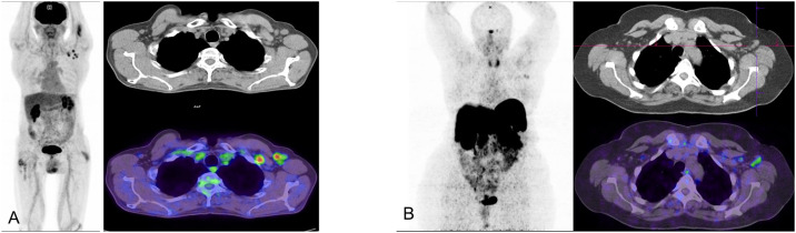
In recent months, much of the scientific efforts have focused on research on SARSCoV-2 infection and its consequences in humans. Still, many aspects remain unknown. It is known that the damage caused by SARS-CoV-2 is multifactorial and that its extension goes beyond lung inflammation and the acute phase, with the appearance of numerous complications and sequelae. To date, knowledge about the usefulness of 18F-FDG-PET/CT in the acute phase has been limited to the incidental detection of SARS-CoV-2 unsuspected pneumonia. Recent studies have been appearing collecting the findings of 18F-FDG-PET/CT in long COVID-19 or persistent COVID-19 state as well as the alterations caused after mass vaccination of the population in the metabolic studies. This work aims to review the existing literature focusing on these three issues and to briefly present our own preliminary experience.
Durante los últimos meses gran parte de los esfuerzos científicos se han centrado en la investigación sobre el SARS-CoV-2 y las consecuencias de su infección en humanos. Aun así, muchos aspectos siguen siendo desconocidos. Se sabe que la afectación por SARS-CoV-2 es multifactorial y que su extensión va más allá del daño pulmonar y del momento agudo, con aparición de numerosas de complicaciones y secuelas. El conocimiento de la utilidad de la 18F-FDG-PET/TC en el momento agudo se ha limitado, hasta la fecha, a la detección incidental de afectación pulmonar por SARS-CoV-2. En los últimos meses han ido apareciendo trabajos que recogen los hallazgos de la 18F-FDG-PET/TC en el estado post-COVID, asícomo las alteraciones provocadas en la imagen metabólica tras la vacunación masiva de la población. Este trabajo pretende revisar la literatura existente sobre estas tres cuestiones y exponer de manera breve la experiencia preliminar propia.
Keywords: (18)F-FDG-PET/CT; (18)F-FDG-PET/TC; COVID-19 persistente; Inflamación; Inflammation; Long COVID-19; SARS-CoV-2; Vaccine; Vacuna.
Copyright © 2021 Sociedad Española de Medicina Nuclear e Imagen Molecular. Published by Elsevier España, S.L.U. All rights reserved.
Figures
Similar articles
-
[18F-FDG-PET/CT in SARS-CoV-2 infection and its sequelae].Rev Esp Med Nucl Imagen Mol. 2021 Sep-Oct;40(5):299-309. doi: 10.1016/j.remn.2021.07.002. Epub 2021 Jul 12. Rev Esp Med Nucl Imagen Mol. 2021. PMID: 35368611 Free PMC article. Spanish.
-
Assessment of extra-parenchymal lung involvement in asymptomatic cancer patients with COVID-19 pneumonia detected on 18F-FDG PET-CT studies.Eur J Nucl Med Mol Imaging. 2021 Mar;48(3):768-776. doi: 10.1007/s00259-020-05019-y. Epub 2020 Sep 8. Eur J Nucl Med Mol Imaging. 2021. PMID: 32901353 Free PMC article.
-
Incidental Findings Suggestive of COVID-19 Pneumonia in Oncologic Patients Undergoing 18F-FDG PET/CT Studies: Association Between Metabolic and Structural Lung Changes.J Nucl Med. 2022 Feb;63(2):274-279. doi: 10.2967/jnumed.121.261915. Epub 2021 Jun 4. J Nucl Med. 2022. PMID: 34088776 Free PMC article.
-
18F-FDG-PET Imaging for Post-COVID-19 Brain and Skeletal Muscle Alterations.Viruses. 2021 Nov 15;13(11):2283. doi: 10.3390/v13112283. Viruses. 2021. PMID: 34835088 Free PMC article. Review.
-
FDG-PET/CT of COVID-19 and Other Lung Infections.Semin Nucl Med. 2022 Jan;52(1):61-70. doi: 10.1053/j.semnuclmed.2021.06.017. Epub 2021 Jun 22. Semin Nucl Med. 2022. PMID: 34246449 Free PMC article. Review.
Cited by
-
New-onset sarcoidosis in a patient with long COVID.Clin Case Rep. 2024 Aug 10;12(8):e9186. doi: 10.1002/ccr3.9186. eCollection 2024 Aug. Clin Case Rep. 2024. PMID: 39130813 Free PMC article.
-
Brain Imaging Changes in Patients Recovered From COVID-19: A Narrative Review.Front Neurosci. 2022 Apr 22;16:855868. doi: 10.3389/fnins.2022.855868. eCollection 2022. Front Neurosci. 2022. PMID: 35527821 Free PMC article.
-
Post-COVID-19 Brain [18F] FDG-PET Findings: A Retrospective Single-Center Study in the United States.AJNR Am J Neuroradiol. 2023 May;44(5):517-522. doi: 10.3174/ajnr.A7863. Epub 2023 Apr 27. AJNR Am J Neuroradiol. 2023. PMID: 37105680 Free PMC article.
-
Recent Advancements in Radiopharmaceuticals for Infection Imaging.Methods Mol Biol. 2024;2813:205-217. doi: 10.1007/978-1-0716-3890-3_14. Methods Mol Biol. 2024. PMID: 38888780 Review.
-
Follow-Up of a Cohort of Patients with Post-Acute COVID-19 Syndrome in a Belgian Family Practice.Viruses. 2022 Sep 9;14(9):2000. doi: 10.3390/v14092000. Viruses. 2022. PMID: 36146806 Free PMC article.
References
-
- WHO Coronavirus (COVID-19) Dashboard | WHO Coronavirus (COVID-19) Dashboard with Vaccination Data [Internet]. [cited 2021 May 31]. Available from: https://covid19.who.int/.
Publication types
MeSH terms
Substances
LinkOut - more resources
Full Text Sources
Medical
Miscellaneous
