. 2021 Aug 19;184(17):4531-4546.e26.
doi: 10.1016/j.cell.2021.07.005. Epub 2021 Jul 26.

Ribosome ADP-ribosylation inhibits translation and maintains proteostasis in cancers

Affiliations

Ribosome ADP-ribosylation inhibits translation and maintains proteostasis in cancers

Sridevi Challa et al. Cell. .

Abstract

Defects in translation lead to changes in the expression of proteins that can serve as drivers of cancer formation. Here, we show that cytosolic NAD+ synthesis plays an essential role in ovarian cancer by regulating translation and maintaining protein homeostasis. Expression of NMNAT-2, a cytosolic NAD+ synthase, is highly upregulated in ovarian cancers. NMNAT-2 supports the catalytic activity of the mono(ADP-ribosyl) transferase (MART) PARP-16, which mono(ADP-ribosyl)ates (MARylates) ribosomal proteins. Depletion of NMNAT-2 or PARP-16 leads to inhibition of MARylation, increased polysome association and enhanced translation of specific mRNAs, aggregation of their translated protein products, and reduced growth of ovarian cancer cells. Furthermore, MARylation of the ribosomal proteins, such as RPL24 and RPS6, inhibits polysome assembly by stabilizing eIF6 binding to ribosomes. Collectively, our results demonstrate that ribosome MARylation promotes protein homeostasis in cancers by fine-tuning the levels of protein synthesis and preventing toxic protein aggregation.

Keywords: ADP-ribosylation; MARylation; NAD(+); NAD(+) sensor; NAD(+) synthesis; NMNAT-2; PARP-16; cancer; mRNA translation; mono(ADP-ribose); mono(ADP-ribosyl)ation; ovarian cancer; protein aggregation; protein synthesis; ribosomes; translation.


Conflict of interest statement

Declaration of interests W.L.K. is a founder and consultant for Ribon Therapeutics, Inc. and ARase Therapeutics, Inc. He is also coholder of U.S. Patent 9,599,606 covering the ADP-ribose detection reagent used herein, which has been licensed to and is sold by EMD Millipore.

Figures
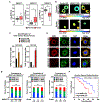
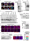
Figure 1.
Figure 1.. NAD+ synthesis and ADPRylation are compartmentalized in ovarian cancer cells.
(A) RNA-seq expression data for NMNAT1, NMNAT2, and NMNAT3 mRNAs in ovarian cancer tissues, expressed as transcripts per million (TPM). (TCGA ovarian cancer samples, n = 426) compared to normal ovarian tissues (GTEx data, n = 88) (* p < 0.05). (B and C) NMNAT-1 and NMNAT-2 regulate compartment-specific NAD+ levels in OVCAR3 cells with NMNAT1 or NMNAT2 knockdown (KD). The fluorescence images in (B) were generated using cytosolic and nuclear NAD+ sensors. The scale bar shows the inverse relationship between fluorescence and NAD+ level. Each bar in the graph in (C) represents the mean ± SEM of the NAD+ concentrations calculated using sensor(488/405 nm)/control(488/405 nm) fluorescence ratios determined by live cell imaging using a standard curve. (n = 3, ANOVA, * p < 0.05, ** p < 0.001). (D) Co-localization of MAR and RPS6, a ribosomal protein, in OVCAR3 cells as determined by immunofluorescent staining. Representative images are shown. Scale bar = 10 μm. (E) MAR levels positively correlate with NMNAT-2 expression in ovarian cancer patient samples, and high grade ovarian cancers have higher levels of NMNAT-2 and MAR. IHC analysis for MAR and NMNAT-2 using ovarian cancer tissue microarrays. The number of patients in each group are indicated below the graphs (Chi-square test, *** p<0.001, **** p < 0.0001). (F) High MAR levels by IHC are a predictor of poor survival in ovarian cancer patients. Analysis of progression free survival using the immunohistochemistry staining for MAR in high grade serous ovarian cancer tissues (n = 49). HR: Hazard ratio. See also Figure S1.
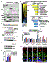
Figure 2.
Figure 2.. NMNAT-2-dependent MARylation of ribosomal proteins inhibits protein synthesis.
(A) MARylation, but not PARylation, is detected in ribosomal fractions. Western blot analysis for MAR and PAR of ribosomal fractions or whole cell extracts prepared from OVCAR3 cells. RPS6 and SNRP70 were used as the markers for ribosomal and nuclear fractions, respectively. (B) FK866 treatment reduces both MARylation and PARylation in OVCAR3 cells. Western blot analysis of ribosomal fractions or whole cell extracts isolated from OVCAR3 cells treated with 20 nM FK866 for 48 hours. (C) NMNAT-2 depletion inhibits ribosomal protein MARylation. Western blot analysis of ribosomal fractions or whole cell extracts isolated from OVCAR3 cells subjected to NMNAT2 knockdown. (D) NMNAT-2 catalytic activity is required for ribosomal protein MARylation. Western blot analysis of ribosomal fractions or whole cell extracts prepared from OVCAR3 cells subjected to Dox-induced expression of wild-type (Wt) or catalytically dead (H24D) mouse NMNAT-2 (Nmnat2; siRNA-resistant) followed by siRNA-mediated knockdown of NMNAT2. (E) NMNAT-2 depletion enhances protein synthesis in OVCAR3 cells. Western blot analysis of puromycin incorporation assays from OVCAR3 cells subjected to NMNAT1 or NMNAT2 knockdown. (F) Ectopic expression of NMNAT-2 overexpression inhibits protein synthesis. Western blot analysis of puromycin incorporation assays prepared from OVCAR3 cells subjected to Dox-induced expression of wild-type (Wt) or catalytically dead (H24D) mouse NMNAT-2 (Nmnat2; siRNA-resistant) followed by siRNA-mediated knockdown of NMNAT2. See also Figure S1.
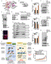
Figure 3.
Figure 3.. PARP-16 and NMNAT-2 regulates ribosomal protein MARylation-dependent protein homeostasis.
(A and B) PARP-16 mediates ribosomal protein MARylation. OVCAR3 cells were subjected to knockdown with two different siRNAs targeting each of the expressed cytosolic PARP monoenzymes. (A) Representative images from immunofluorescent staining for MAR, RPS6, and DNA (DAPI). The results from siRNA #2 targeting each PARP monoenzymes are shown. Scale bar = 10 μm. (B) Western blot analysis of ribosomal fractions from cells treated as described in (A). (C) PARP-16 knockdown reduces ribosomal protein MARylation. Western blot analysis for MAR and PARP-16 of ribosomal fractions prepared from OVCAR3 cells subjected to shRNA-mediated knockdown of PARP16. RPS6 was used as the marker for ribosomal fractions. (D) PARP-16 depletion enhances protein synthesis in OVCAR3 cells. Western blot analysis of puromycin incorporation assays from OVCAR3 cells subjected to PARP16 knockdown. (E) PARP-16 associates with ribosomes. Cell fractionation and Western blot analysis of PARP-16 in whole cell extracts and ribosomal fractions prepared from OVCAR3 cells. RPL10 and tubulin were used as markers/loading controls for the ribosomal fractions and whole cell extracts, respectively. (F) NMNAT-2 regulates PARP-16 activity. PARP-16 was immunoprecipitated from 293T cells ectopically expressing Flag-tagged wild-type (Wt) or catalytically dead (W92G) NMNAT-2 and subjected to Western blotting for MAR and Flag. (G) Depletion of PARP-16 or NMNAT-2 promotes the accumulation of protein aggregates. Staining of protein aggregates using Proteostat aggresome detection reagent in OVCAR3 cells subjected to PARP16 or NMNAT2 knockdown. Treatment with a low dose of cycloheximide (10 μg/mL) for 16 hours inhibits the accumulation of the aggregates. Scale bar = 25 μm. (H) Depletion of PARP-16 or NMNAT-2 causes proteotoxicity. OVCAR3 cells subjected to PARP16 or NMNAT2 knockdown were assayed for eIF2α phosphorylation and cleaved caspase-3 by Western blotting. Inhibition of translation by cycloheximide blocks the phosphorylation of eIF2α and caspase-3 cleavage. (I and J) MAR levels negatively correlate with protein aggregation in ovarian cancer patient samples. (I) Representative images of IHC analysis for Proteostat aggresome detection reagent staining using ovarian cancer tissue microarrays. (J) IHC analysis for MAR and Proteostat aggresome detection reagent staining using ovarian cancer tissue microarrays. The number of patients in each group are indicated below the graphs (Chi-square test, * p<0.05). See also Figures S2, S3, and S4.
Figure 4.
Figure 4.. NMNAT-2 and PARP-16 support ovarian cancer cell growth through ribosomal protein MARylation
(A) Depletion of PARP-16 or NMNAT-2 inhibits the anchorage-independent growth of OVCAR3 cells. Soft agar assay of OVCAR3 cells subjected to PARP16 or NMNAT2 knockdown. Each bar in the graph represents the mean ± SEM of the relative number of colonies (n = 3, one-way ANOVA, ** p < 0.01). (B) Depletion of PARP-16 or NMNAT-2 inhibits the in vivo growth of xenograft tumors formed from OVCAR3 cells subjected to PARP16 or NMNAT2 knockdown (n = 8 per group, ANOVA, * p<0.05). (C) Weights of tumors formed from OVCAR3 cells subjected to PARP16 or NMNAT2 knockdown (n = 8, one-way ANOVA, ** p<0.01, *** p<0.001, **** p<0.0001). (D) Depletion of PARP-16 or NMNAT-2 enhances protein synthesis and protein aggregation in vivo. Each bar in the graph in (D) represents the mean ± SEM of the relative ratios of Western blot signals of puromycin to tubulin (n = 3, t-test with Holm-Sidak correction, *** p<0.001). (E) Depletion of PARP-16 or NMNAT-2 causes proteotoxicity in vivo. Analysis of xenograft tumors described in (B) with Proteostat aggresome detection reagent staining and IHC using an antibody that recognizes cleaved caspase-3. See also Figure S5.
Figure 5.
Figure 5.. Ribosomal protein MARylation regulates polysome function through 3’ UTR stem-loop structures in mRNAs.
(A) Ribosomal protein MARylation is enriched in the monosome and polysome fractions of OVCAR3 cells. Western blot analysis for MAR and PARP-16 of the sucrose density gradient fractions prepared from OVCAR3 cells. RPS6 and RPL10 were used as markers for the small and large ribosomal subunits, respectively. (B and C) Depletion of NMNAT-2 or PARP-16 alters mRNA loading on polysomes. RNA-sequencing assay of mRNAs associated with polysomes isolated from OVCAR3 cells subjected to NMNAT2 or PARP16 knockdown. (B) Heatmap representation of mRNAs that exhibited altered loading on the polysomes when NMNAT-2 or PARP-16 were depleted. (C) Gene GO analysis of these mRNAs. (D) Identification of a transferable stem-loop motif in the 3’UTRs of mRNAs enriched on polysomes after NMNAT2 or PARP16 knockdown. (Top panel) Sequence of the motif with the highest score. (Bottom panels) The stem-loop motif in the 3’UTR of Flag-luciferase mRNA is required for translational regulation by PARP-16 and NMNAT-2. Western blot analysis for Flag-luciferase of lysates from PARP16 or NMNAT2 knockdown OVCAR3 cells that were transfected with the indicated Flag-luciferase constructs. (E) Addition of the stem-loop motif to the 3’UTR regulates polysome loading of Flag-luciferase mRNA. RT-qPCR analysis of Flag-luciferase mRNA isolated from the density gradient fractions corresponding to free ribosomal subunits, monosomes, and polysomes from PARP-16 or NMNAT-2 depleted OVCAR3 cells. Each bar in the graph represents the mean ± SEM of the relative Flag-luciferase mRNA levels (n = 3, two-way ANOVA, * p < 0.05, ** p < 0.01, *** p < 0.001, ****p < 0.0001). (F and G) Depletion of PARP-16 or NMNAT-2 enhances COX20 protein levels. (F) Western blot analysis for COX20 in OVCAR3 cells subjected to PARP16 or NMNAT2 knockdown. Each bar in the graph in (G) represents the mean ± SEM of the ratio of the levels of COX20 to tubulin (n = 3, two-way ANOVA, * p < 0.05). (H) Depletion of PARP-16 or NMNAT-2 promotes the accumulation of COX20 protein aggregates in OVCAR3 cells. Co-staining of protein aggregates using Proteostat aggresome detection reagent and COX20 in OVCAR3 cells subjected to PARP16 or NMNAT2 knockdown. Scale bar = 25 μm. See also Figure S6.
Figure 6.
Figure 6.. Role of NMNAT-2 and PARP-16 in the regulation of translation in normal fallopian tube cells.
(A) NMNAT-2 and PARP-16 levels are higher in ovarian cancer cells. (Top panel) Representative images of Western blot analysis of lysates prepared from a panel of fallopian tube cells and ovarian cancer cells. (Bottom panel) Violin plot of average expression of PARP-16 obtained from three independent biological replicates (t-test, ** p < 0.01). (B) Depletion of NMNAT-2 suppresses protein synthesis in fallopian tube cells. Western blot analysis of puromycin incorporation assays from FT194 and FT282 cells subjected to siRNA-mediated NMNAT2 knockdown. (C) NMNAT-2 expression enhances protein synthesis in FT194 cells. Western blot analysis of puromycin incorporation assays from FT194 cells subjected to Dox-induced expression of NMNAT-2. (D) Ectopic expression of PARP-16 inhibits protein synthesis in FT194 cells. Western blot analysis of puromycin incorporation assays from FT194 cells transfected with GFP-epitope tagged PARP-16 and Dox-induced expression of NMNAT-2. (E) Ectopic expression of NMNAT-2 alters mRNA loading on polysomes. Heatmaps showing the results of RNA-sequencing assay of mRNAs associated with polysomes isolated from OVCAR3 cells subjected to Dox-induced expression of wild-type mouse NMNAT-2 (Nmnat2) followed by siRNA-mediated knockdown of NMNAT2. (F) Ectopic expression of NMNAT-2 alters mRNA loading on polysomes. Heatmaps showing the results of RNA-sequencing assay of mRNAs associated with polysomes isolated from FT194 cells subjected to Dox-induced expression of wild-type NMNAT-2. (G) Ectopic expression of NMNAT-2 partially reverses the loading of mRNA onto polysomes of genes whose polysome loading is altered with depletion of NMNAT-2 and PARP-16 (Fig. 5B) with ectopic expression of NMNAT-2 in OVCAR3 and FT194 cells (p-value < 2.2e-16).
Figure 7.
Figure 7.. Site-specific MARylation of RPL24 at Glu4 inhibits polysome formation.
(A) Spatial distribution of the proteins modified by MARylation in the 80S ribosome (PDB ID: 4V6X). RPL24, which is located at the 60S-40S interface, is MARylated. (B) RPL24 is MARylated at Glu 4. HA-tagged RPL24 was immunoprecipitated from OVCAR3 cells ectopically expressing wild-type (Wt) or MARylation deficient (E4Q) RPL24 and subjected to Western blotting for MAR and HA. (C) RPL24-E4Q expression enhances protein synthesis in OVCAR3 cells. Western blot analysis of puromycin incorporation assays from OVCAR3 cells subjected to Dox-induced expression of RPL24. (D) RPL24-E4Q expression promotes the accumulation of protein aggregates. Staining of protein aggregates using Proteostat aggresome detection reagent in OVCAR3 cells subjected to Dox-induced expression of RPL24. (E) RPL24-E4Q expression enhances COX20 protein levels. Western blot analysis for COX20 in OVCAR3 cells subjected to Dox-induced expression of RPL24. (F) Loss of RPL24 MARylation induces apoptosis. OVCAR3 cells subjected to Dox-induced expression of RPL24 were assayed for caspase 3 cleavage by Western blotting. Inhibition of translation by cycloheximide blocks the cleavage of caspase 3. (G - J) Loss of RPL24 MARylation induces polysome formation. (G) Western blot analysis for HA-tagged RPL24, eIF6 and RPS6 of the sucrose density gradient fractions prepared from OVCAR3 cells subjected to Dox-induced expression of RPL24. Each bar in the graph in (H) represents the mean ± SEM of the relative abundance of RPL24, eIF6, and RPS6 in monosomes or polysomes (n = 4, Student’s t-test, * p < 0.05 and ** p<0.01). (K) Loss of Glu4 MARylation inhibits RPL24 interaction with eIF6. HA-tagged RPL24 was immunoprecipitated from OVCAR3 cells with Dox-induced expression of RPL24 and subjected to Western blotting for eIF6, RPS6, and HA. (L) MARylation of RPS6 at Glu 35 inhibits binding to RPL24. Flag-tagged RPS6 was immunoprecipitated from OVCAR3 cells subjected to Dox-induced expression of wild-type (Wt) or MARylation deficient (E35Q) RPS6 and subjected to Western blotting for MAR, RPL24 and Flag. (M) RPS6-E35Q expression enhances protein synthesis in OVCAR3 cells. Western blot analysis of puromycin incorporation assays from OVCAR3 cells subjected to Dox-induced expression of RPS6. (N) RPL24-E4Q expression inhibits cell growth. OVCAR3 cells subjected to Dox-induced knockdown and re-expression of RPL24 for 7 days and crystal violet staining was performed, (n = 4, one-way ANOVA, * p < 0.01). (O) Schematic of the mechanisms by which NMNAT-2/NAD+ and PARP-16/MAR regulate protein homeostasis and ovarian cancer growth. Additional details are provided in the text. See also Figure S7 and Table S1.

Similar articles

Cited by

References

    1. Ali YO, Allen HM, Yu L, Li-Kroeger D, Bakhshizadehmahmoudi D, Hatcher A, McCabe C, Xu J, Bjorklund N, Taglialatela G, et al. (2016). NMNAT2:HSP90 Complex Mediates Proteostasis in Proteinopathies. PLoS Biol 14, e1002472. - PMC - PubMed
    1. Andrews S (2010). FASTQC. A quality control tool for high throughput sequence data.
    1. Bai Y, Zhou K, and Doudna JA (2013). Hepatitis C virus 3′UTR regulates viral translation through direct interactions with the host translation machinery. Nucleic Acids Res 41, 7861–7874. - PMC - PubMed
    1. Bailey TL, Boden M, Buske FA, Frith M, Grant CE, Clementi L, Ren J, Li WW, and Noble WS (2009). MEME SUITE: tools for motif discovery and searching. Nucleic Acids Res 37, W202–208. - PMC - PubMed
    1. Berger F, Lau C, Dahlmann M, and Ziegler M (2005). Subcellular compartmentation and differential catalytic properties of the three human nicotinamide mononucleotide adenylyltransferase isoforms. J Biol Chem 280, 36334–36341. - PubMed

Publication types

MeSH terms