

Mir-106b Cluster Regulates Primordial Germ Cells Differentiation from Human Mesenchymal Stem Cells
- PMID: 34308572
- PMCID: PMC8286458
- DOI: 10.22074/cellj.2021.6836
Mir-106b Cluster Regulates Primordial Germ Cells Differentiation from Human Mesenchymal Stem Cells
Abstract
Objective: Numerous evidence indicates that microRNAs (miRNAs) are critical regulators in the spermatogenesis process. The aim of this study was to investigate miR-106b cluster regulates primordial germ cells (PGCs) differentiation from human mesenchymal stem cells (MSCs).
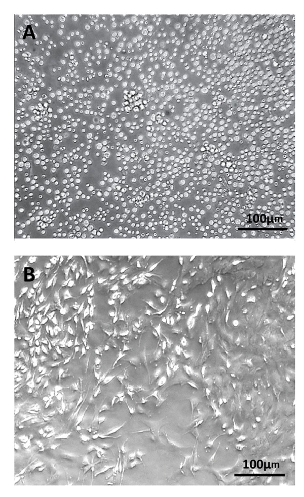
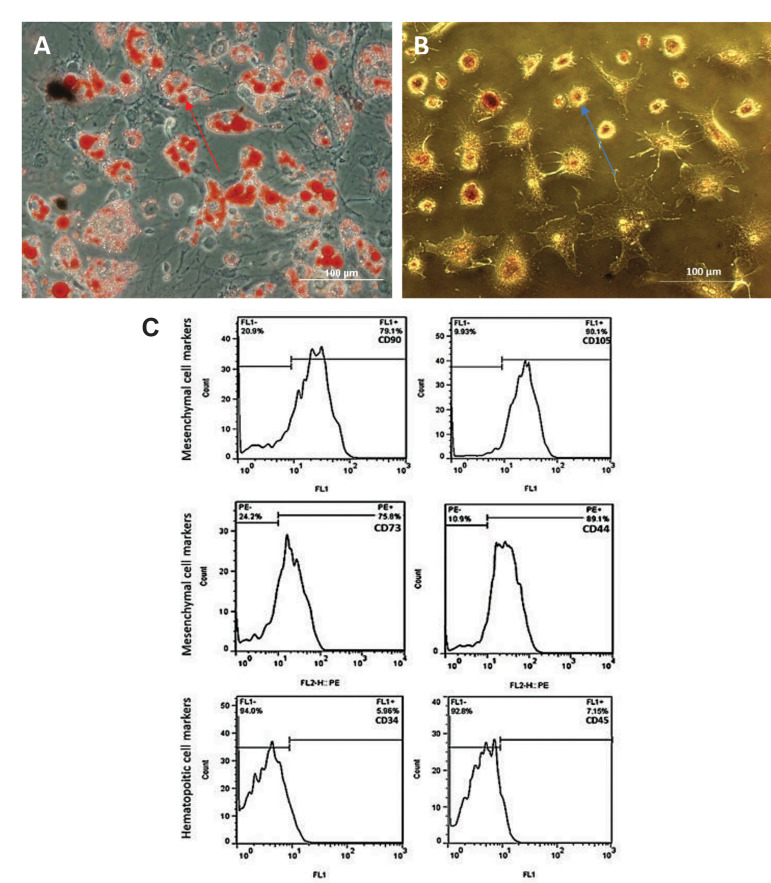
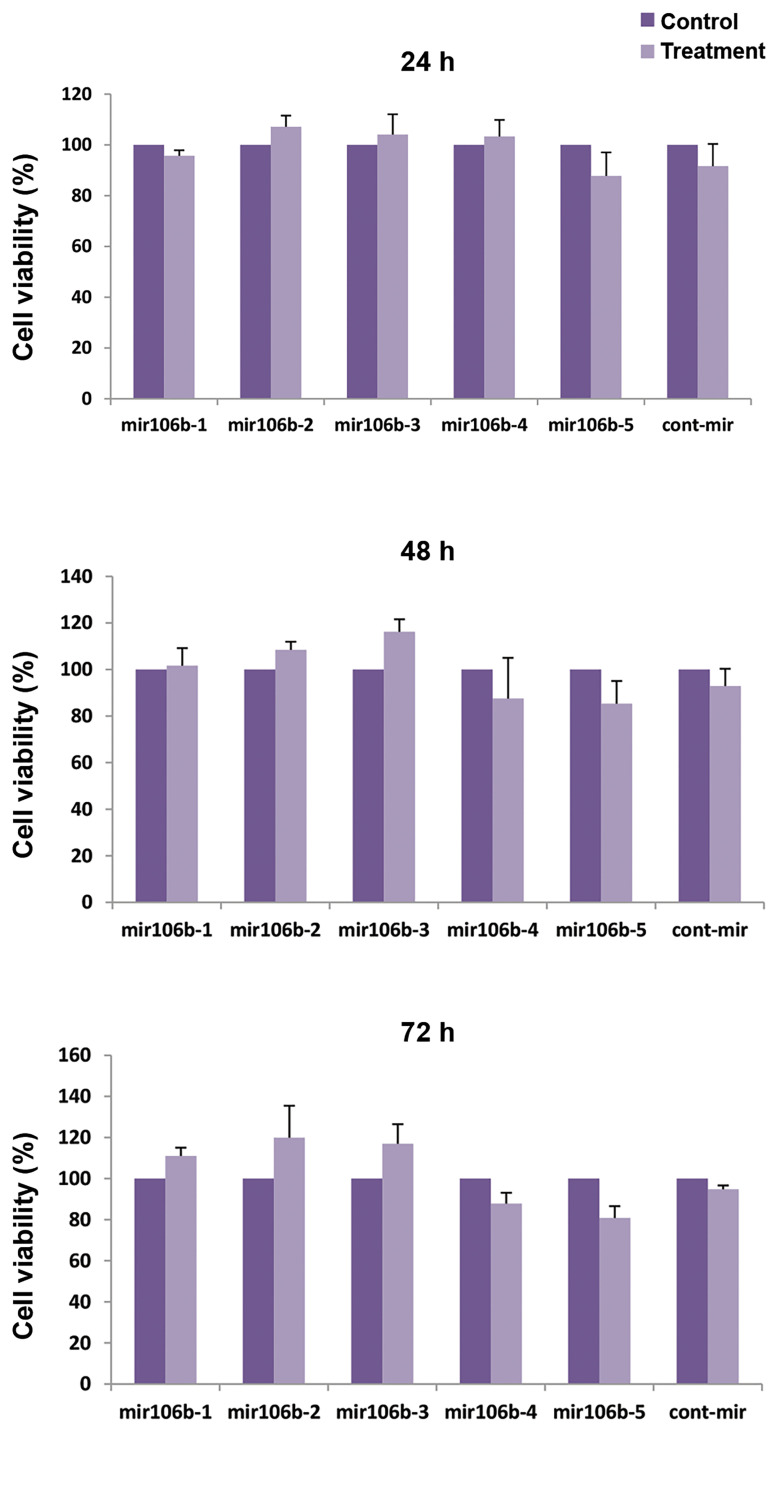
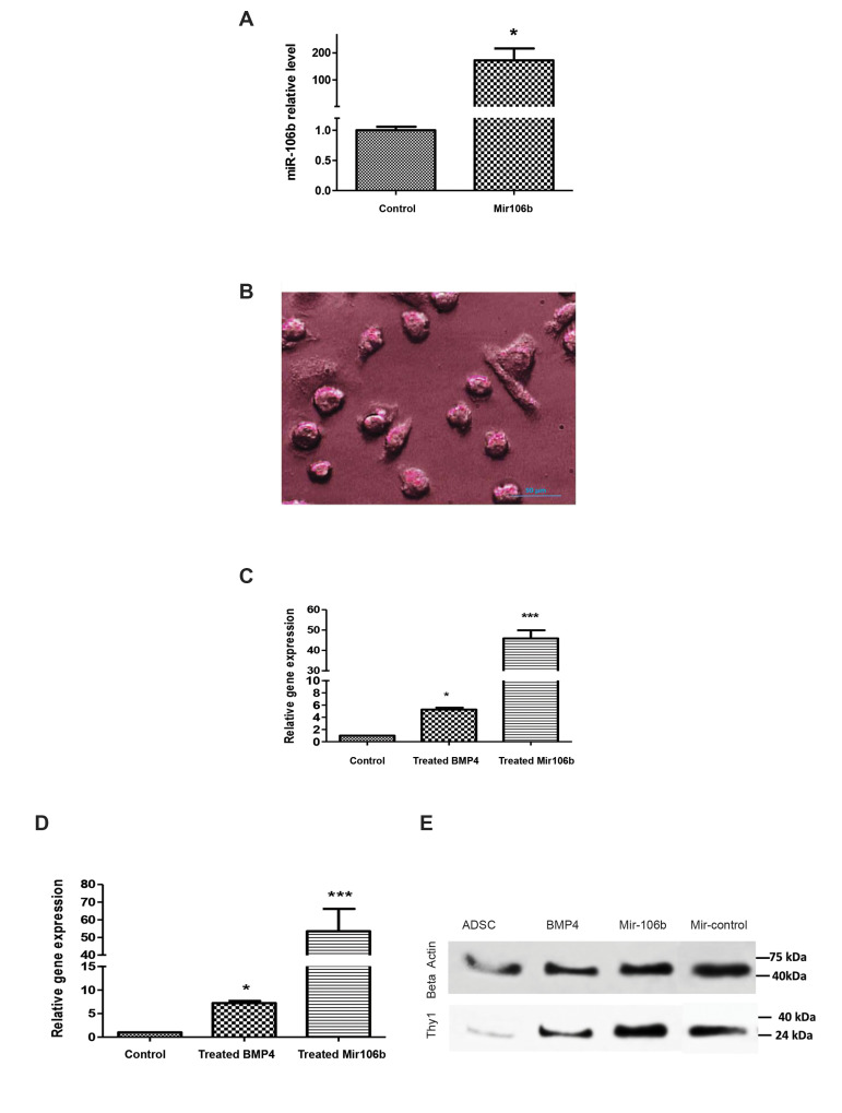
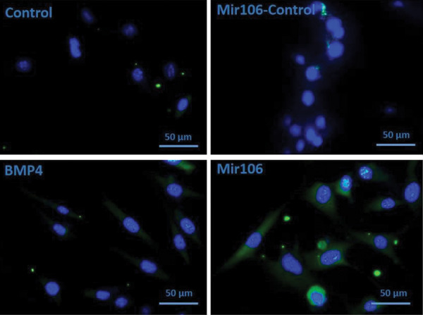
Materials and methods: In this experimental study, samples containing male adipose (n: 9 samples- age: 25-40 years) were obtained from cosmetic surgeries performed for the liposuction in Imam Khomeini Hospital. The differentiation of MSCs into PGCs was accomplished by transfection of a lentivector expressing miR-106b. The transfection of miR-106b was also confirmed by the detection of a clear green fluorescent protein (GFP) signal in MSCs. MSCs were treated with bone morphogenic factor 4 (BMP4) protein, as a putative inducer of PGCs differentiation, to induce the differentiation of MSCs into PGCs (positive control). After 4 days of transfection, the expression of miR-106b, STELLA, and FRAGILIS genes was evaluated by real-time polymerase chain reaction (PCR). Also, the levels of thymocyte differentiation antigen 1 (Thy1) protein was assessed by the western blot analysis. The cell surface expression of CD90 was also determined by immunocytochemistry method. The cytotoxicity of miR-106b was examined in MSCs after 24, 48, and 72 hours using the MTT assay.
Results: MSCs treated with BMP4 or transfected by miR-106b were successfully differentiated into PGCs. The results of this study also showed that the expression of miR-106b was significantly increased after 48 hours from transfection. Also, we showed STELLA, FARGILIS, as well as the protein expression of Thy1, was significantly higher in MSCs transfected by lentivector expressing miR-106b in comparison with MSCs treated with BMP4 (P≤0.05). MTT assay showed miR-106b was no toxic during 72 hours in 1 μg/ml dose, that this amount could elevated germ cells marker significantly higher than other experimental groups (P≤0.05).
Conclusion: According to this findings, it appears that miR-106b plays an essential role in the differentiation of MSCs into PGCs.
Keywords: Mesenchymal Stem Cells; MiR-106b; MicroRNA.
Copyright© by Royan Institute. All rights reserved.
Conflict of interest statement
There is no conflict of interest in this study.
Figures
Similar articles
-
miR-106b enhances human mesenchymal stem cell differentiation to spermatogonial stem cells under germ cell profile genes involved in TGF-b signaling pathways.In Vitro Cell Dev Biol Anim. 2022 Aug;58(7):539-548. doi: 10.1007/s11626-022-00688-5. Epub 2022 Aug 8. In Vitro Cell Dev Biol Anim. 2022. PMID: 35939226
-
Upregulation of miR-210 promotes differentiation of mesenchymal stem cells (MSCs) into osteoblasts.Bosn J Basic Med Sci. 2018 Nov 7;18(4):328-335. doi: 10.17305/bjbms.2018.2633. Bosn J Basic Med Sci. 2018. PMID: 30054999 Free PMC article.
-
Functional Concentrations of BMP4 on Differentiation of Mouse Embryonic Stem Cells to Primordial Germ Cells.Int J Fertil Steril. 2011 Jul;5(2):104-9. Epub 2011 Sep 23. Int J Fertil Steril. 2011. PMID: 24963367 Free PMC article.
-
miR-106b promotes cancer progression in hepatitis B virus-associated hepatocellular carcinoma.World J Gastroenterol. 2016 Jun 14;22(22):5183-92. doi: 10.3748/wjg.v22.i22.5183. World J Gastroenterol. 2016. PMID: 27298561 Free PMC article.
-
The emerging roles of the polycistronic miR-106b∼25 cluster in cancer - A comprehensive review.Biomed Pharmacother. 2018 Nov;107:1183-1195. doi: 10.1016/j.biopha.2018.08.097. Epub 2018 Aug 29. Biomed Pharmacother. 2018. PMID: 30257332 Review.
Cited by
-
Germline stem cells in human.Signal Transduct Target Ther. 2022 Oct 2;7(1):345. doi: 10.1038/s41392-022-01197-3. Signal Transduct Target Ther. 2022. PMID: 36184610 Free PMC article. Review.
-
Current Progress in Stem Cell Therapy for Male Infertility.Stem Cell Rev Rep. 2023 Oct;19(7):2073-2093. doi: 10.1007/s12015-023-10577-3. Epub 2023 Jul 13. Stem Cell Rev Rep. 2023. PMID: 37440145 Review.
-
Early Gonadal Development and Sex Determination in Mammal.Int J Mol Sci. 2022 Jul 6;23(14):7500. doi: 10.3390/ijms23147500. Int J Mol Sci. 2022. PMID: 35886859 Free PMC article. Review.
-
Reprogramming Human Female Adipose Mesenchymal Stem Cells into Primordial Germ Cell-Like Cells.Stem Cell Rev Rep. 2023 Oct;19(7):2274-2283. doi: 10.1007/s12015-023-10561-x. Epub 2023 Jun 20. Stem Cell Rev Rep. 2023. PMID: 37338786 Free PMC article.
-
Strategies for Mammalian Mesenchymal Stem Cells Differentiation into Primordial Germ Cell-Like Cells: A Review.Cell J. 2022 Aug 28;24(8):434-441. doi: 10.22074/cellj.2022.8087. Cell J. 2022. PMID: 36093802 Free PMC article.
References
-
- Huang P, Lin LM, Wu XY, Tang QL, Feng XY, Lin GY, et al. Differentiation of human umbilical cord Wharton’s jelly‐derived mesenchymal stem cells into germ‐like cells in vitro. J Cell Biochem. 2010;109(4):747–754. - PubMed
-
- Gnecchi M, Melo LG. Bone marrow-derived mesenchymal stem cells: isolation, expansion, characterization, viral transduction, and production of conditioned medium. Methods Mol Biol. 2009;482:281–294. - PubMed
LinkOut - more resources
Full Text Sources
Miscellaneous