. 2021 Sep 1;321(3):H542-H557.
doi: 10.1152/ajpheart.00125.2021. Epub 2021 Jul 23.

Notch2 suppression mimicking changes in human pulmonary hypertension modulates Notch1 and promotes endothelial cell proliferation

Affiliations

Notch2 suppression mimicking changes in human pulmonary hypertension modulates Notch1 and promotes endothelial cell proliferation

Sanghamitra Sahoo et al. Am J Physiol Heart Circ Physiol. .

Abstract

Pulmonary arterial hypertension (PAH) is a fatal cardiopulmonary disease characterized by increased vascular cell proliferation with apoptosis resistance and occlusive remodeling of the small pulmonary arteries. The Notch family of proteins subserves proximal signaling of an evolutionarily conserved pathway that effects cell proliferation, fate determination, and development. In endothelial cells (ECs), Notch receptor 2 (Notch2) was shown to promote endothelial apoptosis. However, a pro- or antiproliferative role for Notch2 in pulmonary endothelial proliferation and ensuing PAH is unknown. We postulated that suppressed Notch2 signaling drives pulmonary endothelial proliferation in the context of PAH. We observed that levels of Notch2 are ablated in lungs from PAH subjects compared with non-PAH controls. Notch2 expression was attenuated in human pulmonary artery endothelial cells (hPAECs) exposed to vasoactive stimuli including hypoxia, TGF-β, ET-1, and IGF-1. Notch2-deficient hPAECs activated Akt, Erk1/2, and antiapoptotic protein Bcl-2 and reduced levels of p21cip and Bax associated with increased EC proliferation and reduced apoptosis. In addition, Notch2 suppression elicited a paradoxical activation of Notch1 and canonical Notch target gene Hes1, Hey1, and Hey2 transcription. Furthermore, reduction in Rb and increased E2F1 binding to the Notch1 promoter appear to explain the Notch1 upregulation. Yet, when Notch1 was decreased in Notch2-suppressed cells, the wound injury response was augmented. In aggregate, our results demonstrate that loss of Notch2 in hPAECs derepresses Notch1 and elicits EC hallmarks of PAH. Augmented EC proliferation upon Notch1 knockdown points to a context-dependent role for Notch1 and 2 in endothelial cell homeostasis.NEW & NOTEWORTHY This study demonstrates a previously unidentified role for Notch2 in the maintenance of lung vascular endothelial cell quiescence and pulmonary artery hypertension (PAH). A key novel finding is that Notch2 suppression activates Notch1 via Rb-E2F1-mediated signaling and induces proliferation and apoptosis resistance in human pulmonary artery endothelial cells. Notably, PAH patients show reduced levels of endothelial Notch2 in their pulmonary arteries, supporting Notch2 as a fundamental driver of PAH pathogenesis.

Keywords: Notch; Rb; endothelial cell; proliferation; pulmonary arterial hypertension.


Figures
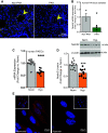
Figure 1.
Figure 1.
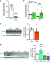
AE: expression of Notch2 is reduced in the pulmonary vasculature in pulmonary arterial (PA) hypertension (PAH) and in human pulmonary artery endothelial cells (hPAECs) exposed to hypoxia vs. normoxia. A and B: Notch2 expression is reduced in lung arteries of subjects with PAH. A: Notch2 localization and abundance in human PAH and non-PAH lung samples by immunofluorescence (IF). Expression of Notch2 (white) and nuclei (DAPI, blue) are shown (n = 3). B: quantification of transcript levels of Notch2 in pulmonary artery tissue homogenates of human non-PAH and PAH samples (n = 3). CE: Notch2 expression in commercially obtained hPAECs (Lonza) exposed to hypoxia to mimic cell conditions of pulmonary hypertension in vitro. C: mRNA expression of Notch2 upon 24 h hypoxia (Hyp) vs. normoxia (Norm) quantified by quantitative RT-PCR (n = 12). D: representative immunoblot and cumulative quantification (bar graph) of Notch2 in hPAECs in response to 24 h hypoxia (Hyp) vs. normoxia (Norm) (n = 12). E: IF labelling of Notch2 (red) in hPAECs upon 24 h hypoxia vs. normoxia. Nuclei stained with DAPI are shown in blue. Insets allow visualization of greater cell numbers (lower magnification). Dots represent individual data points of biological replicates. Bar graphs indicate means ± SE. P values for paired comparisons were calculated using Student’s t test. *P <0.05, ***P <0.001.
Figure 2.
Figure 2.
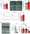
A and B: hypoxia inducible factor-1α (HIF-1α) gene silencing rescues hypoxia-mediated suppression of Notch2 in human pulmonary artery endothelial cells (hPAECs). A and B: hPAECs were transfected with HIF-1α siRNA or scrambled control (Scrmb). Transfected cells were exposed to hypoxia (Hyp) vs. normoxia (Norm), and samples for quantitative RT-PCR analysis were collected 24 h postexposure. Gene expression analyses for HIF-1α and Notch2 performed in hPAECs are shown (n = 9). C and D: vasoactive peptide endothelin-1 (ET-1), growth factors, and cytokines differentially regulate Notch2 expression in hPAECs. hPAECs were serum-starved overnight followed by stimulation with TGFβ1 (10 ng/mL), TNFα (10 ng/mL), ET-1 (10 ng/mL), and IGF-1 (200 ng/mL) for 24 h. Cells were lysed in RIPA buffer containing protease inhibitors and subjected to Western blot. Representative immunoblots and quantitative bar graphs depicting Notch2 in hPAECs (n = 6–9) are shown. P values for paired comparisons were calculated using Student’s t test. *P < 0.05, **P < 0.01, ***P <0.001. For B, values were determined using ANOVA. Post hoc tests applied the Bonferroni correction to allow for multiple comparisons. *P < 0.05 hypoxia vs. normoxia scrambled control, ###P < 0.001 HIF-1α siRNA vs. scrambled control hypoxia.
Figure 3.
Figure 3.
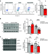
AD: Notch2 silencing in control pulmonary artery endothelial cells (PAECs) increases EC proliferation and migration, activates phosphorylation of Erk1/2 and Akt, and reduces cyclin-dependent kinase inhibitor p21cip levels. AC: human (h)PAECs were transfected with Notch2 siRNA or scrambled control (Scrmb) and assays performed 72 h posttransfection. A: transfected hPAECs were seeded and synchronized overnight, and proliferation was measured in a plate-based fluorescence assay using CyQuant (n = 12). B: gap-wound injury was made swiping a sterile 1,000-µl pipette tip across a lawn of transfected hPAECs. Brightfield images of the wound were captured and percent wound closure in cells treated with Notch2 siRNA was measured as the change in wound size between t = 0 and 24 h vs. the change occurring in Scrmb control-treated cells (n = 12). C: analysis of cell cycle distribution of transfected hPAECs harvested and stained with propidium iodide (PI) was performed by flow cytometry. Representative flow cytometry histograms show G1, S, and G2 + M phases of cell cycle distribution in Notch2 gene silenced vs. scrambled control hPAECs. Bar graphs depict proliferating cells quantified as total number of cells in S + G2/M phases of cell cycle as a function of control (n = 9). D: representative immunoblot and quantitative bar graphs of expression levels of Notch2, phospho-Erk1/2 vs. total Erk1/2 (p-42/44 MAPK vs. total 42/44 MAPK), phospho-Akt ser473 vs. total Akt, and cell cycle protein cyclin-dependent kinase inhibitor 1 (p21cip) in lysates from Notch2 siRNA-transfected vs. scrambled control hPAECs (n = 6–9). Dots represent individual biological replicates. Bar graphs indicate means ± SE. P values for paired comparisons were calculated using Student’s t test. *P < 0.05, **P < 0.01, ***P < 0.001.
Figure 4.
Figure 4.
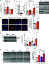
AC: Notch2 gene silencing in control human pulmonary artery endothelial cells (hPAECs) inhibits endothelial cell apoptosis. hPAECs were transfected with Notch2 siRNA or scrambled control (Scrmb), synchronized overnight, and analyzed 72 h posttransfection. A: annexin V-propidium iodide apoptosis assay was performed to study cell apoptosis. Living cells are double negative for annexin V and propidium iodide (Q4), early apoptotic cells are annexin V positive and propidium iodide negative (Q3), late apoptotic cells are double positive for annexin V and propidium iodide (Q2), and necrotic/dead cells are propidium iodide-positive only (Q1). Values in Q2 and Q3 represent all apoptotic cells. Reduced numbers in Q2 and Q3 are representative of apoptosis resistance. Representative scatterplot and quantitative bar graph of total apoptosis is presented as fold change (n = 7–8). B and C: representative immunoblots and quantitative bar graphs of Bcl-2 and Bax (B) and phospho-SAPK and p38 MAPK (C) levels in Notch2 siRNA-transfected vs. scrambled control hPAEC lysates (n = 9). Dots represent individual biological replicates. Bar graphs indicate means ± SE. P values for paired comparisons were calculated using Student’s t test. *P < 0.05, **P < 0.01, ***P < 0.001.
Figure 5.
Figure 5.
AE: Notch2 negatively regulates Notch1. Hypoxia augments Notch2 suppression-induced Notch1 levels. A and B: human pulmonary artery endothelial cells (hPAECs) transfected with Notch2 siRNA or scrambled control (Scrmb), synchronized overnight with serum-reduced media, and assayed 72 h posttransfection for gene expression of endothelial Notch1 and Notch4 (n = 8) (A) and immunoblot analysis for Notch2, Notch1, and cleaved Notch1 (B). Representative immunoblot and quantitative bar graphs are provided (n = 3–11). C: immunofluorescence (IF) labelling of hPAECs seeded onto gelatin-coated glass-coverslips and transfected with Notch2 siRNA or scrambled control (Scrmb), 72 h posttransfection. Representative images of cleaved Notch1 (red) with nuclei stained with DAPI (blue). Quantitative bar graphs depicting total fluorescent intensity of cleaved Notch1 protein in Notch2 siRNA-transfected hPAECs vs. scrambled control are provided (n = 5). D: representative immunoblot and quantitative bar graphs of cleaved Notch1 protein in lysates of hPAECs transfected with Notch 1 or 2 siRNA or scrambled control (Scrmb) exposed to hypoxia or normoxia for 24 h. (n = 6). E: representative images of gap-wound injury made on ECs transfected with Notch 1 siRNA and Notch2 siRNA either alone or in combination and scrambled control (Scrmb) as described in Fig. 3. Brightfield images of the wound were captured and percent wound closure in cells was measured as the change in wound size between t = 0 and 24 h vs. the change occurring in Scrmb control-treated cells. Representative brightfield images of the wound at both time points are provided (n = 8). Dots represent individual biological replicates. Bar graphs indicate means ± SE. P values for paired comparisons were calculated using Student’s t test. **P < 0.01, ***P < 0.001. For D, P values were calculated using two-way ANOVA. Post hoc tests applied Bonferroni correction to allow for multiple comparisons. *P < 0.05 vs. scrambled control within group, #P < 0.05 between groups. For E, P values were calculated using one-way ANOVA. Post hoc tests used Bonferroni correction to allow for multiple comparisons. *P < 0.05 vs. Notch2 siRNA, ***P < 0.001 vs. scrambled control or Notch1 siRNA.
Figure 6.
Figure 6.
AD: Notch2 induces nuclear Notch1 and canonical Notch effectors, independent of changes in Notch ligands in human pulmonary artery endothelial cells (hPAECs). hPAECs transfected with Notch2 siRNA or scrambled control (Scrmb), synchronized overnight with serum-reduced media, and assayed 72 h posttransfection for quantitative (q)RT-PCR or Western blot analysis. A: representative immunoblot and quantitative bar graphs of nuclear cleaved Notch1 in nuclear lysates of Notch2 siRNA-treated cells vs. scrambled control 72 h posttransfection (n = 3). TBP, TATA-binding protein. B: gene expression analysis for Hes1, Hey1, and Hey2 was performed in transfected hPAECs 72 h posttransfection by qRT-PCR (n = 8). C: gene expression analysis for Notch ligands Dll1, DLL4, Jag1, and Jag2 was performed in transfected hPAECs 72 h posttransfection (n = 6–11). D: representative immunoblot and quantitative bar graphs depicting level of endothelial DLL4 and Jag1 in hPAECs transfected with Notch2 siRNA (siRNA) or scrambled control (Scrmb) are provided (n = 5–8). Dots represent individual biological replicates. Bar graphs indicate means ± SE. P values for paired comparisons were calculated using Student’s t test. *P < 0.05; **P < 0.01.
Figure 7.
Figure 7.
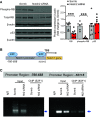
AC: Notch2 gene silencing abrogates Rb protein expression and modulates E2F1 binding to the Notch1 promoter. A: representative immunoblot and quantitative bar graphs for tumor suppressors Rb (total and phospho) and p53 performed in human pulmonary artery endothelial cells (hPAECs) transfected with Notch2 siRNA or scrambled control (Scrmb) (n = 6–9). B: schematic diagram of the human Notch1 promoter illustrating the E2F binding sites in the regulatory region; upstream regions were numbered in relation to the transcription start site (TSS). C: for chromatin immunoprecipitation (ChIP) assay, hPAECs transfected with Notch2 (Nt2) siRNA or scrambled control (Scrmb) were fixed with formaldehyde and cross-linked chromatin was subjected to immunoprecipitation with E2F1 antibody. The amount of chromatin bound to E2F1 was assayed by PCR using primers flanking specific regions on Notch1 promoter (arrowheads). Controls include PCR reactions performed with no DNA, input DNA and DNA immunoprecipitated by normal species-matched IgG antibody. Dots represent individual biological replicates. Bar graphs indicate means ± SE. P values for paired comparisons were calculated using Student’s t test. ***P < 0.001.
Figure 8.
Figure 8.
A and B: proposed protective function of Notch2 signaling axis in pulmonary arterial hypertension. A: in the normal endothelium, abundance of Notch2 maintains quiescence via balancing the rate of endothelial cell proliferation and apoptosis, and thus supports preservation of pulmonary artery endothelium integrity. B: perturbed endothelia, however, express decreased levels of Notch2 resulting in increased active Notch1 and increased rates of proliferation and apoptosis-resistant pulmonary artery endothelial cells (EC), leading to endothelial dysfunction and subsequent vascular remodeling associated with pulmonary arterial hypertension.

Similar articles

Cited by

References

    1. Hopper RK, Feinstein JA, Manning MA, Benitz W, Hudgins L. Neonatal pulmonary arterial hypertension and Noonan syndrome: two fatal cases with a specific RAF1 mutation. Am J Med Genet A 167A: 882–885, 2015. doi:10.1002/ajmg.a.37024. - DOI - PubMed
    1. Tonelli AR, Arelli V, Minai OA, Newman J, Bair N, Heresi GA, Dweik RA. Causes and circumstances of death in pulmonary arterial hypertension. Am J Respir Crit Care Med 188: 365–369, 2013. doi:10.1164/rccm.201209-1640OC. - DOI - PMC - PubMed
    1. Humbert M, Morrell NW, Archer SL, Stenmark KR, MacLean MR, Lang IM, Christman BW, Weir EK, Eickelberg O, Voelkel NF, Rabinovitch M. Cellular and molecular pathobiology of pulmonary arterial hypertension. J Am Coll Cardiol 43: 13S–24S, 2004. doi:10.1016/j.jacc.2004.02.029. - DOI - PubMed
    1. Morrell NW, Adnot S, Archer SL, Dupuis J, Lloyd Jones P, MacLean MR, McMurtry IF, Stenmark KR, Thistlethwaite PA, Weissmann N, Yuan JX, Weir EK. Cellular and molecular basis of pulmonary arterial hypertension. J Am Coll Cardiol 54: S20–31, 2009. doi:10.1016/j.jacc.2009.04.018. - DOI - PMC - PubMed
    1. Rabinovitch M. Molecular pathogenesis of pulmonary arterial hypertension. J Clin Invest 122: 4306–4313, 2012. doi:10.1172/JCI60658. - DOI - PMC - PubMed

Publication types

MeSH terms