. 2021 Jul 27;118(30):e2103393118.
doi: 10.1073/pnas.2103393118.

Membrane association of importin α facilitates viral entry into salivary gland cells of vector insects

Affiliations

Membrane association of importin α facilitates viral entry into salivary gland cells of vector insects

Yonghuan Ma et al. Proc Natl Acad Sci U S A. .

Abstract

The importin α family belongs to the conserved nuclear transport pathway in eukaryotes. However, the biological functions of importin α in the plasma membrane are still elusive. Here, we report that importin α, as a plasma membrane-associated protein, is exploited by the rice stripe virus (RSV) to enter vector insect cells, especially salivary gland cells. When the expression of three importin α genes was simultaneously knocked down, few virions entered the salivary glands of the small brown planthopper, Laodelphax striatellus Through hemocoel inoculation of virions, only importin α2 was found to efficiently regulate viral entry into insect salivary-gland cells. Importin α2 bound the nucleocapsid protein of RSV with a relatively high affinity through its importin β-binding (IBB) domain, with a dissociation constant KD of 9.1 μM. Furthermore, importin α2 and its IBB domain showed a distinct distribution in the plasma membrane through binding to heparin in heparan sulfate proteoglycan. When the expression of importin α2 was knocked down in viruliferous planthoppers or in nonviruliferous planthoppers before they acquired virions, the viral transmission efficiency of the vector insects in terms of the viral amount and disease incidence in rice was dramatically decreased. These findings not only reveal the specific function of the importin α family in the plasma membrane utilized by viruses, but also provide a promising target gene in vector insects for manipulation to efficiently control outbreaks of rice stripe disease.

Keywords: importin α; plasma membrane; rice stripe virus; salivary gland; small brown planthopper.


Conflict of interest statement

The authors declare no competing interest.

Figures
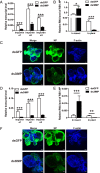
Fig. 1.
Fig. 1.
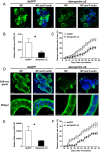
Effect of the importin α family on RSV transmission by L. striatellus. (A) Relative transcript levels of three importin α genes in viruliferous small brown planthoppers at 8 d after the injection of a mixture of dsRNAs for the three importin α genes (ds3IMP) as measured by RT-qPCR. The transcript levels of these genes were normalized to that of EF2. The control group was injected with dsRNA for GFP (dsGFP). (B) Relative RNA level of RSV NP in viruliferous planthoppers at 8 d after the injection of ds3IMP and in the rice fed to these insects for 1 d as measured by RT-qPCR. The transcript level of planthopper EF2 or rice tubulin was quantified to normalize the cDNA templates. (C) Immunohistochemistry showing the variation in RSV amounts in salivary glands of viruliferous planthoppers at 8 d after injecting ds3IMP compared to that of injecting dsGFP. RSV NP was observed using Alexa Fluor 488-conjugated anti-NP monoclonal antibody (green). F-actin was stained with phalloidin (blue). (Scale bars, 40 μm.) (D) Relative transcript levels of three importin α genes in the nonviruliferous planthoppers after injection with ds3IMP and feeding on RSV-infected rice plants for 8 d measured by RT-qPCR. (E) Relative RNA level of RSV NP in nonviruliferous planthoppers after injection with ds3IMP and feeding on RSV-infected rice plants for 8 d and in the rice fed to these insects for 2 d measured by RT-qPCR. (F) Immunohistochemistry showing the variation in RSV amounts in salivary glands of nonviruliferous planthoppers after injection with ds3IMP and feeding on RSV-infected rice plants for 8 d using Alexa Fluor 488-conjugated anti-NP monoclonal antibody (green). Insects injected with dsGFP were used as controls. F-actin was stained with phalloidin (blue). (Scale bars, 40 μm.) *P < 0.05, **P < 0.01, ***P < 0.001.
Fig. 2.
Fig. 2.
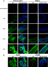
Effect of the importin α family on RSV entry into salivary glands of L. striatellus through hemocoel inoculation with RSV. (A) Immunohistochemistry showing RSV virions in salivary glands and midguts of nonviruliferous small brown planthoppers from 1 to 4 dpi of RSV crude extracts from viruliferous planthoppers in the hemolymph. RSV NP was observed using Alexa Fluor 488-conjugated anti-NP monoclonal antibody (green). F-actin was stained with phalloidin (blue). The nonviruliferous insects were used as negative controls. (Scale bars, 40 μm.) (B) Immunohistochemistry showing the variation in RSV amounts in salivary glands of nonviruliferous planthoppers at 4 d after injection of dsRNAs for the three importin α genes (ds3IMP) and RSV crude extracts. The control group was injected with dsGFP and RSV crude extracts. (Scale bars, 40 μm.)
Fig. 3.
Fig. 3.
Importin α2 regulates RSV entry into salivary glands of L. striatellus. (A) Immunohistochemistry showing the variation in RSV amounts in salivary glands of nonviruliferous small brown planthoppers at 4 d after injection of RSV crude extracts and dsRNA of each importin α into the hemolymph. The control group was injected with dsGFP and RSV crude extracts. RSV NP was observed using Alexa Fluor 488-conjugated anti-NP monoclonal antibody (green). F-actin was stained with phalloidin (blue). (Scale bars, 40 μm.) (B) Relative transcript levels of importin α genes and relative RNA level of RSV NP to that of EF2 in nonviruliferous planthoppers at 6 d after injection of RSV crude extracts and dsRNA of each importin α as measured by RT-qPCR. The control group was injected with dsGFP and RSV crude extracts. *P < 0.05, ***P < 0.001.
Fig. 4.
Fig. 4.
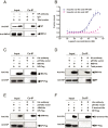
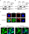
Importin α2 binds viral nucleocapsid protein with a relatively high affinity through the IBB domain. (A) NP and importin α2 (IMPα2) were coprecipitated from total protein of viruliferous small brown planthoppers in the Co-IP and Western blot assay using the anti-NP monoclonal antibody and anti–importin α2 polyclonal antibody. Mouse IgG was used as negative control. (B) MST assay to reveal specific binding between recombinant importin α2–His and NP-GST. The NP-GST (ligand) was gradient-diluted. The GST protein was used instead of NP-GST in the control group. The solid curve was fit to the standard KD-fit function. Bars represent SE. (C, E, and F) Interactions between recombinantly expressed Flag-tagged RSV NP and His-tagged IBB domain, Arm domain, and carboxyl-terminal domain of importin α2 in the Co-IP and Western blot assay using anti-His and anti-NP monoclonal antibodies. The expression products from the pET28a vector were used as a negative control. (D) The recombinantly expressed IBB-His of importin α2 pulled down the NP from total protein of viruliferous planthoppers in the Co-IP and Western blot assay using anti-His and anti-NP monoclonal antibodies. The expression products from the pET28a vector were used as a negative control.
Fig. 5.
Fig. 5.
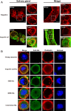
Importin α2 localizes to the plasma membrane of L. striatellus as assayed by immunohistochemistry. (A) Localization of importin α2 in salivary glands and midguts of viruliferous small brown planthoppers. The boxed region was enlarged and is shown in two panels on the right side. Importin α2 was observed using Alexa Fluor 594-conjugated anti–importin α2 polyclonal antibody (green). F-actin was stained with phalloidin (red). The negative control did not include the primary antibodies. (Scale bars, 40 μm.) (B) Immunofluorescence labeling of recombinantly expressed importin α2–His, IBB-His, Arm-His, and carboxyl-terminal–His of importin α2 in Drosophila S2 cells. The His-tagged proteins were labeled with an Alexa Fluor 488-conjugated anti-His monoclonal antibody (green). F-actin was labeled with phalloidin (red). Nuclei were stained with Hoechst (blue). Cells transfected with the empty pAc-5.1/V5-HisB plasmid were used as negative controls. (Scale bars, 2 μm.)
Fig. 6.
Fig. 6.
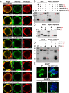
Importin α2 and RSV nucleocapsid proteins bind heparin in HSPG to localize to the plasma membrane of L. striatellus. (A) Localization of recombinantly expressed importin α2–His and the IBB-His domain in Drosophila S2 cells treated with different concentrations of Wnt-C59 revealed by immunofluorescence. The His-tagged proteins were labeled with an Alexa Fluor 488-conjugated anti-His monoclonal antibody (green). F-actin was labeled with phalloidin (red). (Scale bars, 2 μm.) (B) Heparin-binding and Western blot assays for the recombinantly expressed importin α2–His, IBB-His, Arm-His, or carboxyl-terminal–His of importin α2. After elution from heparin beads, the supernatant was used for Western blotting using an anti-His monoclonal antibody. M, marker. (C) Heparin-binding and Western blot assays for recombinantly expressed RSV NP-His using an anti-His monoclonal antibody. The Arm-His was used as a negative control. (D) Heparin-binding and Western blot assays for IBB-His and NP-His using anti-His monoclonal antibody. The two proteins were mixed together before being loaded on heparin beads. A mixture of Arm-His and NP-His was used as a negative control. (E) Immunohistochemistry showing the variation in RSV amounts in salivary glands of nonviruliferous small brown planthoppers at 4 d after injection of RSV crude extracts and dsRNA of HSPG (dsHSPG) into the hemolymph. The control group was injected with dsRNA of GFP (dsGFP) and RSV crude extracts. RSV NP was observed using Alexa Fluor 488-conjugated anti-NP monoclonal antibody (green). F-actin was stained with phalloidin (blue). (Scale bars, 40 μm.)
Fig. 7.
Fig. 7.
Importin β has no obvious effects on RSV entry into salivary glands of L. striatellus. (A and B) Interactions between recombinantly expressed importin β-HA (IMPβ-HA) and His-tagged IBB domain of importin α2 or Flag-tagged RSV NP in the Co-IP and Western blot assay using anti-His, anti-HA, and anti-Flag monoclonal antibodies. The expression products from the pET28a vector were used as a negative control. (C) Immunofluorescence labeling of recombinantly expressed importin β-His in Drosophila S2 cells. Importin β-His was labeled with an Alexa Fluor 488-conjugated anti-His monoclonal antibody (green). F-actin was labeled with phalloidin (red). Nuclei were stained with Hoechst (blue). Cells transfected with the empty pAc-5.1/V5-HisB plasmid were used as a negative control. (Scale bars, 2 μm.) (D) Immunohistochemistry showing the variation in RSV amounts in salivary glands of nonviruliferous small brown planthoppers at 4 d after injection of RSV crude extracts and dsRNA of importin β (dsimportin β) into the hemolymph. The control group was injected with dsRNA of GFP (dsGFP) and RSV crude extracts. RSV NP was observed using Alexa Fluor 488-conjugated anti-NP monoclonal antibody (green). F-actin was stained with phalloidin (blue). (Scale bars, 40 μm.)
Fig. 8.
Fig. 8.
Effect of importin α2 on the viral transmission efficiency of L. striatellus. (A) Immunohistochemistry showing the variation in RSV amounts in salivary glands of viruliferous small brown planthoppers at 8 d after injection of dsRNA of importin α2 (dsimportin α2) or (D) in salivary glands and midguts of nonviruliferous planthoppers after injection with dsimportin α2 and 7 d of feeding on RSV-infected rice. The control group was injected with dsRNA of GFP (dsGFP). RSV NP was observed using Alexa Fluor 488-conjugated anti-NP monoclonal antibody (green). F-actin was stained with phalloidin (blue). (Scale bars, 40 μm.) (B) Relative RNA level of RSV NP to that of tubulin in rice fed to dsimportin α2–injected viruliferous planthoppers for 2 d measured by RT-qPCR or (E) in rice fed to dsimportin α2–injected nonviruliferous planthoppers for 3 d after acquiring RSV from infected rice for 7 d. The control group was fed to dsGFP-injected planthoppers. (C) The disease incidence of rice fed to dsimportin α2–injected viruliferous planthoppers for 2 d or (F) rice fed to dsimportin α2–injected nonviruliferous planthoppers for 3 d after acquiring RSV from infected rice for 7 d. The control group was fed to dsGFP-injected planthoppers. A total of 10 rice seedlings per replicate and six replicates were applied. The values were reported as the mean ± SE. *P < 0.05.

Similar articles

Cited by

References

    1. Miyamoto Y., Yamada K., Yoneda Y., Importin α: A key molecule in nuclear transport and non-transport functions. J. Biochem. 160, 69–75 (2016). - PubMed
    1. Miyamoto Y., Boag P. R., Hime G. R., Loveland K. L., Regulated nucleocytoplasmic transport during gametogenesis. Biochim. Biophys. Acta 1819, 616–630 (2012). - PubMed
    1. Goldfarb D. S., Corbett A. H., Mason D. A., Harreman M. T., Adam S. A., Importin α: A multipurpose nuclear-transport receptor. Trends Cell Biol. 14, 505–514 (2004). - PubMed
    1. Yasuda Y., et al. ., Nuclear retention of importin α coordinates cell fate through changes in gene expression. EMBO J. 31, 83–94 (2012). - PMC - PubMed
    1. Chan C. K., Jans D. A., Synergy of importin alpha recognition and DNA binding by the yeast transcriptional activator GAL4. FEBS Lett. 462, 221–224 (1999). - PubMed

Publication types

LinkOut - more resources