. 2021 Aug 31;12(4):e0079521.
doi: 10.1128/mBio.00795-21. Epub 2021 Jul 20.

FKBP3 Induces Human Immunodeficiency Virus Type 1 Latency by Recruiting Histone Deacetylase 1/2 to the Viral Long Terminal Repeat

Affiliations

FKBP3 Induces Human Immunodeficiency Virus Type 1 Latency by Recruiting Histone Deacetylase 1/2 to the Viral Long Terminal Repeat

Xinyi Yang et al. mBio. .

Abstract

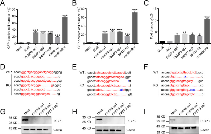
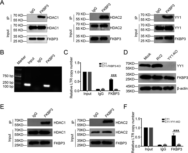
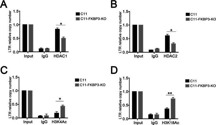
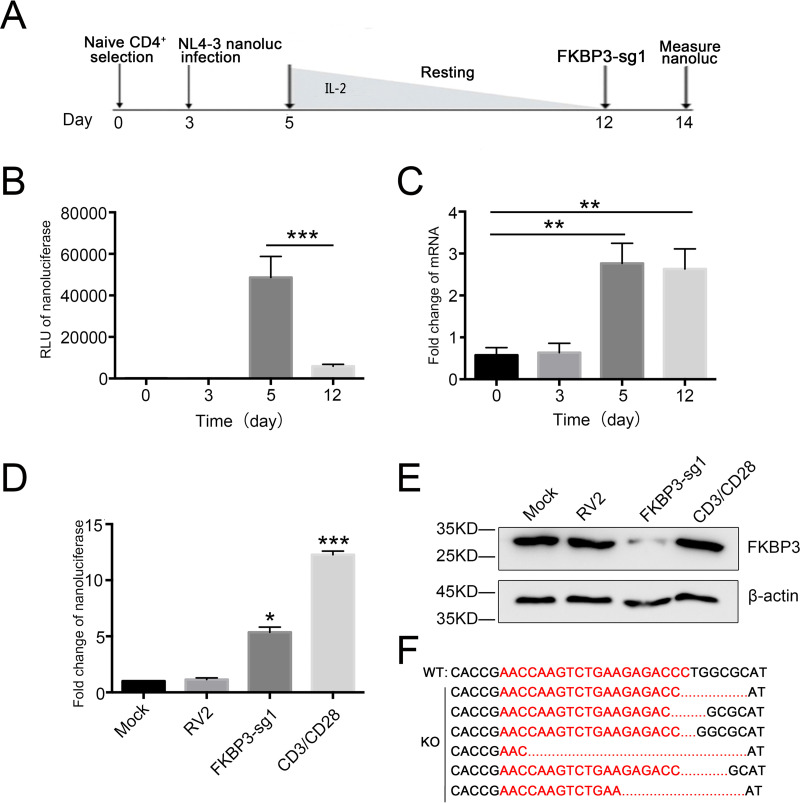
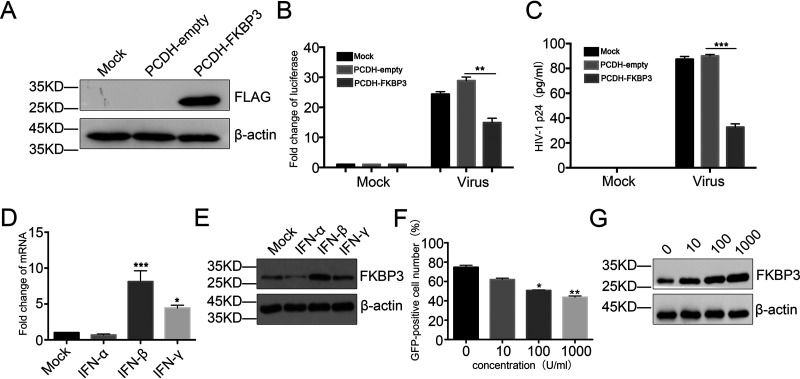
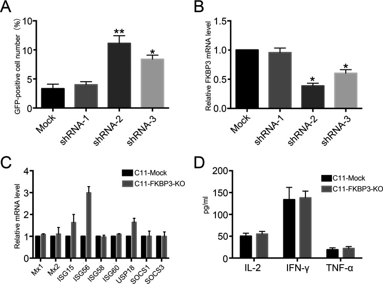
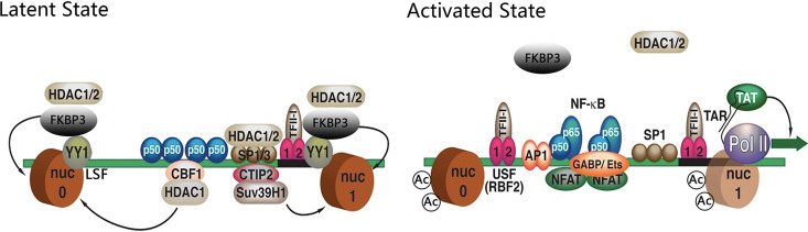
Human immunodeficiency virus type 1 (HIV-1) cannot be completely eliminated because of existence of the latent HIV-1 reservoir. However, the facts of HIV-1 latency, including its establishment and maintenance, are incomplete. FKBP3, encoded by the FKBP3 gene, belongs to the immunophilin family of proteins and is involved in immunoregulation and such cellular processes as protein folding. In a previous study, we found that FKBP3 may be related to HIV-1 latency using CRISPR screening. In this study, we knocked out the FKBP3 gene in multiple latently infected cell lines to promote latent HIV-1 activation. We found that FKBP3 could indirectly bind to the HIV-1 long terminal repeat through interaction with YY1, thereby recruiting histone deacetylase 1/2 to it. This promotes histone deacetylation and induces HIV-1 latency. Finally, in a primary latent cell model, we confirmed the effect of FKBP3 knockout on the latent activation of HIV-1. Our results suggest a new mechanism for the epigenetic regulation of HIV-1 latency and a new potential target for activating latent HIV-1. IMPORTANCE The primary reason why AIDS cannot be completely cured is the existence of a latent HIV-1 reservoir. Currently, the facts of HIV-1 latency, including its establishment and maintenance, are incomplete. Using a CRISPR library in our earlier screening of genes related to HIV-1 latency, we identified FBKP3 as a candidate gene related to HIV-1 latency. Therefore, in this mechanistic study, we first confirmed the HIV-1 latency-promoting effect of FKBP3 and determined that FKBP3 promotes histone deacetylation by recruiting histone deacetylase 1/2 to the HIV-1 long terminal repeat. We also confirmed, for the first time, that FKBP3 can act as a transcription factor (TF) recruitment scaffold and participate in epigenetic regulation of HIV-1 latency. These findings suggest a new mechanism for the epigenetic regulation of HIV-1 latency and a new potential target for activating latent HIV-1.

Keywords: FKBP3; HDAC1/2; HIV-1; HIV-1 latency; acetylation.


Figures

FIG 1
FIG 1
Knocking out the FKBP3 gene can promote HIV-1 latent reversal in HIV-1 latently infected cell lines. (A and B) Knocking out the FKBP3 gene can promote HIV-1 latent reversal in C11 and J-Lat 10.6 cell lines. C11 and J-Lat 10.6 cell lines were infected by lentiCRISPR v2.0 packaged lentiviruses with sgRNA, followed by screening for 14 days with purinomycin 2 μg/ml. The percentage of GFP-positive cells was measured by flow cytometry to determine the level of HIV-1 reactivation. Each datum represented the mean ± SD of three independent experiments (n = 3) and was analyzed with t test. *, P < 0.05; **, P < 0.01; ***, P < 0.001. (C) The effect of knocking out the FKBP3 gene on HIV latency was further verified in ACH2 models of HIV latency. The expression levels of p24 in ACH2 cells were detected by HIV-1 p24 ELISA. Each datum represented the mean ± SD of three independent experiments. (n = 3) and was analyzed with t test. *, P < 0.05; **, P < 0.01; ***, P < 0.001. (D to F) Sequencing FKBP3 PCR products after clone screening. The PCR products of FKBP3 gene were cloned and then sequenced. FKBP3-sg1 (D), FKBP3-sg2 (E), and FKBP3-sg3 (F) target sites are shown in red letters. Dashes indicate the deleted bases relative to the wild-type sequence. (G to I) FKBP3 protein levels were measured by Western blotting after knockout in C11 (G), J-Lat 10.6 (H), and ACH2 (I) cells by LvFKBP3-sg1, LvFKBP3-sg2, LvFKBP3-sg3, and mock C11 cells, which served as control.
FIG 2
FIG 2
FKBP3 could interact with YY1 and HDAC1/2 and indirectly bind to the HIV-1 LTR site through YY1 in an HIV-1 latently infected cell line. (A) The immunoprecipitation assay was performed in C11 cell lysates with anti-FKBP3 antibody, followed by Western blotting with anti-YY1, anti-HDAC1, or anti-HDAC2 antibodies. (B and C) The interaction between FKBP3 and HIV-1 LTR was detected by ChIP. Chromatin fragments from C11 cells and C11-FKBP3-KO cells were immunoprecipitated with anti-FKBP3 antibodies or control normal rabbit serum (IgG). After ChIP, the binding of FKBP3 to HIV LTR was detected by PCR (B) and qPCR (C) with specific primers for HIV LTR. The number of copies was normalized to input group. Each datum represented the mean ± SD of three independent experiments (n = 3) and was analyzed with t test. ***, P < 0.001. (D) Identification of the lack of YY1 and FKBP3 protein expression in C11-YY1-KO cells. YY1 and FKBP3 protein level was measured by Western blotting after knockout in C11 cells by lentivirus sgRNA-YY1. Mock C11 cells and lentiCRISPR v2.0 without sgRNA (RV2)-infected C11 cells served as mock control. (E) The interaction between FKBP3 and HIV-1 LTR was detected by ChIP in the C11-YY1-KO cell line. Chromatin fragments from C11 cells and C11-YY1-KO cells were immunoprecipitated with anti-FKBP3 antibodies or control normal rabbit serum (IgG). After ChIP, the binding of FKBP3 to HIV LTR was detected by qPCR with specific primers for HIV LTR. The number of copies was normalized to input group. Each datum represented the mean ± SD of three independent experiments and was analyzed with t test. ***, P < 0.001.
FIG 3
FIG 3
FKBP3 promotes histone deacetylation by recruiting HDAC1/2 to HIV-1 LTR. (A and B) The effect of FKBP3 knockout on HDAC1 (A) or HDAC2 (B) protein recruitment into HIV LTR was detected by ChIP. Chromatin fragments from C11 cells and C11-FKBP3-KO cells were immunoprecipitated with HDAC1 (A) or HDAC2 (B) antibodies or control normal rabbit serum (IgG). After ChIP, the binding of HDAC1 (A) or HDAC2 (B) to HIV LTR was detected by qPCR with specific primers for HIV LTR. The number of copies was normalized to input group. Each datum represented the mean ± SD of three independent experiments and was analyzed with t test. *, P < 0.05. (C and D) The effect of FKBP3 knockout on histone acetylation of HIV LTR was detected by ChIP. Chromatin fragments from C11 cells and C11-FKBP3-KO cells were immunoprecipitated with H3K4Ac (C) or H3K18Ac (D) antibodies or control normal rabbit serum (IgG). After ChIP, the histone acetylation of H3K4Ac (C) or H3K18Ac (D) of HIV LTR was detected by qPCR with specific primers for HIV LTR. The number of copies was normalized to input group. Each datum represent the mean ± SD of three independent experiments (n = 3) and was analyzed with t test. *, P < 0.05; **, P < 0.01.
FIG 4
FIG 4
Knockout FKBP3 reactivates latent HIV-1 in the primary CD4+ T model of latency (3 donors). (A) Outline of the protocol to establish HIV latency. Human primary CD4+ T cells were activated and expanded with α-CD3/CD28 beads on day 1. The α-CD3/CD28 beads were removed on day 3. Cells were then infected with HIV-1 NL4.3-NanoLuc on the 3rd day after expansion and maintained over 7 days with a decreasing concentration of IL-2 to establish latency until day 12. On the 12th day, cells were transduced with Cas9 and FKBP3-sgRNA plasmid by electroporation. (B) During HIV-1 infection, the transcription of HIV-1 in the primary CD4+ T cells was determined by NanoLuc luciferase assay at different time points. Each datum represented the mean ± SD of three independent experiments and was analyzed with t test. ***, P < 0.001, compared with the cells 5 days postinfection. (C) After infection with vesicular stomatitis virus glycoprotein G (VSV-G)-pseudotyped HIV-1 NL4.3-NanoLuc, the mRNA expression of FKBP3 was measured by qPCR. Each datum represented the mean ± SD of three independent experiments and was analyzed with t test. **, P < 0.01, compared with uninfected mock cells. (D) The role of FKBP3 in the primary CD4+ T cell model of latency. The expression of HIV-1 was measured by NanoLuc in the primary CD4+ T cells after gene knockout by mock, RV2, and FKBP3-sg1 with or without α-CD3/CD28 stimulation. Each datum represented the mean ± SD of three independent experiments and was analyzed with t test. *, P < 0.05; ***, P < 0.001, compared with cells after Lv-RV2 mock knockout. (E) FKBP3 protein was determined by Western blot of whole-cell lysate from primary CD4+ T cells. (F) Sequencing FKBP3 PCR products after Lv-FKBP3-sg1 knockout in primary CD4+ T cells. The PCR products of FKBP3 were cloned and then sequenced. FKBP3-sg1 target sites are shown in red letters. Dashes indicate deleted bases relative to the wild-type sequence.
FIG 5
FIG 5
Effects of interferons on FKBP3 induction and FKBP3 inhibition of virus infection and replication. (A) The expression of FKBP3 protein was measured by Western blotting in TZM-bl cells infected with mock, Lv-PCDH-empty, or Lv-PCDH-PEBP1 virus. (B and C) The effect of overexpression of FKBP3 on HIV-1 replication. The cells were infected with supernatants of patient blood for 12 h and then washed three times. The transcription of HIV-1 was evaluated 72 h postinfection by luciferase activity (B) and levels of p24 released from supernatants (C). Each datum represented the mean ± SD of three independent experiments (n = 3) and was analyzed with t test. **, P < 0.01; ***, P < 0.001. (D and E) After treatment of IFN-α, IFN-β, and IFN-γ in Jurkat CD4+ T cells for 24 h, the mRNA (D) or protein (E) expression levels of FKBP3 were detected by qPCR or Western blot analysis. Each datum represented the mean ± SD of three independent experiments (n = 3) and was analyzed with t test. *, P < 0.05; ***, P < 0.001. (F) Ya cell lines were treated with different concentration of IFN-β for 48 h. The percentage of GFP-positive cells was measured by flow cytometry to determine the level of HIV-1 reactivation. Each datum represented the mean ± SD of three independent experiments (n = 3) and was analyzed with t-test. *, P < 0.05; **, P < 0.01. (E) FKBP3 protein was determined by Western blot of whole-cell lysate from Ya cells treated with IFN-β for 48 h.
FIG 6
FIG 6
FKBP3 knockout does not cause significant changes in immune response. (A) Knocking down FKBP3 could promote HIV-1 latent reversal in the C11 cell line. C11 cells were infected by pLKO.1 packaged lentivirus with shRNA targeting FKBP3. The percentage of GFP-positive cells was measured by flow cytometry to determine the level of HIV-1 reactivation. Each datum represented the mean ± SD of three independent experiments (n = 3) and was analyzed with t test. *, P < 0.05; **, P < 0.01; ***, P < 0.001. (B) FKBP3 expression was measured by qPCR in C11 cells infected with lentivirus with or without shRNA targeting FKBP3. Each datum represented the mean ± SD of three independent experiments (n = 3) and was analyzed with t test. *, P < 0.05; **, P < 0.01; ***, P < 0.001. (C) Expression of ISGs was measured by qPCR in C11 and C11-FKBP3-KO cells. Each datum represented the mean ± SD of three independent experiments (n = 3) and was analyzed with t test. *, P < 0.05; **, P < 0.01; ***, P < 0.001. (D) The levels of IL-2, tumor necrosis factor alpha (TNF-α), and IFN-γ were evaluated by ELISA in the supernatants of C11 and C11-FKBP3-KO cells.
FIG 7
FIG 7
Working model of the role of FKBP3 in the establishment of HIV latency.

Similar articles

Cited by

References

    1. Ruelas DS, Greene WC. 2013. An integrated overview of HIV-1 latency. Cell 155:519–529. doi:10.1016/j.cell.2013.09.044. - DOI - PMC - PubMed
    1. Margolis DM, Garcia JV, Hazuda DJ, Haynes BF. 2016. Latency reversal and viral clearance to cure HIV-1. Science 353:aaf6517. doi:10.1126/science.aaf6517. - DOI - PMC - PubMed
    1. Davey RT, Bhat N, Yoder C, Chun TW, Metcalf JA, Dewar R, Natarajan V, Lempicki RA, Adelsberger JW, Miller KD, Kovacs JA, Polis MA, Walker RE, Falloon J, Masur H, Gee D, Baseler M, Dimitrov DS, Fauci AS, Lane HC. 1999. HIV-1 and T cell dynamics after interruption of highly active antiretroviral therapy (HAART) in patients with a history of sustained viral suppression. Proc Natl Acad Sci U S A 96:15109–15114. doi:10.1073/pnas.96.26.15109. - DOI - PMC - PubMed
    1. Dahabieh MS, Battivelli E, Verdin E. 2015. Understanding HIV latency: the road to an HIV cure. Annu Rev Med 66:407–421. doi:10.1146/annurev-med-092112-152941. - DOI - PMC - PubMed
    1. Finzi D, Hermankova M, Pierson T, Carruth LM, Buck C, Chaisson RE, Quinn TC, Chadwick K, Margolick J, Brookmeyer R, Gallant J, Markowitz M, Ho DD, Richman DD, Siliciano RF. 1997. Identification of a reservoir for HIV-1 in patients on highly active antiretroviral therapy. Science 278:1295–1300. doi:10.1126/science.278.5341.1295. - DOI - PubMed

Publication types

MeSH terms

LinkOut - more resources