HIV-1 Transactivator of Transcription (Tat) Co-operates With AP-1 Factors to Enhance c-MYC Transcription
- PMID: 34277639
- PMCID: PMC8278106
- DOI: 10.3389/fcell.2021.693706
HIV-1 Transactivator of Transcription (Tat) Co-operates With AP-1 Factors to Enhance c-MYC Transcription
Abstract
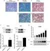
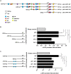
HIV-1 infection often leads to the development of co-morbidities including cancer. Burkitt lymphoma (BL) is one of the most over-represented non-Hodgkin lymphoma among HIV-infected individuals, and displays a highly aggressive phenotype in this population group, with comparatively poorer outcomes, despite these patients being on anti-retroviral therapy. Accumulating evidence indicates that the molecular pathogenesis of HIV-associated malignancies is unique, with components of the virus playing an active role in driving oncogenesis, and in order to improve patient prognosis and treatment, a better understanding of disease pathobiology and progression is needed. In this study, we found HIV-1 Tat to be localized within the tumor cells of BL patients, and enhanced expression of oncogenic c-MYC in these cells. Using luciferase reporter assays we show that HIV-1 Tat enhances the c-MYC gene promoter activity and that this is partially mediated via two AP-1 binding elements located at positions -1128 and -1375 bp, as revealed by mutagenesis experiments. We further demonstrate, using pull-down assays, that Tat can exist within a protein complex with the AP-1 factor JunB, and that this complex can bind these AP-1 sites within the c-MYC promoter, as shown by in vivo chromatin immunoprecipitation assays. Therefore, these findings show that in HIV-infected individuals, Tat infiltrates B-cells, where it can enhance the expression of oncogenic factors, which contributes toward the more aggressive disease phenotype observed in these patients.
Keywords: AP-1; HIV-1; c-MYC; non-Hodgkin lymphoma; transactivator of transcription.
Copyright © 2021 Alves de Souza Rios, Mapekula, Mdletshe, Chetty and Mowla.
Conflict of interest statement
The authors declare that the research was conducted in the absence of any commercial or financial relationships that could be construed as a potential conflict of interest.
Figures


Similar articles
-
HIV-1 Tat protein can transactivate a heterologous TATAA element independent of viral promoter sequences and the trans-activation response element.AIDS. 1997 Feb;11(2):139-46. doi: 10.1097/00002030-199702000-00002. AIDS. 1997. PMID: 9030359
-
The human immunodeficiency virus type 1 Tat protein up-regulates the promoter activity of the beta-chemokine monocyte chemoattractant protein 1 in the human astrocytoma cell line U-87 MG: role of SP-1, AP-1, and NF-kappaB consensus sites.J Virol. 2000 Feb;74(4):1632-40. doi: 10.1128/jvi.74.4.1632-1640.2000. J Virol. 2000. PMID: 10644332 Free PMC article.
-
The HIV-Tat protein interacts with Sp3 transcription factor and inhibits its binding to a distal site of the sod2 promoter in human pulmonary artery endothelial cells.Free Radic Biol Med. 2020 Feb 1;147:102-113. doi: 10.1016/j.freeradbiomed.2019.12.015. Epub 2019 Dec 19. Free Radic Biol Med. 2020. PMID: 31863909 Free PMC article.
-
MYC-associated and double-hit lymphomas: a review of pathobiology, prognosis, and therapeutic approaches.Cancer. 2014 Dec 15;120(24):3884-95. doi: 10.1002/cncr.28899. Epub 2014 Jul 24. Cancer. 2014. PMID: 25060588 Review.
-
Multiple modes of transcriptional regulation by the HIV-1 Tat transactivator.IUBMB Life. 2001 Mar;51(3):175-81. doi: 10.1080/152165401753544241. IUBMB Life. 2001. PMID: 11547919 Review.
Cited by
-
JUND Promotes Tumorigenesis via Specifically Binding on Enhancers of Multiple Oncogenes in Cervical Cancer.Onco Targets Ther. 2023 May 31;16:347-357. doi: 10.2147/OTT.S405027. eCollection 2023. Onco Targets Ther. 2023. PMID: 37283647 Free PMC article.
-
Identification of viral protein R of human immunodeficiency virus-1 (HIV) and interleukin-6 as risk factors for malignancies in HIV-infected individuals: A cohort study.PLoS One. 2024 Jan 2;19(1):e0296502. doi: 10.1371/journal.pone.0296502. eCollection 2024. PLoS One. 2024. PMID: 38166062 Free PMC article.
-
Modulation of Cellular MicroRNA by HIV-1 in Burkitt Lymphoma Cells-A Pathway to Promoting Oncogenesis.Genes (Basel). 2021 Aug 24;12(9):1302. doi: 10.3390/genes12091302. Genes (Basel). 2021. PMID: 34573283 Free PMC article.
-
Immune Characteristics and Immunotherapy of HIV-Associated Lymphoma.Curr Issues Mol Biol. 2024 Sep 10;46(9):9984-9997. doi: 10.3390/cimb46090596. Curr Issues Mol Biol. 2024. PMID: 39329948 Free PMC article. Review.
-
Ectopic expression of HIV-1 Tat modifies gene expression in cultured B cells: implications for the development of B-cell lymphomas in HIV-1-infected patients.PeerJ. 2022 Oct 18;10:e13986. doi: 10.7717/peerj.13986. eCollection 2022. PeerJ. 2022. PMID: 36275462 Free PMC article.
References
-
- Abayomi E. A., Somers A., Grewal R., Sissolak G., Bassa F., Maartens D., et al. (2011). Impact of the HIV epidemic and anti-retroviral treatment policy on lymphoma incidence and subtypes seen in the western cape of South Africa, 2002–2009: preliminary findings of the tygerberg lymphoma study group. Transfus. Apher. Sci. 44 161–166. 10.1016/j.transci.2011.01.007 - DOI - PMC - PubMed
-
- Abudulai L. N., Fernandez S., Corscadden K., Hunter M., Kirkham L.-A. S., Post J. J., et al. (2016). Chronic HIV-1 infection induces B-cell dysfunction that is incompletely resolved by long-term antiretroviral therapy. J. Acquir. Immune Defic. Syndr. 71 381–389. 10.1097/QAI.0000000000000869 - DOI - PubMed
Grants and funding
LinkOut - more resources
Full Text Sources

