Review
. 2021 Jun 21:12:676702.
doi: 10.3389/fimmu.2021.676702. eCollection 2021.

PTX3 Regulation of Inflammation, Hemostatic Response, Tissue Repair, and Resolution of Fibrosis Favors a Role in Limiting Idiopathic Pulmonary Fibrosis

Affiliations
Review

PTX3 Regulation of Inflammation, Hemostatic Response, Tissue Repair, and Resolution of Fibrosis Favors a Role in Limiting Idiopathic Pulmonary Fibrosis

Andrea Doni et al. Front Immunol. .

Abstract

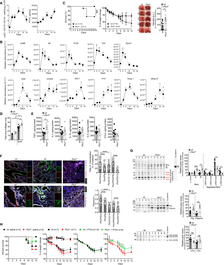
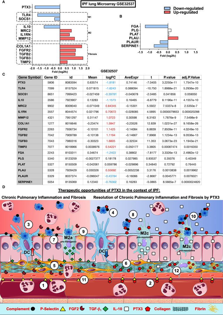
PTX3 is a soluble pattern recognition molecule (PRM) belonging to the humoral innate immune system, rapidly produced at inflammatory sites by phagocytes and stromal cells in response to infection or tissue injury. PTX3 interacts with microbial moieties and selected pathogens, with molecules of the complement and hemostatic systems, and with extracellular matrix (ECM) components. In wound sites, PTX3 interacts with fibrin and plasminogen and favors a timely removal of fibrin-rich ECM for an efficient tissue repair. Idiopathic Pulmonary Fibrosis (IPF) is a chronic and progressive interstitial lung disease of unknown origin, associated with excessive ECM deposition affecting tissue architecture, with irreversible loss of lung function and impact on the patient's life quality. Maccarinelli et al. recently demonstrated a protective role of PTX3 using the bleomycin (BLM)-induced experimental model of lung fibrosis, in line with the reported role of PTX3 in tissue repair. However, the mechanisms and therapeutic potential of PTX3 in IPF remained to be investigated. Herein, we provide new insights on the possible role of PTX3 in the development of IPF and BLM-induced lung fibrosis. In mice, PTX3-deficiency was associated with worsening of the disease and with impaired fibrin removal and subsequently increased collagen deposition. In IPF patients, microarray data indicated a down-regulation of PTX3 expression, thus suggesting a potential rational underlying the development of disease. Therefore, we provide new insights for considering PTX3 as a possible target molecule underlying therapeutic intervention in IPF.

Keywords: IPF; PTX3; fibrosis; hemostasis; humoral immunity; inflammation; resolution.


Conflict of interest statement

AM and BB are inventors of patents on pentraxin-3 and obtain royalties on related reagents. The remaining authors declare that the research was conducted in the absence of any commercial or financial relationships that could be construed as a potential conflict of interest.

Figures

Figure 1
Figure 1
PTX3 protects mice from BLM-induced pulmonary fibrosis. A model of lung fibrosis was induced by BLM instillation (3.75 mg/Kg, i.n.) in Wild Type (WT) and ptx3-/- mice (A-E). (A, left), kinetic of macrophage influx in the lungs of WT mice after BLM by FACS analysis. (A, right), kinetic of PTX3 lung content assessed by ELISA (PTX3 DuoSet® Kit ELISA DY2166, R&D Systems) in tissue homogenates. (B), Transcription analysis of M1 genes (Cd86-Mm00444543_m1, Il6-Mm99999064_m1, Il12a-Mm99999066_m1, Tlr4-Mm00445274_m1, Socs1-Mm00782550_s1) and M2 genes (Arg1-Mm00475988_m1, Chi3l3-Mm00657889_mH, Mrc1-Mm01329362_m1, Il1rn-Mm00446185_m1, Timp1-Mm00441818_m1, Mmp12-Mm00500554_m1) markers of macrophage polarization by TaqMan probes (Applied Biosystems) at different days after BLM treatment. (C, D), Susceptibility of Ptx3-/- mice to lung fibrosis induced by BLM instillation. (C, left) survival curve of mice (100% of WT and 54.5% of Ptx3-/- mice), as defined by humane end-points (e.g., weight loss of more than 25% of initial body weight, anorexia, excessive decrease in activity, shaggy hair, diarrhea, urinary retention, breathing difficulties). **P=0.01; Log-rank test. (C, middle) monitoring of weight loss. *P < 0.05; unpaired t-test). (C, right) representative photographs showing the appearance of lung parenchyma and quantification of autofluorescence intensity (excitation 405nm; emission collection at 550-60nm; CLARIOstar Microplate Reader, BMG Labtech) typically associated to hemoglobin in lung lysates of wt (n=9) and Ptx3-/- (n=7) mice (day 14). *P=0.05, unpaired t-test. (D) lung collagen content after 21 days assessed by Sircol assay. (E), measurement of TGF-β1, IL-4, IL-10, IL-6, and CCL2 in lung lysates of wt (n=7) and Ptx3-/- (n=5) mice (day 14 after BLM treatment) by ELISA (R&D Systems). *P=0.05, unpaired t-test. (F), confocal microscopy analysis on lung specimens (10µm) from WT mice (n=7) 14 days after BLM treatment. (F, upper panels) localization of PTX3 (green), Collagen I (blue), PDGFRα+ (red) mesenchymal cells and CD11b+ (white) immune cells. Representative localization of PTX3 around blood vessels (F, upper, left) or associated with fibrotic ECM and damaged epithelium (F, upper, middle). (F, lower panels) colocalization of PTX3 (green) with fibrin (white) and plasminogen (red) in fibrotic lung associated with blood vessels (F, lower, left) or ECM and damaged epithelium (F, lower, middle). Blue, nuclei. Lungs obtained from Ptx3-/- mice were used as control (Upper and lower panels, right). Bar, 100µm. The following antibodies were used: collagen I, rabbit polyclonal (5µg/ml; AbCam); PTX3, goat polyclonal (0.5µg/ml; R&D Systems); PDGFRα, BV421 rat (APA5, 1.5µg/ml; BD Horizon); CD11b, APC-Cy7™ rat (M1/70, 2µg/ml; BD Pharmingen); plasminogen, rat monoclonal (1µg/ml; Cell Sciences); fibrinogen, rabbit polyclonal (4µg/ml; Dako); species-specific Alexa Fluor 488/568/647- conjugated secondary antibodies were used. (F, right) Rate of colocalization (% of material; Fiji software) of PTX3 signal with fibrin, plasminogen, collagen I (COL1A), PDGFRα, and CD11b and relative Pearson’s correlation coefficient. Mean ± SEM of 5-8 images acquired for each mouse (n=7). *P < 0.0001, unpaired t-test. (G, upper), Western blot analysis of fibrin in lung lysates (10µg total proteins per lane on 10% SDS-PAGE) of WT (n=5) and Ptx3-/- (n=5) mice at day 7. A polyclonal rabbit anti-fibrinogen was used (3µg/ml; Agilent/DAKO). 1µl of basal mouse plasma in ACD-A (Anticoagulant Citrate Dextrose Solution) was used as a control for fibrinogen; 1µl of mouse plasma-ACD incubated with thrombin (1U/ml; 1h) was used as a control for fibrin. A typical band pattern of fibrin (Aα; Bβ, γ-γ dimer) is indicated in the fibrin control and lung lysates. Red arrows, lower molecular weight bands corresponding to degraded fibrin. (G, upper, right), quantification of fibrin bands as relative gray values (Fiji software) on Ponceau red staining. ***P < 0.005, *P < 0.5; unpaired t-test. (G, middle), Western blot analysis of plasminogen and relative band quantification as gray values (Fiji software) in same lysates (50µg total proteins per lane on 10% SDS-PAGE, G, middle, right). A polyclonal goat anti-plasminogen was used (0.5µg/ml; R&D Systems). The molecular weight of plasminogen and plasmin activation bands are indicated. (G, lower), Western blot analysis of the complement component C3 in the same lysates (10µg total proteins per lane on 10% SDS-PAGE) and relative band quantification as gray values (Fiji software, G, lower, right). A polyclonal goat anti-human/mouse C3 and activation fragments (1:3000; Merck-Millipore) was used. (H) Effect of PTX3 administration in BLM-induced lung fibrosis. One experiment was performed. Human recombinant PTX3 (50µg/mouse) was injected i.p. one day after BLM (5 mg/Kg, i.n.) treatment in WT mice. Survival (H, left) (Ptx3-/- mice from 42.8% to 77.8%, and WT mice from 57.1 to 80% of survival) and body weight (H, 3 panels right) were recorded until day 14. Curves referring to weight loss are shown compared to untreated WT and Ptx3-/- mice (H, first left) or separated by genotype and compared with the correspondent treated group (H, right). *P < 0.05; unpaired t-test. All results were expressed as mean ± SEM. Normalized data were analyzed by One-Way ANOVA with Tukey post-test, using the software GraphPad Prism 8.0. Differences were considered significant at P < 0.05.
Figure 2
Figure 2
Impact of PTX3 in Idiopathic Pulmonary Fibrosis and therapeutic opportunities. Microarray analysis of lung samples from IPF (n=119) and healthy (n=50) individuals from GEO database: GSE32537 were analyzed by Phantasus (58) (https://genome.ifmo.ru/phantasus). (A) Lung expression of PTX3, fibrogenic markers as COL1A1, FGFR2, TGFB2, TGFB3 and TIMP-2, M1 (SOCS1 and TLR4) and M2 (IL-10, MRC2, IL1RN, and MMP-12) macrophage polarization markers (B) Expression of coagulation cascade FGA (Fibrinogen alpha-chain precursor), PLG (Plasminogen precursor), PLAT (Tissue-type Plasminogen Activator precursor), PLAU (Urokinase-type Plasminogen Activator precursor), PLAUR (Urokinase Plasminogen Activator Receptor) and SERPINE1 (PAI-1 Plasminogen Activator Inhibitor-1) in IPF and health samples. (C) Data table with analysis from GEO database GSE32537 analyzed by Phantasus according to the instructions for the use of the application. Differences were considered significant at P value <0,05. (D) Possible mechanisms and therapeutic opportunities of PTX3 in the context of IPF. IPF is characterized by a reduced PTX3 production (red background), however PTX3 may act as an anti-inflammatory as well as a pro-resolutive modulator of chronic pulmonary inflammation and fibrosis in IPF (blue background) at different levels: (1) Neutrophil influx is facilitated through the interaction with P-selectin expressed on the surface of ECs, (2) PTX3 could antagonize endothelial P-selectin, dampening neutrophil influx during chronic pulmonary inflammation; (3) the abundance of apoptotic cells in airways from IPF is related to DC phagocytosis and activation that sustain chronic lung inflammation, (4) PTX3 may block apoptotic cell internalization and consequent inflammation; (5) FGF2 activates fibroblasts and ECs, (6) however PTX3 interacts with FGF2 reducing its availability for binding to FGFR2 on fibroblasts and consequent fibrosis; (7) complement and apoptotic cell deposition in the lungs lead to chronic inflammation, (8) on the other side PTX3 may act as a scavenger preventing the excessive deposition of both complement components and apoptotic cells in lungs and consequent attenuation of tissue damage and inflammation; (9) Alveolar macrophages from IPF display defective efferocytosis and increased TGF-β1 production, contributing to tissue fibrogenesis, (10) while PTX3 may enhance macrophage efferocytosis and M2 polarization and resolution of inflammation by IL-10; (11) Finally, defective PTX3 production in IPF may increase fibrin deposition and fibrosis, (12) but PTX3 could contribute to the resolution of fibrosis, interacting with fibrin-clots and disorganized collagen fibers in the lung parenchyma, supporting fibrinolysis and clearance of ECM debris by macrophage phagocytosis, promoting lung tissue healing and repair.

Similar articles

Cited by

References

    1. Garlanda C, Bottazzi B, Magrini E, Inforzato A, Mantovani A. PTX3, a Humoral Pattern Recognition Molecule, in Innate Immunity, Tissue Repair, and Cancer. Physiol Rev (2018) 98:623–39. 10.1152/physrev.00016.2017 - DOI - PMC - PubMed
    1. Magrini E, Mantovani A, Garlanda C. The Dual Complexity of PTX3 in Health and Disease: A Balancing Act? Trends Mol Med (2016) 22:497–510. 10.1016/j.molmed.2016.04.007 - DOI - PMC - PubMed
    1. Doni A, Stravalaci M, Inforzato A, Magrini E, Mantovani A, Garlanda C, et al. . The Long Pentraxin PTX3 as a Link Between Innate Immunity, Tissue Remodeling, and Cancer. Front Immunol (2019) 10:712. 10.3389/fimmu.2019.00712 - DOI - PMC - PubMed
    1. Manfredi AA, Rovere-Querini P, Bottazzi B, Garlanda C, Mantovani A. Pentraxins, Humoral Innate Immunity and Tissue Injury. Curr Opin Immunol (2008) 20:538–44. 10.1016/j.coi.2008.05.004 - DOI - PubMed
    1. Doni A, Garlanda C, Mantovani A. Innate Immunity, Hemostasis and Matrix Remodeling: PTX3 as a Link. Semin Immunol (2016) 28:570–7. 10.1016/j.smim.2016.10.012 - DOI - PMC - PubMed

Publication types

MeSH terms