Human umbilical cord mesenchymal stromal cells attenuate pulmonary fibrosis via regulatory T cell through interaction with macrophage
- PMID: 34256845
- PMCID: PMC8278716
- DOI: 10.1186/s13287-021-02469-5
Human umbilical cord mesenchymal stromal cells attenuate pulmonary fibrosis via regulatory T cell through interaction with macrophage
Abstract
Background: Pulmonary fibrosis (PF) is a growing clinical problem with limited therapeutic options. Human umbilical cord mesenchymal stromal cell (hucMSC) therapy is being investigated in clinical trials for the treatment of PF patients. However, little is known about the underlying molecular and cellular mechanisms of hucMSC therapy on PF. In this study, the molecular and cellular behavior of hucMSC was investigated in a bleomycin-induced mouse PF model.
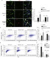
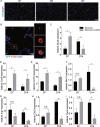
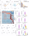
Methods: The effect of hucMSCs on mouse lung regeneration was determined by detecting Ki67 expression and EdU incorporation in alveolar type 2 (AT2) and lung fibroblast cells. hucMSCs were transfected to express the membrane localized GFP before transplant into the mouse lung. The cellular behavior of hucMSCs in mouse lung was tracked by GFP staining. Single cell RNA sequencing was performed to investigate the effects of hucMSCs on gene expression profiles of macrophages after bleomycin treatment.
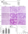
Results: hucMSCs could alleviate collagen accumulation in lung and decrease the mortality of mouse induced by bleomycin. hucMSC transplantation promoted AT2 cell proliferation and inhibited lung fibroblast cell proliferation. By using single cell RNA sequencing, a subcluster of interferon-sensitive macrophages (IFNSMs) were identified after hucMSC infusion. These IFNSMs elevate the secretion of CXCL9 and CXCL10 following hucMSC infusion and recruit more Treg cells to the injured lung.
Conclusions: Our study establishes a link between hucMSCs, macrophage, Treg, and PF. It provides new insights into how hucMSCs interact with macrophage during the repair process of bleomycin-induced PF and play its immunoregulation function.
Keywords: Human umbilical cord mesenchymal stromal cell; Macrophage; Regulatory T cell; Single cell RNA sequencing.
© 2021. The Author(s).
Conflict of interest statement
The authors declare that they have no competing interests.
Figures
Similar articles
-
Human umbilical cord mesenchymal stem cell-derived extracellular vesicles alleviated silica induced lung inflammation and fibrosis in mice via circPWWP2A/miR-223-3p/NLRP3 axis.Ecotoxicol Environ Saf. 2023 Feb;251:114537. doi: 10.1016/j.ecoenv.2023.114537. Epub 2023 Jan 14. Ecotoxicol Environ Saf. 2023. PMID: 36646008
-
Melatonin-pretreated human umbilical cord mesenchymal stem cells improved endometrium regeneration and fertility recovery through macrophage immunomodulation in rats with intrauterine adhesions†.Biol Reprod. 2023 Dec 11;109(6):918-937. doi: 10.1093/biolre/ioad102. Biol Reprod. 2023. PMID: 37672216
-
Exosomal let-7i-5p from three-dimensional cultured human umbilical cord mesenchymal stem cells inhibits fibroblast activation in silicosis through targeting TGFBR1.Ecotoxicol Environ Saf. 2022 Mar 15;233:113302. doi: 10.1016/j.ecoenv.2022.113302. Epub 2022 Feb 18. Ecotoxicol Environ Saf. 2022. PMID: 35189518
-
Human pluripotent stem cell-derived macrophages and macrophage-derived exosomes: therapeutic potential in pulmonary fibrosis.Stem Cell Res Ther. 2022 Sep 2;13(1):433. doi: 10.1186/s13287-022-03136-z. Stem Cell Res Ther. 2022. PMID: 36056418 Free PMC article. Review.
-
The novel molecular mechanism of pulmonary fibrosis: insight into lipid metabolism from reanalysis of single-cell RNA-seq databases.Lipids Health Dis. 2024 Apr 3;23(1):98. doi: 10.1186/s12944-024-02062-8. Lipids Health Dis. 2024. PMID: 38570797 Free PMC article. Review.
Cited by
-
Three-dimensional cultured human umbilical cord mesenchymal stem cells attenuate pulmonary fibrosis by improving the balance of mitochondrial fusion and fission.Stem Cells Transl Med. 2024 Sep 10;13(9):912-926. doi: 10.1093/stcltm/szae051. Stem Cells Transl Med. 2024. PMID: 39077914 Free PMC article.
-
Stem cell-based therapy for fibrotic diseases: mechanisms and pathways.Stem Cell Res Ther. 2024 Jun 18;15(1):170. doi: 10.1186/s13287-024-03782-5. Stem Cell Res Ther. 2024. PMID: 38886859 Free PMC article. Review.
-
Influenza, SARS-CoV-2, and Their Impact on Chronic Lung Diseases and Fibrosis: Exploring Therapeutic Options.Am J Pathol. 2024 Oct;194(10):1807-1822. doi: 10.1016/j.ajpath.2024.06.004. Epub 2024 Jul 18. Am J Pathol. 2024. PMID: 39032604 Review.
-
Stem cell-based therapy for pulmonary fibrosis.Stem Cell Res Ther. 2022 Oct 4;13(1):492. doi: 10.1186/s13287-022-03181-8. Stem Cell Res Ther. 2022. PMID: 36195893 Free PMC article. Review.
-
Immunomodulatory effects of mesenchymal stem cells in peripheral nerve injury.Stem Cell Res Ther. 2022 Jan 15;13(1):18. doi: 10.1186/s13287-021-02690-2. Stem Cell Res Ther. 2022. PMID: 35033187 Free PMC article. Review.
References
-
- Aran D, Looney AP, Liu L, Wu E, Fong V, Hsu A, Chak S, Naikawadi RP, Wolters PJ, Abate AR, Butte AJ, Bhattacharya M. Reference-based analysis of lung single-cell sequencing reveals a transitional profibrotic macrophage. Nat Immunol. 2019;20(2):163–172. doi: 10.1038/s41590-018-0276-y. - DOI - PMC - PubMed
Publication types
MeSH terms
LinkOut - more resources
Full Text Sources
Medical
Research Materials

