. 2021 Jul 13;17(7):e1009311.
doi: 10.1371/journal.ppat.1009311. eCollection 2021 Jul.

Treponema denticola dentilisin triggered TLR2/MyD88 activation upregulates a tissue destructive program involving MMPs via Sp1 in human oral cells

Affiliations

Treponema denticola dentilisin triggered TLR2/MyD88 activation upregulates a tissue destructive program involving MMPs via Sp1 in human oral cells

Sean Ganther et al. PLoS Pathog. .

Abstract

Periodontal disease is driven by dysbiosis in the oral microbiome, resulting in over-representation of species that induce the release of pro-inflammatory cytokines, chemokines, and tissue-remodeling matrix metalloproteinases (MMPs) in the periodontium. These chronic tissue-destructive inflammatory responses result in gradual loss of tooth-supporting alveolar bone. The oral spirochete Treponema denticola, is consistently found at significantly elevated levels in periodontal lesions. Host-expressed Toll-Like Receptor 2 (TLR2) senses a variety of bacterial ligands, including acylated lipopolysaccharides and lipoproteins. T. denticola dentilisin, a surface-expressed protease complex comprised of three lipoproteins has been implicated as a virulence factor in periodontal disease, primarily due to its proteolytic activity. While the role of acylated bacterial components in induction of inflammation is well-studied, little attention has been given to the potential role of the acylated nature of dentilisin. The purpose of this study was to test the hypothesis that T. denticola dentilisin activates a TLR2-dependent mechanism, leading to upregulation of tissue-destructive genes in periodontal tissue. RNA-sequencing of periodontal ligament cells challenged with T. denticola bacteria revealed significant upregulation of genes associated with extracellular matrix organization and degradation including potentially tissue-specific inducible MMPs that may play novel roles in modulating host immune responses that have yet to be characterized within the context of oral disease. The Gram-negative oral commensal, Veillonella parvula, failed to upregulate these same MMPs. Dentilisin-induced upregulation of MMPs was mediated via TLR2 and MyD88 activation, since knockdown of expression of either abrogated these effects. Challenge with purified dentilisin upregulated the same MMPs while a dentilisin-deficient T. denticola mutant had no effect. Finally, T. denticola-mediated activation of TLR2/MyD88 lead to the nuclear translocation of the transcription factor Sp1, which was shown to be a critical regulator of all T. denticola-dependent MMP expression. Taken together, these data suggest that T. denticola dentilisin stimulates tissue-destructive cellular processes in a TLR2/MyD88/Sp1-dependent fashion.


Conflict of interest statement

The authors have declared that no competing interests exist.

Figures

Fig 1
Fig 1. Differential expression analysis of T. denticola challenged hPDL cells.
Total RNA was extracted from healthy patient-derived hPDL cells challenged with Td-WT bacteria at a MOI of 50 for 2-hours in media free of supplements followed by 3 and 22-hours in media supplemented with 10% FBS, 1% Pen Strep and 1% Amphotericin B. Mean FPKM values were used for downstream analysis (n = 3 patient replicates). A) Overlapping and differentially expressed genes between control, 5-hour and 24-hour incubation groups visualized using a Venn diagram. B) Hierarchical clustering analysis was used to determine similarity of transcriptome profiles based on differential expression as a heatmap. Red (Upregulation) to blue (Downregulation) color gradient of heatmap represents normalized gene expression as row Z-scores. C) Top 20 enriched Gene Ontology terms of hPDL cells challenged for 2-hours followed by a 22-hour incubation using the Reactome nomenclature. Statistical significance was assessed using a Kolmogorov-Smirnov test followed by Benjamini-Hochberg correction (p<0.05). D) Top 20 enriched signaling pathways of hPDL cells challenged for 2-hours followed by a 22-hour incubation using the Kyoto Encyclopedia of Genes and Genomes (KEGG) database. Statistical significance was assessed using a Kolmogorov-Smirnov test followed Benjamini-Hochberg correction (p<0.05).
Fig 2
Fig 2. T. denticola upregulates MMPs in hPDL cells while V. parvula does not.
(A-E) RT-qPCR for MMP-2, MMP-11, MMP-14, MMP-17 and MMP-28 mRNA expression of healthy hPDL cells challenged with V. parvula (ATCC 10790) and Td-WT bacteria at a MOI of 50 for 2-hours, followed by a 22-hour incubation in media supplemented with 10% FBS and 1% Pen/Strep and 1% Amphotericin B. Expression of each gene was normalized to that of GAPDH. Statistical significance was determined using a One-Way ANOVA followed by Post-Hoc Tukey’s multiple comparisons. Bars represent mean ± SEM (n = 5). *p < .05 versus control. **p < .01 versus control. ***p < .001 versus control.
Fig 3
Fig 3. T. denticola surface-expressed dentilisin mediates the upregulation of MMP 2, 11, 14, 17 and 28 mRNA levels in hPDL cells.
(A-E) RT-qPCR for MMP-2, MMP-11, MMP-14, MMP-17 and MMP-28 mRNA expression of healthy hPDL cells challenged with isogenic Td-CF522 bacteria, Td-WT bacteria and purified dentilisin. Cells were stimulated at an MOI of 50 and a final concentration of of 1 μg/mL. Cells were challenged for 2-hours in alpha-MEM media supplemented with 10% FBS followed by a 22-hour incubation in alpha-MEM media with 10% FBS, 1% PenStrep and 1% Amphotericin B. The expression of each gene was normalized to that of GAPDH. Statistical significance was determined using a One-Way ANOVA followed by Tukey’s Post-Hoc multiple comparisons. Bars represent mean ± SEM (n = 4). **p < .01 versus control. ***p < .001 versus control.
Fig 4
Fig 4. Suppression of TLR2 inhibits T. denticola-stimulated upregulation of MMPs 2, 14, 17 and 28 while exacerbating MMP 11 expression in hPDL cells.
A) RT-qPCR validation of stable gene suppression using shRNA vectors targeted against TLR2 in healthy hPDL cells. Cells transduced with scrambled shRNA vectors were used as a control. Statistical significance was determined using an unpaired t-test. Bars represent ± SEM of mean value (n = 3 clones). *p < .05 versus control. B-F) RT-qPCR for MMP-2, MMP-11, MMP-14, MMP-17 and MMP-28 mRNA expression of scrambled shRNA control and TLR2 shRNA hPDL cells challenged or stimulated with Td-CF522, purified dentilisin or Td-WT at an MOI of 50 and concentration of 1 μg/mL for 2-hours in alpha-MEM media with no supplementation followed by a 22-hour incubation in alpha-MEM media supplemented with 10% FBS, 1% Pen/Strep and 1% Amphotericin B. The expression of each gene was normalized to that of GAPDH. Statistical significance was determined using a Two-Way ANOVA followed by post-hoc Tukey’s multiple comparisons. Bars represent mean ± SEM (n = 3). #p < .05 versus scramble control group. ##p < .01 versus scramble control group. ###p < .001 versus scramble control group. *p < .05 versus TLR2 shRNA equivalent group. ***p < .001 versus TLR2 equivalent shRNA group.
Fig 5
Fig 5. Suppression of MyD88 inhibits T. denticola-stimulated upregulation of MMP 2, 11, 14, 17 and 28 in hPDL cells.
A) RT-qPCR validation of stable gene suppression using shRNA vectors targeted against MyD88 in healthy hPDL cells. Cells transduced with scrambled shRNA vectors were used as a control. Statistical significance was determined using an unpaired t-test. Bars represent ± SEM of mean values (n = 3 clones). *p < .05 versus control. B-F) RT-qPCR for MMP-2, MMP-11, MMP-14, MMP-17 and MMP-28 mRNA expression of scrambled shRNA control and MyD88 shRNA hPDL cells challenged or stimulated with Td-CF522, purified dentilisin or Td-WT at an MOI of 50 and concentration of 1 μg/mL for 2-hours in alpha-MEM media with no supplementation followed by a 22-hour incubation in alpha-MEM media supplemented with 10% FBS, 1% Pen/Strep and 1% Amphotericin B. The expression of each gene was normalized to that of GAPDH. Statistical significance was determined using a Two-Way ANOVA followed by post-hoc Tukey’s multiple comparisons. Bars represent mean ± SEM (n = 3). #p < .05 versus scramble control group. ###p < .001 versus scramble control group *p < .05 versus MyD88 shRNA equivalent group. **p < .01 versus MyD88 shRNA equivalent group. ***p < .001 versus MyD88 shRNA equivalent group.
Fig 6
Fig 6
Healthy hPDL cells were challenged with A) Td-WT and B) isogenic Td-CF522 bacteria at an MOI of 50 as previously described. Whole cell lysates were generated and used for Western Blot analysis utilizing total anti-Sp1 antibodies. Total Sp1 protein expression was normalized against GAPDH protein expression as a loading control. Statistical significance was determined using a paired t-test. Bars represent mean ± SD (n = 3). **p < .01 versus control.
Fig 7
Fig 7. Knockdown of TLR2-dependent signaling inhibits translocation of transcription factor Sp1 in both T. denticola- and dentilisin-stimulated hPDL cells.
A-B) Healthy hPDL cells were challenged with Td-WT and isogenic Td-CF522 bacteria at an MOI of 50 as previously described. Whole cell lysates were generated and used for Western blot analysis utilizing total Sp1-specific antibodies. Total Sp1 protein expression was normalized using GAPDH as a loading control. Statistical significance was determined using a paired t-test. Bars represent mean ± SD (n = 3). **p < .01 versus control. C) Scrambled shRNA control and TLR2 shRNA hPDL cells were challenged or stimulated with Td-CF522, purified dentilisin or Td-WT at an MOI of 50 and concentration of 1 μg/mL for 2-hours in alpha-MEM media with no supplementation followed by a 22-hour incubation in alpha-MEM media supplemented with 10% FBS, 1% Pen/Strep and 1% Amphotericin B. Cells were stained with Hoescht 33342 (Blue) and total Sp1-specific antibodies (Green), and subjected to confocal microscopy. Scale bar represents 10 μm.
Fig 8
Fig 8. Knockdown of MyD88-dependent signaling inhibits translocation of transcription factor Sp1 in both T. denticola- and dentilisin-stimulated hPDL cells.
A) Scrambled shRNA control and MyD88 shRNA hPDL cells were challenged or stimulated with Td-CF522, purified dentilisin or Td-WT at an MOI of 50 and concentration of 1 μg/mL for 2-hours in alpha-MEM media with no supplementation followed by a 22-hour incubation in alpha-MEM media supplemented with 10% FBS, 1% Pen/Strep and 1% Amphotericin B. Cells were stained with Hoescht 33342 (Blue) and total Sp1-specific antibodies (Green) and subjected to confocal microscopy. Scale bar represents 10 μm. Scale bar represents 10 μm.
Fig 9
Fig 9. Stable Suppression of Sp1 inhibits T. denticola-stimulated upregulation of MMP 2, 11, 14, 17 and 28 in hPDL cells.
A) RT-qPCR validation of stable gene suppression using shRNA vectors targeted against Sp1 in healthy hPDL cells. Cells transduced with scrambled shRNA vectors were used as a control. Statistical significance was determined using an unpaired t-test. Bars represent ± SEM of mean values (n = 3 clones). **p < .01 versus control. B-F) RT-qPCR for MMP-2, MMP-11, MMP-14, MMP-17 and MMP-28 mRNA expression of scrambled shRNA control and Sp1 shRNA hPDL cells challenged or stimulated with purified dentilisin at a concentration of 1 μg/mL or Td-WT at an MOI of 50 for 2-hours in alpha-MEM media supplemented with 10% FBS and no antibiotics followed by a 22-hour incubation in alpha-MEM media supplemented with 10% FBS, 1% Pen/Strep and 1% Amphotericin B. The expression of each gene was normalized to that of GAPDH. Statistical significance was determined using a Two-Way ANOVA followed by post-hoc Tukey’s multiple comparisons. Bars represent mean ± SEM (n = 3). #p < .05 versus scramble control group. ##p < .01 versus scramble control group. ###p < .001 versus scramble control group. *p < .05 versus Sp1 shRNA equivalent group. **p < .01 versus Sp1 shRNA equivalent group. ***p < .001 versus Sp1 shRNA equivalent group.
Fig 10
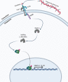
Fig 10. Model of proposed mechanism. T. denticola activates host expressed TLR2 receptors via surface-expressed dentilisin.
Subsequent downstream activation of MyD88 and translocation of the transcription factor Sp1 lead to the upregulation of MMPs 2, 11, 14, 17 and 28 in human periodontal ligament cells. Grey colored icons represent genes that were identified through RNA-sequencing and analysis but were not directly validated.

Similar articles

Cited by

References

    1. Eke P.I., et al.., Prevalence of Periodontitis in Adults in the United States: 2009 and 2010. Journal of Dental Research, 2012. 91(10): p. 914–920. doi: 10.1177/0022034512457373 - DOI - PubMed
    1. Kebschull M., Demmer R.T., and Papapanou P.N., "Gum bug, leave my heart alone!"—epidemiologic and mechanistic evidence linking periodontal infections and atherosclerosis. Journal of dental research, 2010. 89(9): p. 879–902. doi: 10.1177/0022034510375281 - DOI - PMC - PubMed
    1. Madianos P.N., Bobetsis Y.A., and Offenbacher S., Adverse pregnancy outcomes (APOs) and periodontal disease: pathogenic mechanisms. Journal of Periodontology, 2013. 84(4S): p. S170–S180. - PubMed
    1. Lundberg K., et al.., Periodontitis in RA—the citrullinated enolase connection. Nature Reviews Rheumatology, 2010. 6(12): p. 727–730. doi: 10.1038/nrrheum.2010.139 - DOI - PubMed
    1. Karpinski T.M., Role of Oral Microbiota in Cancer Development. Microorganisms, 2019. 7(1). doi: 10.3390/microorganisms7010020 - DOI - PMC - PubMed

Publication types

MeSH terms

LinkOut - more resources