. 2021 Oct;27(10):1241-1256.
doi: 10.1261/rna.078858.121. Epub 2021 Jul 8.

Recruitment of endoplasmic reticulum-targeted and cytosolic mRNAs into membrane-associated stress granules

Affiliations

Recruitment of endoplasmic reticulum-targeted and cytosolic mRNAs into membrane-associated stress granules

Jessica R Child et al. RNA. 2021 Oct.

Abstract

Stress granules (SGs) are membraneless organelles composed of mRNAs and RNA binding proteins which undergo assembly in response to stress-induced inactivation of translation initiation. In general, SG recruitment is limited to a subpopulation of a given mRNA species and RNA-seq analyses of purified SGs revealed that signal sequence-encoding (i.e., endoplasmic reticulum [ER]-targeted) transcripts are significantly underrepresented, consistent with prior reports that ER localization can protect mRNAs from SG recruitment. Using translational profiling, cell fractionation, and single molecule mRNA imaging, we examined SG biogenesis following activation of the unfolded protein response (UPR) by 1,4-dithiothreitol (DTT) and report that gene-specific subsets of cytosolic and ER-targeted mRNAs can be recruited into SGs. Furthermore, we demonstrate that SGs form in close proximity to or directly associated with the ER membrane. ER-associated SG assembly was also observed during arsenite stress, suggesting broad roles for the ER in SG biogenesis. Recruitment of a given mRNA into SGs required stress-induced translational repression, though translational inhibition was not solely predictive of an mRNA's propensity for SG recruitment. SG formation was prevented by the transcriptional inhibitors actinomycin D or triptolide, suggesting a functional link between gene transcriptional state and SG biogenesis. Collectively these data demonstrate that ER-targeted and cytosolic mRNAs can be recruited into ER-associated SGs and this recruitment is sensitive to transcriptional inhibition. We propose that newly transcribed mRNAs exported under conditions of suppressed translation initiation are primary SG substrates, with the ER serving as the central subcellular site of SG formation.

Keywords: endoplasmic reticulum; mRNA; oxidative stress; stress granule; translational regulation; unfolded protein response.


Figures

FIGURE 1.
FIGURE 1.
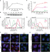
Selective recruitment of ER-targeted mRNAs into UPR-induced stress granules. (A) Representative time course of UPR-induced inhibition of protein synthesis assayed by [35S]Met/Cys incorporation. HeLa cell cultures were treated with DTT and protein synthesis rates assayed at the the indicated time points. (B) Immunoblot analysis of eIF2α and phospho-eIF2α levels following DTT treatment of cell cultures for the indicated times. (C) Representative time course of UPR-elicited transcriptional activation of the UPR response genes GRP94 and GRP78 and the ER-targeted gene B2M. Cell cultures were treated with DTT and total RNA was extracted for RT-qPCR analysis of transcript levels at the indicated time points. Data points are mean log2 fold-change ± SD, normalized to GAPDH levels. (D,E) Polyribosome profiling of GRP94, GRP78, and B2M translational status by sucrose density gradient velocity sedimentation. HeLa cell cultures at time zero (D) or following DTT treatment (E) were detergent extracted, and total polyribosome profiles were determined by the A254 nm absorbance traces (gray). mRNA distributions were determined by RT-qPCR analysis of GRP78 (red), GRP94 (magenta), and B2M (green) mRNAs extracted from the gradient fractions. Data are representative of three biological replicates; RT-qPCR data are mean fraction of total mRNA for the given gene across all gradient fractions ± SD. (F) Representative smFISH visualization of GRP94 (magenta), GRP78 (red), and B2M (green) mRNAs in untreated and DTT-treated (60 min) HeLa cells. (G) As in F but treatment with sodium arsenite (60 min). DAPI nuclear stain (blue) is indicated for all images. Scale bar = 20 µm.
FIGURE 2.
FIGURE 2.
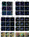
UPR activation elicits ER-associated stress granules. (A) Representative GRP94 smFISH (magenta) with immunofluorescence costaining for the stress granule protein markers HuR, G3BP1, and PABP (cyan) in untreated and DTT-treated HeLa cell cultures. Dotted boxes indicate regions of grayscale insets for mRNA and protein channels, as well as color merge, for DTT-treated cells (right). (B) Representative GRP94 smFISH (magenta) and G3BP1 immunofluorescence (cyan) costaining in DTT-treated cells. Following DTT treatment, cells were permeabilized with digitonin-supplemented buffer to release cytosolic contents (digitonin) or sequentially treated with digitonin and n-dodecyl-β-d-maltoside (DDM) buffers to solubilize organelle membranes. See also Supplemental Figure S1 for detergent permeabilization protocol validation. (C) As in B but with arsenite stress. (D) Representative micrographs of ER membrane protein TRAPα immunofluorescence (yellow) with GRP94 smFISH (magenta) and/or G3BP1 immunofluorescence (cyan) costaining in unfractionated and cytosol-depleted (digitonin-permeabilized) cells following DTT treatment or untreated control. Boxes indicate regions of interest for (E). (E) 3D renderings of representative granules from (D) in unfractionated (Supplemental Movie 1) and digitonin-permeabilized (Supplemental Movie 2) cells. DAPI staining (blue) is indicated for all images. Full cell scale bars = 20 µm, inset and 3D scale bars = 4 µm.
FIGURE 3.
FIGURE 3.
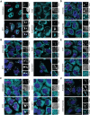
Transcriptional inhibitors actinomycin D and triptolide prevent RNA recruitment into stress granules. (A) Representative time course analysis of GRP94 (magenta) and B2M (green) smFISH staining patterns over the course of maximal inhibition of eIF2α activity (see Fig. 1A). (B) Sucrose density velocity sedimentation gradients and RT-qPCR analysis as in Figure 1D at the indicated time points following DTT addition. Data are representative of three biological replicates. (C) Representative GRP94 smFISH in control (untreated, ActD, triptolide) and stressed (treatment with DTT, with and without treatment with indicated transcriptional inhibitor, ActD or triptolide) conditions. Boxes indicate regions of grayscale insets of mRNA distributions (right). (D) RT-qPCR analysis of spliced and unspliced XBP1 mRNA in control (untreated or ActD) and stressed (treatment with DTT, with and without ActD) conditions. Data are expression level relative to 0 min without ActD after normalization to GAPDH ± SEM from three biological replicates. (E) RT-qPCR analysis of GRP94 mRNA levels over a time course of DTT treatment with or without ActD addition. Data are mean log2 fold change of expression relative to GAPDH ± SEM from three biological replicates. (F) Representative GRP94 smFISH in untreated and sodium arsenite-treated cells with or without ActD addition. Dotted boxes indicate regions of grayscale insets of mRNA distribution (right). DAPI staining (blue) is included for all images. Full cell scale bar = 20 µm, inset scale bar = 4 µm.
FIGURE 4.
FIGURE 4.
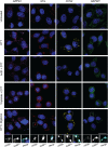
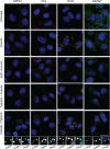
Actinomycin D inhibits RNA binding protein granulation during the UPR. (A) Representative GRP94 smFISH (magenta) with immunofluorescence costaining for HuR (cyan) in control (untreated or ActD) and stressed (treatment with DTT, with and without ActD) HeLa cell cultures. Note redistribution of HuR from the nucleoplasm to the cytoplasm in response to ActD treatment. Boxes indicate regions of grayscale insets for mRNA and protein channels, as well as color merge, for each condition (right). (B) As in A but immunofluorescence staining for G3BP1 (cyan). (C) As in A but immunofluorescence staining for PABP (cyan). (D) Representative GRP94 smFISH (magenta) with immunofluorescence costaining for PABP (cyan) in cells treated with arsenite with and without cycloheximide (CHX). Boxes indicate regions of grayscale insets for mRNA and protein channels, as well as color merge, for each condition (right). (E) As in D but with DTT stress. (F) Representative GRP94 smFISH (magenta) with immunofluorescence costaining for G3BP1 (cyan) in cells treated with arsenite with and without ActD. Boxes indicate regions of grayscale insets for mRNA and protein channels, as well as color merge, for each condition (right). DAPI nuclear stain (blue) is included in all images. Full cell scale bar = 20 µm, inset scale bar = 4 µm.
FIGURE 5.
FIGURE 5.
Transcript length and transcriptional state influence mRNA recruitment into ER-associated stress granules following DTT stress. Representative smFISH of long transcript size-paired genes GRP94 (magenta, 2412 nt) and NCL (red, 2133 nt), and short transcript size-paired genes CCN2 (yellow, 1050 nt) and GAPDH (green, 1008 nt) mRNAs in untreated and DTT-treated HeLa cell cultures. Where indicated, cell cultures were treated with ActD or triptolide in addition to DTT. To evaluate the ER-association and presence of the SG protein marker G3BP1 in observed RNA granules, cells were permeabilized in digitonin-supplemented buffers to extract cytosolic contents while leaving the ER membrane intact, and costained for G3BP1 by immunofluorescence. Dotted boxes indicate regions of grayscale insets for mRNA and G3BP1 protein channels, as well as color merge, for digitonin permeabilized cells (below). DAPI nuclear stain (blue) included in all images. Full cell scale bars = 20 µm, inset scale bars = 4 µm.
FIGURE 6.
FIGURE 6.
Transcript length and transcriptional state influence mRNA recruitment into ER-associated stress granules following arsenite stress. Representative smFISH of long transcript size-paired genes GRP94 (magenta, 2412 nt) and NCL (red, 2133 nt), and short transcript size-paired genes CCN2 (yellow, 1050 nt) and GAPDH (green, 1008 nt) mRNAs in untreated and arsenite-treated HeLa cell cultures. Where indicated, cell cultures were treated with ActD or triptolide in addition to arsenite. To evaluate the ER-association and presence of the SG protein marker G3BP1 in observed RNA granules, cells were permeabilized in digitonin-supplemented buffers to extract cytosolic contents while leaving the ER membrane intact, and costained for G3BP1 by immunofluorescence. Dotted boxes indicate regions of grayscale insets for mRNA and G3BP1 protein channels, as well as color merge, for digitonin permeabilized cells (below). DAPI nuclear stain (blue) included in all images. Full cell scale bars = 20 µm, inset scale bars = 4 µm.
FIGURE 7.
FIGURE 7.
Model depicting partitioning of newly exported mRNAs between translation-engaged and stress granule-associated states in response to UPR activation. A working model depicting the functional states of newly exported mRNAs in homeostatic (active translation, low phospo-eIF2α) or stress-activated (reduced translation, elevated phospo-eIF2α) cellular states. This model highlights a role for translation-dependent RNA binding protein remodeling of newly exported mRNAs in determining mRNA recruitment into polyribosomes or SGs. As illustrated, under conditions of stress-induced translational inhibition, the nuclear RNA binding protein signature of newly exported mRNAs would be relatively long-lived and thus serve as a signal for ribonucleoprotein recruitment into stress granules, which could include both cytoplasmic and ER-associated forms. Once eIF2α phosphorylation is resolved, stress granule-associated mRNAs would resume translation, undergo RNA binding protein remodeling, and enter the polyribosome-associated pool. In this way, stress granules could serve as a triage station for newly exported mRNAs and undergo rapid mobilization following stress adaptation.

Similar articles

Cited by

References

    1. Andreev DE, O'Connor PB, Fahey C, Kenny EM, Terenin IM, Dmitriev SE, Cormican P, Morris DW, Shatsky IN, Baranov PV. 2015. Translation of 5′ leaders is pervasive in genes resistant to eIF2 repression. Elife 4: e03971. 10.7554/eLife.03971 - DOI - PMC - PubMed
    1. Bley N, Lederer M, Pfalz B, Reinke C, Fuchs T, Glass M, Moller B, Huttelmaier S. 2015. Stress granules are dispensable for mRNA stabilization during cellular stress. Nucleic Acids Res 43: e26. 10.1093/nar/gku1275 - DOI - PMC - PubMed
    1. Blower MD. 2013. Molecular insights into intracellular RNA localization. Int Rev Cell Mol Biol 302: 1–39. 10.1016/B978-0-12-407699-0.00001-7 - DOI - PMC - PubMed
    1. Bounedjah O, Desforges B, Wu TD, Pioche-Durieu C, Marco S, Hamon L, Curmi PA, Guerquin-Kern JL, Pietrement O, Pastre D. 2014. Free mRNA in excess upon polysome dissociation is a scaffold for protein multimerization to form stress granules. Nucleic Acids Res 42: 8678–8691. 10.1093/nar/gku582 - DOI - PMC - PubMed
    1. Braakman I, Helenius J, Helenius A. 1992. Manipulating disulfide bond formation and protein folding in the endoplasmic reticulum. EMBO J 11: 1717–1722. 10.1002/j.1460-2075.1992.tb05223.x - DOI - PMC - PubMed

Publication types

MeSH terms