

Resistance of SARS-CoV-2 variants to neutralization by antibodies induced in convalescent patients with COVID-19
- PMID: 34237284
- PMCID: PMC8226103
- DOI: 10.1016/j.celrep.2021.109385
Resistance of SARS-CoV-2 variants to neutralization by antibodies induced in convalescent patients with COVID-19
Abstract
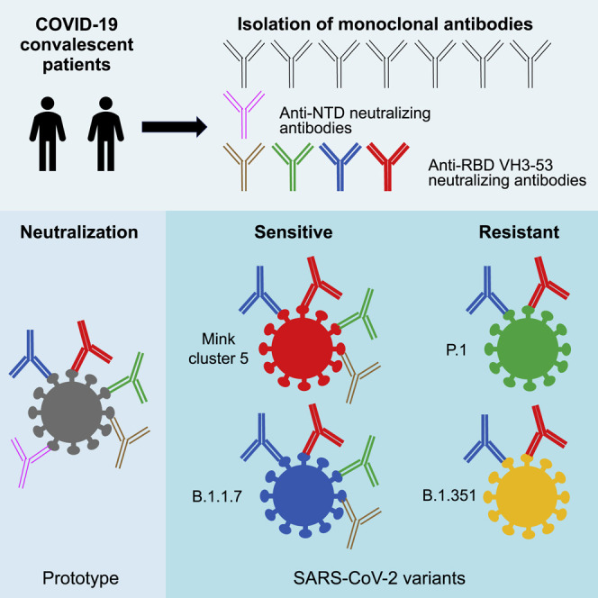
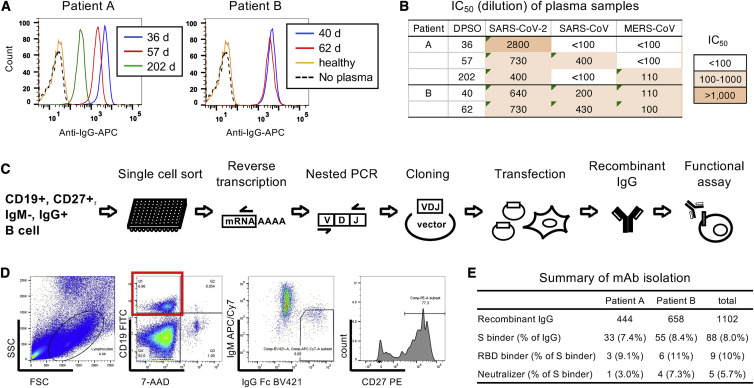
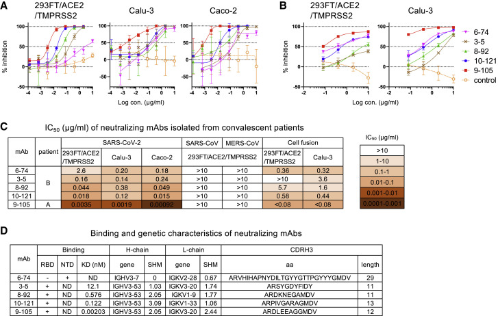
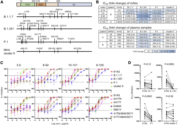
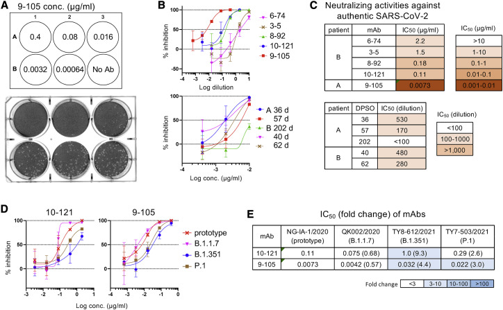
Administration of convalescent plasma or neutralizing monoclonal antibodies (mAbs) is a potent therapeutic option for coronavirus disease 2019 (COVID-19) caused by severe acute respiratory syndrome coronavirus 2 (SARS-CoV-2) infection. However, SARS-CoV-2 variants with mutations in the spike protein have emerged in many countries. To evaluate the efficacy of neutralizing antibodies induced in convalescent patients against emerging variants, we isolate anti-spike mAbs from two convalescent COVID-19 patients infected with prototypic SARS-CoV-2 by single-cell sorting of immunoglobulin-G-positive (IgG+) memory B cells. Anti-spike antibody induction is robust in these patients, and five mAbs have potent neutralizing activities. The efficacy of most neutralizing mAbs and convalescent plasma samples is maintained against B.1.1.7 and mink cluster 5 variants but is significantly decreased against variants B.1.351 from South Africa and P.1 from Brazil. However, mAbs with a high affinity for the receptor-binding domain remain effective against these neutralization-resistant variants. Rapid spread of these variants significantly impacts antibody-based therapies and vaccine strategies against SARS-CoV-2.
Keywords: COVID-19; SARS-CoV-2; mAb; neutralizing antibody; variant.
Copyright © 2021 The Author(s). Published by Elsevier Inc. All rights reserved.
Conflict of interest statement
Declaration of interests Y. Kaku, T.K., and S.M. are listed as inventors on a patent application related to this work. The remaining authors declare no competing interests.
Figures
Similar articles
-
Antibody resistance of SARS-CoV-2 variants B.1.351 and B.1.1.7.Nature. 2021 May;593(7857):130-135. doi: 10.1038/s41586-021-03398-2. Epub 2021 Mar 8. Nature. 2021. PMID: 33684923
-
Escape from neutralizing antibodies by SARS-CoV-2 spike protein variants.Elife. 2020 Oct 28;9:e61312. doi: 10.7554/eLife.61312. Elife. 2020. PMID: 33112236 Free PMC article.
-
Emergence of Multiple SARS-CoV-2 Antibody Escape Variants in an Immunocompromised Host Undergoing Convalescent Plasma Treatment.mSphere. 2021 Aug 25;6(4):e0048021. doi: 10.1128/mSphere.00480-21. Epub 2021 Aug 25. mSphere. 2021. PMID: 34431691 Free PMC article.
-
Structural Analysis of Neutralizing Epitopes of the SARS-CoV-2 Spike to Guide Therapy and Vaccine Design Strategies.Viruses. 2021 Jan 19;13(1):134. doi: 10.3390/v13010134. Viruses. 2021. PMID: 33477902 Free PMC article. Review.
-
Neutralising antibody escape of SARS-CoV-2 spike protein: Risk assessment for antibody-based Covid-19 therapeutics and vaccines.Rev Med Virol. 2021 Nov;31(6):e2231. doi: 10.1002/rmv.2231. Epub 2021 Mar 16. Rev Med Virol. 2021. PMID: 33724631 Free PMC article. Review.
Cited by
-
Factors Associated with Neutralizing Antibody Responses following 2-Dose and 3rd Booster Monovalent COVID-19 Vaccination in Japanese People Living with HIV.Viruses. 2024 Apr 2;16(4):555. doi: 10.3390/v16040555. Viruses. 2024. PMID: 38675897 Free PMC article.
-
Arsenal of nanobodies shows broad-spectrum neutralization against SARS-CoV-2 variants of concern in vitro and in vivo in hamster models.Commun Biol. 2022 Sep 9;5(1):933. doi: 10.1038/s42003-022-03866-z. Commun Biol. 2022. PMID: 36085335 Free PMC article.
-
Efficacy, safety, and immunogenicity of a booster regimen of Ad26.COV2.S vaccine against COVID-19 (ENSEMBLE2): results of a randomised, double-blind, placebo-controlled, phase 3 trial.Lancet Infect Dis. 2022 Dec;22(12):1703-1715. doi: 10.1016/S1473-3099(22)00506-0. Epub 2022 Sep 13. Lancet Infect Dis. 2022. PMID: 36113538 Free PMC article. Clinical Trial.
-
Neutralizing and enhancing monoclonal antibodies in SARS-CoV-2 convalescent patients: lessons from early variant infection and impact on shaping emerging variants.Emerg Microbes Infect. 2024 Dec;13(1):2307510. doi: 10.1080/22221751.2024.2307510. Epub 2024 Jan 30. Emerg Microbes Infect. 2024. PMID: 38240255 Free PMC article.
-
Characteristics of Immunogenicity against SARS-CoV-2 in a Community-Based Model of Care during the Fourth Wave of COVID-19 Outbreak in Ho Chi Minh City.Yonsei Med J. 2024 Sep;65(9):501-510. doi: 10.3349/ymj.2023.0567. Yonsei Med J. 2024. PMID: 39193758 Free PMC article.
References
Publication types
MeSH terms
Substances
LinkOut - more resources
Full Text Sources
Medical
Miscellaneous
