. 2021 Jul 6;4(1):841.
doi: 10.1038/s42003-021-02374-w.

A split protease-E. coli ClpXP system quantifies protein-protein interactions in Escherichia coli cells

Affiliations

A split protease-E. coli ClpXP system quantifies protein-protein interactions in Escherichia coli cells

Shengchen Wang et al. Commun Biol. .

Abstract

Characterizing protein-protein interactions (PPIs) is an effective method to help explore protein function. Here, through integrating a newly identified split human Rhinovirus 3 C (HRV 3 C) protease, super-folder GFP (sfGFP), and ClpXP-SsrA protein degradation machinery, we developed a fluorescence-assisted single-cell methodology (split protease-E. coli ClpXP (SPEC)) to explore protein-protein interactions for both eukaryotic and prokaryotic species in E. coli cells. We firstly identified a highly efficient split HRV 3 C protease with high re-assembly ability and then incorporated it into the SPEC method. The SPEC method could convert the cellular protein-protein interaction to quantitative fluorescence signals through a split HRV 3 C protease-mediated proteolytic reaction with high efficiency and broad temperature adaptability. Using SPEC method, we explored the interactions among effectors of representative type I-E and I-F CRISPR/Cas complexes, which combining with subsequent studies of Cas3 mutations conferred further understanding of the functions and structures of CRISPR/Cas complexes.


Conflict of interest statement

The authors declare no competing interests.

Figures
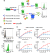
Fig. 1
Fig. 1. Development of the concept of SPEC system.
a Schematic diagram of Split Protease-E. coli ClpXP (SPEC) system. sfGFP is fused with an HRV 3 C protease cleavage sequence (LEVLFQ↓GP) at its C-terminus followed by a SsrA sequence (AANDENYALAA), forming a sfGFP-LEVLFQGP-SsrA cassette. The prey protein and bait protein are fused to the N-terminal and C-terminal split-HRV 3 C proteases, respectively. Through ClpXP-SsrA degradation pathway, highly expressed sfGFP-LEVLFQGP-SsrA cassette is fast degraded in E. coli cells, thus very low green fluorescence is detected. Nevertheless, when prey protein interacts with bait protein, the N-terminal split and C-terminal split-HRV 3 C proteases will re-assemble to a functional HRV 3 C protease, which will cleave off the SsrA peptide from the sfGFP-LEVLFQ↓GP-SsrA cassette, leading to an accumulation of sfGFP for increased fluorescence intensity. b Validation of the ClpXP-SsrA protein degradation machinery mediated sfGFP degradation in E. coli. The total cellular sfGFP fluorescence intensity (sfGFP F. I.) of cells bearing different plasmids were quantitated by flow cytometry (detected with FITC channel, 525/40 nm BP). The experiments were performed under 25 °C with 8 h induction. Upper panel: cells bearing pSPEC-VA vector, Lower panel: cells bearing both pSPEC-VA and pSPEC-VB vectors (Supplementary Table 1, Supplementary Fig. 1). c Comparing TEV protease (blue line, pSPEC-T, Supplementary Table 1) with HRV 3 C protease (red line, pSPEC-VB, Supplementary Table 1) in SPEC system at different temperatures of 37 °C, 30 °C, 25 °C, and 18 °C, respectively. All the vector information can be found in Supplementary Table 1. Data are presented as mean ± SEM (n = 3 independent experiments) with Student’s t test being performed, #P > 0.05, *P ≤ 0.05.
Fig. 2
Fig. 2. Identification of a highly efficient split HRV 3 C protease and its comparison with split TEV protease in the SPEC system.
a Designing split HRV 3 C protease based on its structure (PDB: 2B0F) with three split positions (K82, L94, N107). Blue: N-terminal HRV 3 C protease, Red: C-terminal HRV 3 C protease, Purple: the random coil between N-terminal and C-terminal HRV 3 C protease, Cyan: the designed split sites. b Characterizing different split HRV 3 C proteases in SPEC system using pSPEC-N107/L94/K82-PP and pSPEC-N107/L94/K82-EP vectors, respectively. Cells bearing pSPEC-VB vector containing full length HRV 3 C protease was used as a positive control (right panel), and cells bearing only pSPEC-VA vector was used as a negative control (Fig. 1b). The total cellular sfGFP F. I. of cells bearing different vectors (pSPEC-K82-EP to pSPEC-N107-PP, Supplementary Table 1) were quantitated through flow cytometry. Upper-left panel, representative flow cytometry histograms of the total cellular sfGFP F.I. of cells expressing different split HRV 3 C proteases fused with Z. mobilis Cas1/Cas2-3 protein pair. Lower-left panel, representative flow cytometry histograms of the total cellular sfGFP F.I. of cells expressing different split HRV 3 C proteases fused with S. cerevisiae Yae1/Lto1 protein pair. The experiments were performed at 25 °C with 8 h induction. c Comparison of split HRV 3 C (K82) protease (Wine line, pSPEC-VC1) with split TEV protease (Dark blue line, pSPEC-VC5) using Cas1/Cas2-3 protein pair in SPEC system at 37 °C, 30 °C, 25 °C, and 18 °C, respectively. d Comparison of split HRV 3 C (K82) protease (Yellow line, pSPEC-VC2) with split TEV protease (Cyan line, pSPEC-VC6) using Yae1/Lto1 protein pair in SPEC system at 37 °C, 30 °C, 25 °C, and 18 °C, respectively. All the vector information can be found in Supplementary Table 1. Data are presented as mean ± SEM (n = 3 independent experiments) with Student’s t test being performed, #P > 0.05, *P ≤ 0.05.
Fig. 3
Fig. 3. Development of SPEC system in the engineered BL21(DE3)-SPEC cells.
a Generation of the BL21(DE3)-SPEC cells using no-SCAR incorporated with temperature sensitive plasmid curing strategy. Left panel, scheme of experimental procedure. The lpxM gene fragment in the genome of BL21(DE3) strain is replaced by sfGFP-LEVLFQGP-SsrA fragment using CRISPR/Cas9 assisted λ-Red recombinase technology. The pKD46 based vector is used to prompt the temperature sensitive plasmid curing. Upper-right panel, PCR validation of the inserted sfGFP-LEVLFQGP-SsrA fragment in BL21(DE3)-SPEC strain. Negative control (NC): wild-type BL21(DE3) strain. S: BL21(DE3)-SPEC strain. M: 1Kb DNA ladder. Lower-right panel, Sanger sequencing of BL21(DE3) genome to verify the insertion of the sfGFP-LEVLFQGP-SsrA fragment. b Validation of the SPEC system in the BL21(DE3)-SPEC strain by comparing the HRV 3 C protease (pSPEC-VB) and split HRV 3 C (K82) protease (pSPEC-VC1) with Cas1/Cas2-3 protein pair. The experiments were performed at 25 °C with 8 h induction. c Characterizing split HRV 3 C (K82) protease in BL21(DE3)-SPEC strain with Z. mobilis Cas1/Cas2-3 (pSPEC-VC1) and S. cerevisiae Yae1/Lto1 (pSPEC-VC2) protein pairs at 37 °C, 30 °C, 25 °C, and 18 °C, respectively. Data are presented as mean ± SEM (n = 3 independent experiments). BL21(DE3)-SPEC cells bearing pSPEC-VB plasmid containing HRV 3 C protease was used as a positive control, and the total cellular sfGFP F. I. were quantitated through flow cytometry. All the vector information can be found in Supplementary Table 1.
Fig. 4
Fig. 4. Characterization of protein–protein interactions of effectors in the Type I-E and I-F CRISPR/Cas complex.
a Protein–protein interactions among eight effectors (Cas1, Cas2, Cas3, Cas8, Cas11, Cas7, Cas5, and Cas6) of E. coli K12 Type I-E CRISPR/Cas complex was interpreted using the circular visualization R program. b Protein-protein interactions among six effectors (Cas1, Cas2-3, Csy1, Csy2, Csy3, and Csy4) of Z. mobilis Type I-F CRISPR/Cas complex was interpreted using the circular visualization R program. c The histogram of protein-protein interaction between Cas3 mutants (H74A, D75A, K78A, K320N, D452N, and S483A/T485A, respectively) and seven other effectors (Cas1, Cas2, Cas5, Cas6, Cas7, Cas8, and Cas11) of E. coli K12 Type I-E CRISPR/Cas complex. Cas1/GST was used as a negative control (Supplementary Fig. 2b), and Cas1/Cas2 was used as a positive control. Percentage numbers presented correspondent interaction intensities of protein pairs, which were normalized by the interaction between Cas3 and correspondent effectors (Supplementary Fig. 2b). d The histogram of protein-protein interaction between Cas2-3 mutants (H123A, D124A, K127A, K458N, D608N, and S639A/T641A, respectively) and five other effectors (Cas1, Csy1, Csy2, Csy3, and Csy4) of Z. mobilis Type I-F CRISPR/Cas complex. Cas1/GST was used as a negative control (Supplementary Fig. 3b), and Cas1/Cas2-3 was used as a positive control. Percentage numbers presented correspondent interaction intensities of protein pairs, which were normalized by the interaction between Cas2-3 and correspondent effectors (Supplementary Fig. 3b). All the vector information can be found in Supplementary Table 1. Data are presented as mean ± SEM (n = 3 independent experiments).
Fig. 5
Fig. 5. Simulated structure diagram of Type I-F CRISPR/Cas complex.
Left panel: Model protein structures of six proteins (Cas1, Cas2-3, Csy1, Csy2, Csy3 and Csy4) in CRISPR/Cas complex. Right panel: Location of mutation sites (H123, D124, K127, K458, D608, S639, and T641; red font) in Cas2-3.

Similar articles

Cited by

References

    1. Berggard T, Linse S, James P. Methods for the detection and analysis of protein-protein interactions. Proteomics. 2007;7:2833–2842. doi: 10.1002/pmic.200700131. - DOI - PubMed
    1. Lin J-S, Lai E-M. Protein-protein interactions: yeast two-hybrid system. Methods Mol. Biol. 2017;1615:177–187. doi: 10.1007/978-1-4939-7033-9_14. - DOI - PubMed
    1. Karimova G, Gauliard E, Davi M, Ouellette SP, Ladant D. Protein-protein interaction: bacterial two-hybrid. Methods Mol. Biol. 2017;1615:159–176. doi: 10.1007/978-1-4939-7033-9_13. - DOI - PubMed
    1. Burckstummer T, et al. An efficient tandem affinity purification procedure for interaction proteomics in mammalian cells. Nat. Methods. 2006;3:1013–1019. doi: 10.1038/nmeth968. - DOI - PubMed
    1. Rigaut G, et al. A generic protein purification method for protein complex characterization and proteome exploration. Nat. Biotechnol. 1999;17:1030–1032. doi: 10.1038/13732. - DOI - PubMed

Publication types

MeSH terms

Supplementary concepts