. 2021 Jun 16:12:668937.
doi: 10.3389/fpls.2021.668937. eCollection 2021.

Clathrin Is Important for Virulence Factors Delivery in the Necrotrophic Fungus Botrytis cinerea

Affiliations

Clathrin Is Important for Virulence Factors Delivery in the Necrotrophic Fungus Botrytis cinerea

Eytham Souibgui et al. Front Plant Sci. .

Abstract

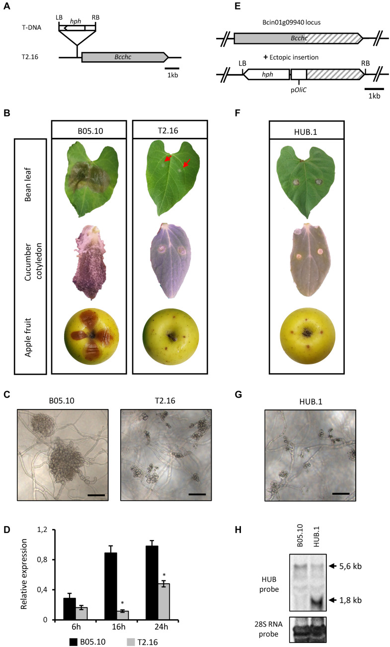
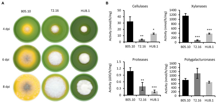
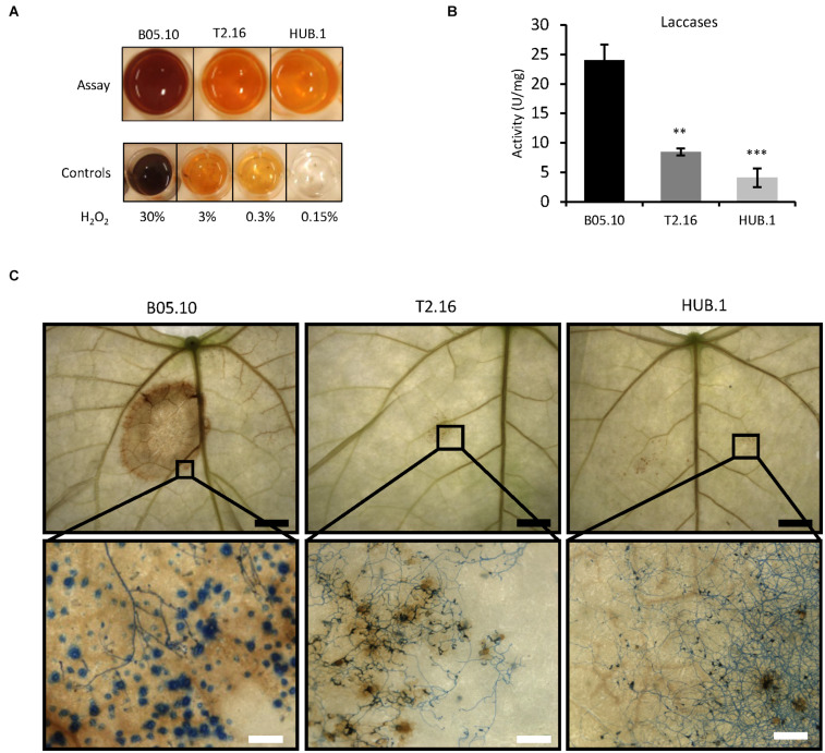
Fungi are the most prevalent plant pathogens, causing annually important damages. To infect and colonize their hosts, they secrete effectors including hydrolytic enzymes able to kill and macerate plant tissues. These secreted proteins are transported from the Endoplasmic Reticulum and the Golgi apparatus to the extracellular space through intracellular vesicles. In pathogenic fungi, intracellular vesicles were described but their biogenesis and their role in virulence remain unclear. In this study, we report the essential role of clathrin heavy chain (CHC) in the pathogenicity of Botrytis cinerea, the agent of gray mold disease. To investigate the importance of this protein involved in coat vesicles formation in eukaryotic cells, a T-DNA insertional mutant reduced in the expression of the CHC-encoding gene, and a mutant expressing a dominant-negative form of CHC were studied. Both mutants were strongly affected in pathogenicity. Characterization of the mutants revealed altered infection cushions and an important defect in protein secretion. This study demonstrates the essential role of clathrin in the infectious process of a plant pathogenic fungus and more particularly its role in virulence factors delivery.

Keywords: Botrytis cinerea; clathrin; infection cushion; secretomics; virulence.


Conflict of interest statement

M-PL was employed by the company Bayer SAS. The remaining authors declare that the research was conducted in the absence of any commercial or financial relationships that could be construed as a potential conflict of interest.

Figures

FIGURE 1
FIGURE 1
Characterization of the T2.16 and HUB mutants of B. cinerea. (A) Schematic representation of the mutated locus in the T2.16 strain generated by A. tumefaciens-mediated transformation. Insertion of the bacterium T-DNA (hph hygromycin B resistance gene flanked by the bacterial left (LB) and right (RB) border regions) occurred 297 bp upstream of open reading frame of the clathrin heavy chain-encoding gene Bcchc and was localized by rescue-PCR analysis. (B,F) The virulence of the T2.16 and HUB mutants was compared to that of the parental strain B05.10 on primary leaves of French bean, cucumber cotyledons and wounded apple fruits. Pictures were taken 4 days post-inoculation. (C,G) Infection cushions were observed by inverted microscopy after 72 h of incubation in liquid PDB 1/4 medium on plastic surfaces (scale bar 50 μm). Photos and data are representative of three independent experiments. (D) Bcchc expression in the parental (B05.10) and the T2.16 mutant strains during a kinetic of fungal growth on Teflon membranes. Mycelia were harvested at 6 h, 16 h and 24 h after inoculation. Bcchc expression is plotted relatively to that of the Bcpda1 housekeeping gene [the same results were obtained with two other reference genes (see Methods)]. Three independent biological replicates were assessed for each experiment. Standard deviations are indicated. Asterisks indicate a significant difference (Student’s t-test, p-value < 0.05). (E) Schematic representation of the DNA construct used to produce the-C-terminal domain of CHC (HUB fragment) in B. cinerea. The A. tumefaciens-compatible plasmid pBHT2 was used to place the HUB DNA sequence (dashed) under the control of the A. nidulans constitutive promoter pOliC. The hygromycin B resistance gene (hph) is shown as well as the left (LB) and right (RB) borders of the T-DNA. (H) Northern blot analysis. RNA extracted from the parental (B05.10) and HUB.1 strains were separated by gel electrophoresis and hybridized with labeled RNA probes corresponding to the HUB DNA sequence (top) or to the 28S RNA (loading control, bottom). Arrows indicate the hybridizing bands corresponding to the HUB fragment RNA (1.8 kb) and to the chc RNA (5.6 kb).
FIGURE 2
FIGURE 2
Endocytosis in the clathrin mutants. (A) Observation of endocytosis by confocal microscopy. Labeling of the plasma membrane by the fluorescent lipophilic marker FM4-64 and its subsequent cellular internalization were monitored over time in young hyphae (16 h culture) of the parental (B05-10) and mutant (T2.16, HUB.1) strains (scale bar: 5 μm). (B) Endocytosis quantification. For 10 different hyphae of each strain, image analysis of the 20 μm apical region led to separate quantifications of the mean total cytoplasmic and mean plasma membrane fluorescence. The plot shows the normalized increase in mean cytoplasmic fluorescence over time (% of plasma membrane fluorescence) in each strain. Standard deviations (10 replicates) are indicated.
FIGURE 3
FIGURE 3
Enzymatic activities and medium acidification in the clathrin mutants. (A) Medium acidification. Agar plugs of parental (B05.10) and mutant (T2.16 and HUB.1) mycelia were used to inoculate solid complete medium adjusted to pH 8.0 and supplemented with 0.01% bromothymol blue (yellow at pH < 6.0). Acidification of the medium was monitored at 4, 6, and 8 days post-inoculation. Photos are representative of three independent experiments. (B) Enzymatic activities measured in the supernatants of liquid cultures in CCPX medium at 72 h post-inoculation for the parental (B05.10) and mutant (T2.16 and HUB.1) strains. Activities are expressed in nmole or ΔDO per hour per mg of mycelium. Three independent biological replicates were performed. Means with standard deviations are indicated, and asterisks indicate significant difference compared to the B05.10 strain (Student’s t-test p-value < 0.05; ∗∗ < 0.01; ∗∗∗ < 0.001).
FIGURE 4
FIGURE 4
Secretion of ROS in the clathrin mutants. (A) Production of ROS in vitro. Equal quantities of mycelia from the parental (B05.10) and mutant (T2.16 and HUB.1) strains were incubated 2 h in the presence of soluble colorless DAB. DAB oxidation into a brown precipitate was monitored and compared to controls using defined quantities of H2O2. (B) Laccase activities measured in the supernatant of liquid cultures in CCPX medium at 72 h for the parental (B05.10) and the mutant (T2.16 and HUB.1) strains. Activities are expressed in arbitrary units (U) per mg of mycelium, three independent biological replicates were performed. Standard deviations are indicated, and asterisks indicate significant difference compared to the B05.10 strain (Student’s t-test p-value ∗∗ < 0.01; ∗∗∗ < 0.001). (C) ROS detection in planta. Primary bean leaves were inoculated with conidia of the parental (B05.10) and mutant (T2.16 & HUB.1) strains and stained with DAB and cotton blue in lactic acid (beta-glucan targeting dye) at 72 h post-infection. DAB oxidation into brown precipitates and hyphal coloration were monitored using a macroscope at different magnifications (black scale bar 5 mm; white scale bar 200 μm). Blue dots in the parental sample could represent callose depositions by the plant (also made of beta-glucans).
FIGURE 5
FIGURE 5
Impact of the T2.16 mutation on the exo-proteome of B. cinerea. Functional categories classification of the secreted proteins that were down- or up-accumulated in comparison to the parental control (B05.10) (fold change > 2), (see Supplementary Table 2 for details). The parental and the T2.16 mutant strains were grown for 3 days in liquid CCPX medium (three independent biological experiments) and a comparative shotgun proteomic analysis was performed on the proteins collected from the cultures supernatants. CAZy, carbohydrate active enzymes; PCWDE, plant cell wall degrading enzymes; HP, Hemicellulose-Pectin; FCWE, fungal cell wall enzymes.

Similar articles

Cited by

References

    1. Adam T., Bouhidel K., Der C., Robert F., Najid A., Simon-Plas F., et al. (2012). Constitutive expression of clathrin hub hinders elicitor-induced clathrin-mediated endocytosis and defense gene expression in plant cells. FEBS Lett. 586 3293–3298. 10.1016/j.febslet.2012.06.053 - DOI - PubMed
    1. Allen C. L., Goulding D., Field M. C. (2003). Clathrin-mediated endocytosis is essential in Trypanosoma brucei. EMBO J. 22 4991–5002. 10.1093/emboj/cdg481 - DOI - PMC - PubMed
    1. Almagro Armenteros J. J., Tsirigos K. D., Sønderby C. K., Petersen T. N., Winther O., Brunak S., et al. (2019). SignalP 5.0 improves signal peptide predictions using deep neural networks. Nat. Biotechnol. 37 420–423. 10.1038/s41587-019-0036-z - DOI - PubMed
    1. Antal Z., Rascle C., Cimerman A., Viaud M., Billon-Grand G., Choquer M., et al. (2012). The homeobox BcHOX8 gene in Botrytis cinerea regulates vegetative growth and morphology. PLoS One 7:e48134. 10.1371/journal.pone.0048134 - DOI - PMC - PubMed
    1. Bairwa G., Caza M., Horianopoulos L., Hu G., Kronstad J. (2019). Role of clathrin-mediated endocytosis in the use of heme and hemoglobin by the fungal pathogen Cryptococcus neoformans. Cell. Microbiol. 21:e12961. 10.1111/cmi.12961 - DOI - PMC - PubMed

LinkOut - more resources