. 2021 Sep 1;81(17):4455-4470.
doi: 10.1158/0008-5472.CAN-21-0772. Epub 2021 Jul 1.

AP-2α-Mediated Activation of E2F and EZH2 Drives Melanoma Metastasis

Affiliations

AP-2α-Mediated Activation of E2F and EZH2 Drives Melanoma Metastasis

Jeffrey R White et al. Cancer Res. .

Abstract

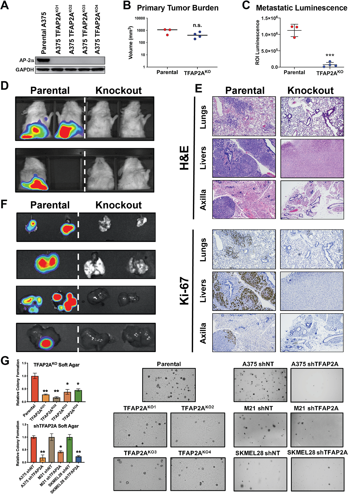
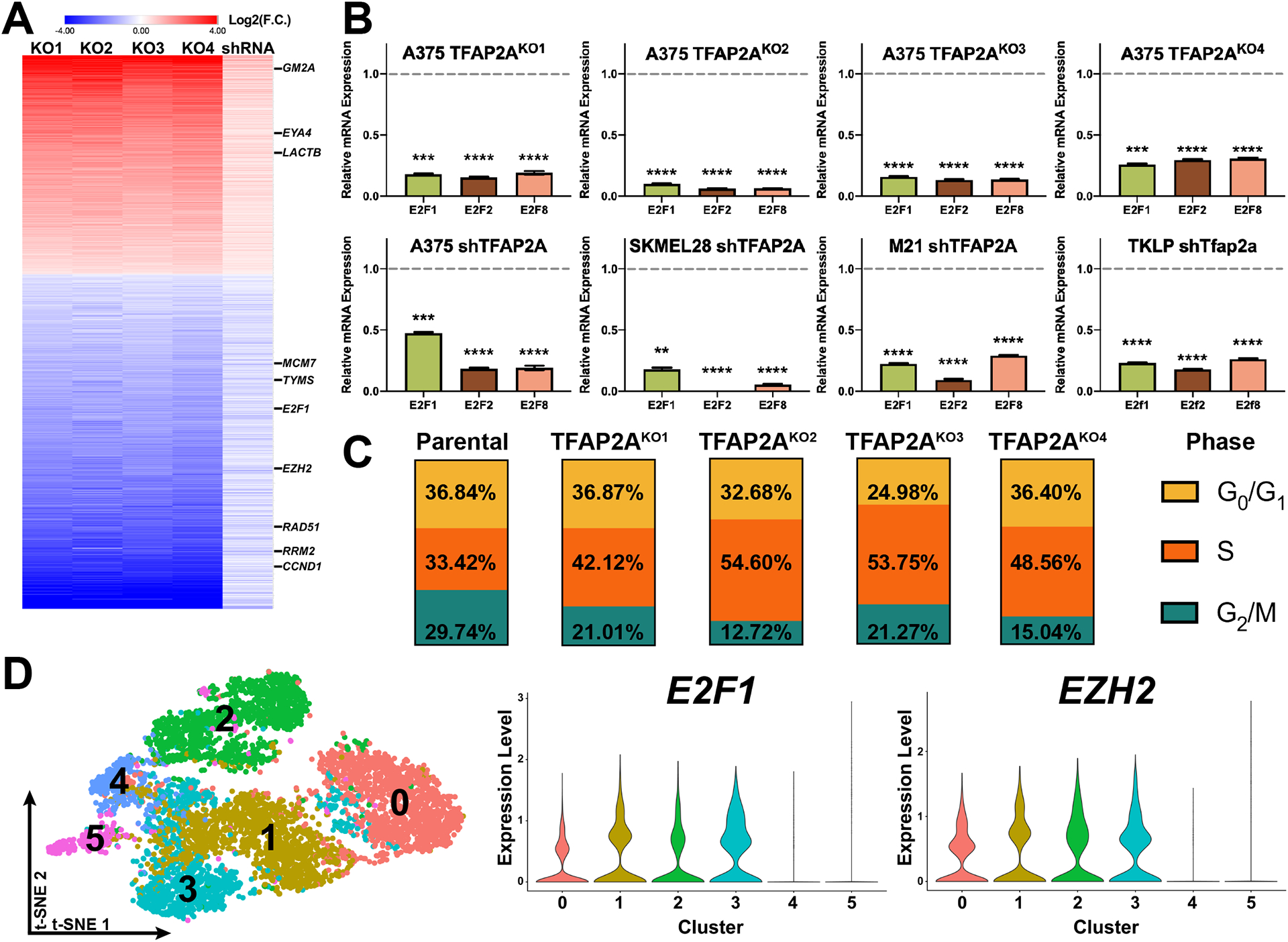
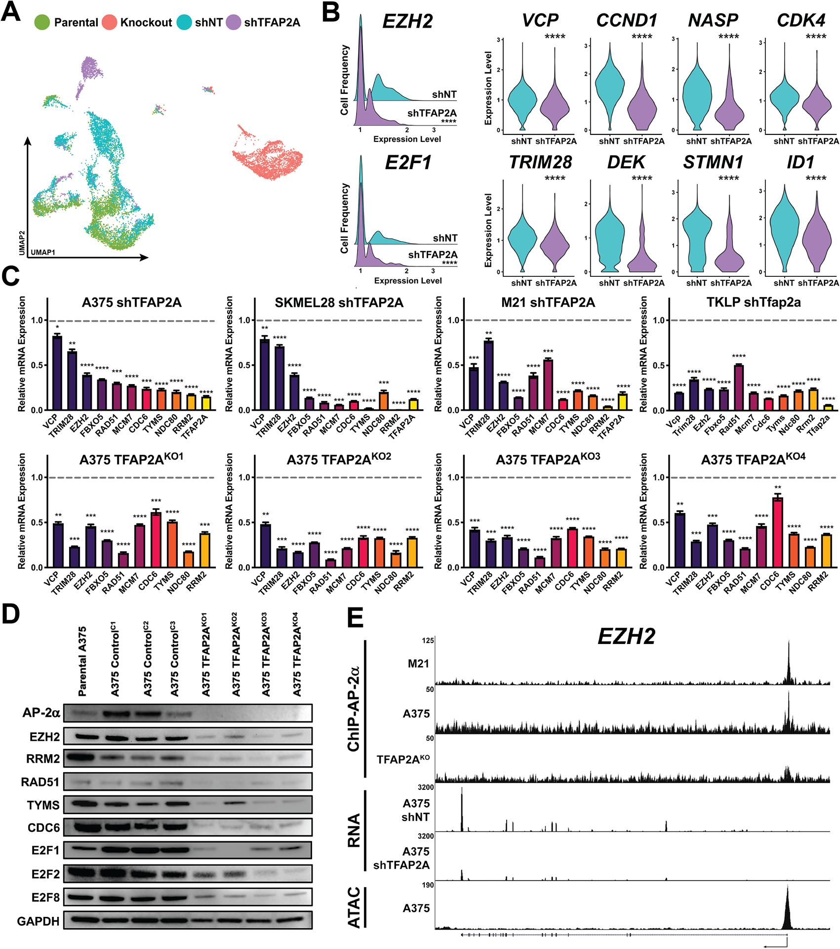
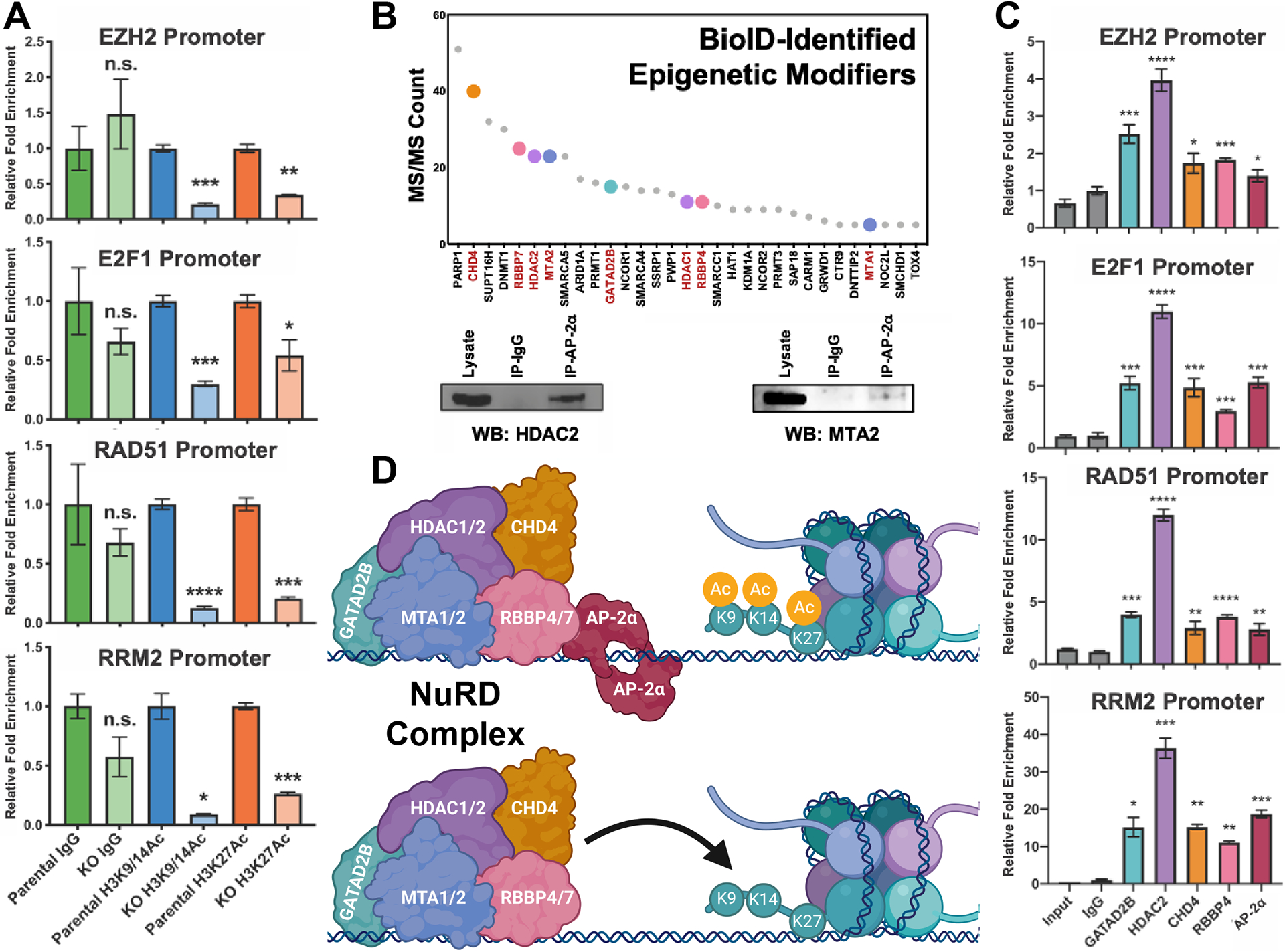
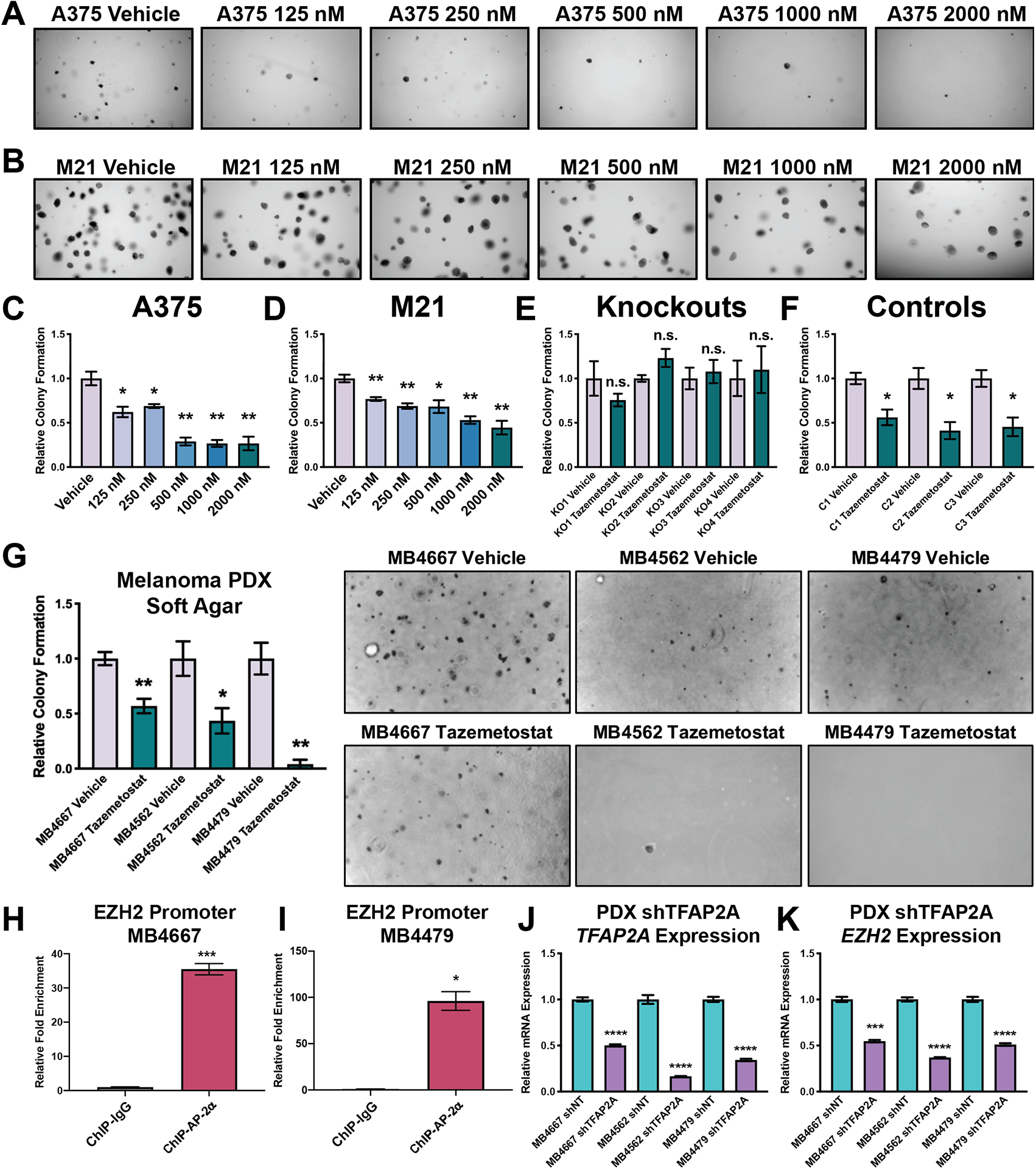
In melanoma metastasis, the role of the AP-2α transcription factor, which is encoded by TFAP2A, is controversial as some findings have suggested tumor suppressor activity while other studies have shown high TFAP2A expression in node-positive melanoma associated with poor prognosis. Here we demonstrate that AP-2α facilitates melanoma metastasis through transcriptional activation of genes within the E2F pathway including EZH2. A BioID screen found that AP-2α interacts with members of the nucleosome remodeling and deacetylase (NuRD) complex. Loss of AP-2α removed activating chromatin marks in the promoters of EZH2 and other E2F target genes through activation of the NuRD repression complex. In melanoma cells, treatment with tazemetostat, an FDA-approved and highly specific EZH2 inhibitor, substantially reduced anchorage-independent colony formation and demonstrated heritable antimetastatic effects, which were dependent on AP-2α. Single-cell RNA sequencing analysis of a metastatic melanoma mouse model revealed hyperexpansion of Tfap2a High/E2F-activated cell populations in transformed melanoma relative to progenitor melanocyte stem cells. These findings demonstrate that melanoma metastasis is driven by the AP-2α/EZH2 pathway and suggest that AP-2α expression can be used as a biomarker to predict responsiveness to EZH2 inhibitors for the treatment of advanced melanomas. SIGNIFICANCE: AP-2α drives melanoma metastasis by upregulating E2F pathway genes including EZH2 through inhibition of the NuRD repression complex, serving as a biomarker to predict responsiveness to EZH2 inhibitors.


Conflict of interest statement

All authors declare there are no competing financial interests in relation to the work described.

Figures

Figure 1.
Figure 1.. Loss of AP-2α Inhibits Melanoma Metastasis and Anchorage Independent Colony Formation.
CRISPR/Cas9 knockout of TFAP2A prevents A375 human melanoma cells from metastasizing in a subcutaneous xenograft model, and either CRISPR/Cas9 knockout or shRNA knockdown of TFAP2A prevents multiple human melanoma cell lines from forming colonies in soft agar. A. Western blot showing successful CRISPR/Cas9 mediated disruption of TFAP2A in four A375 clones results in a complete lack of AP-2α protein. B. Quantification of in vivo bioluminescence imaging of the primary flank tumors of mice xenografted with either the parental A375 cell line (red, n=3) or TFAP2AKO2 (blue, n=4). n.s. denotes p>0.05 (Student’s T-Test). C. Quantification of in vivo bioluminescence imaging of the thoracic/abdominal cavities of mice xenografted with either the parental A375 cell line (red, n=3) or TFAP2AKO2 (blue, n=4). *** denotes p<0.001 (Student’s T-test). D. In situ visualization of thoracic/abdominal metastatic luminescence. E. Representative images of H&E and Ki-67 staining of lungs, livers, and axilla of xenografted mice further confirm loss of AP-2α inhibits metastasis of A375 melanoma cells. F. Ex vivo imaging of livers (above) and lungs (below) of xenografted mice confirms in vivo luminescence corresponds to metastases to these distant organs. G. Soft agar assays show CRISPR/Cas9 mediated knockout or shRNA mediated knockdown substantially reduce the ability of multiple human melanoma cell lines to form anchorage independent colonies in soft agar (1,250 cells per replicate, n=3). Data are represented as mean ± SEM. ** denotes p<0.01, * denotes p<0.05 (Student’s T-test). See also Figure S1.
Figure 2.
Figure 2.. Loss of AP-2α Represses E2F Signaling.
RNA-seq reveals a gene signature shared among four TFAP2A KOs and an shRNA-mediated knockdown that is consistent with inhibition of E2F signaling. A. Heat map illustrating all consistent and statistically significant (per Cuffdiff) differential expression; selected genes within the E2F pathways are highlighted. Differential expression with knockout of TFAP2A was calculated relative to the parental cell line and differential expression with shTFAP2A (denoted shRNA) was calculated relative to a non-targeting (shNT) control. B. RT-qPCRs showing loss of TFAP2A represses E2F1, E2F2, and E2F8 in four TFAP2A KO clones (TFAP2AKO1-4 relative to the parental cell line, dashed line) and three human (A375, SKMEL28, and M21) and one mouse (TKLP) melanoma cell lines by shRNA (relative to a shNT control, dashed line). Data are represented as mean ± SEM. ** denotes p<0.01, *** denotes p<0.001, **** denotes p<0.0001 (Student’s T-Test). C. Propidium iodide staining showing knockout of TFAP2A disrupts cell cycle progression. See also Figure S2. D. 10X Genomics scRNA-seq of the A375 cell line shows a high degree of heterogeneity (left) and violin plots show E2F signaling, as epitomized by EZH2 and E2F1, is active in four out of six clusters (right). Clustering and violin plots were made with the Seurat package for R.
Figure 3.
Figure 3.. scRNA-seq Shows AP-2α-Dependent E2F Pathway Activation.
Loss of AP-2α reduces the proportion and extent to which cells express genes within the E2F pathways as evidenced by scRNA-seq. A. Monocle3 UMAP clustering showing dispersion of four scRNA-seq libraries derived from the A375 cell line. B. (Left) Seurat ridge plots showing the presence of cellular subpopulations highly expressing EZH2 or E2F1; loss of AP-2α causes transcriptional repression in these subpopulations. (Right) Seurat violin plots showing loss of AP-2α drives the transcriptional repression of additional E2F pathway members. **** denotes p<0.0001 (MAST differential expression analysis). See also Figure S3. C. RT-qPCR shows shRNA targeting TFAP2A represses selected E2F pathway members consistently in three human (A375, SKMEL28, M21) and one mouse (TKLP) melanoma cell line (relative to shNT controls, dashed lines), and this repression is reproducible in four A375 TFAP2A knockouts (A375 TFAP2AKO1-4 normalized to expression in the parental cell line, dashed line). Data are represented as mean ± SEM. * denotes p<0.05, ** denotes p<0.01, *** denotes p<0.001, **** denotes p<0.0001 (Student’s T-test). D. Western blots showing repression of E2F pathway members occurs consistently in all four TFAP2A knockout clones (A375 TFAP2AKO1-4), but not AP-2α-positive control clones derived from the A375 cell line (A375 ControlC1–C3). E. ChIP-seq shows AP-2α binds the promoters of E2F pathway genes as exemplified by the EZH2 gene. Occupancy is consistent among both the M21 and A375 human melanoma cell lines. ChIP-seq peak is absent in the TFAP2A knockout negative control. RNA-seq alignment highlights a reduction in reads aligning to E2F pathway genes consistent with repression following shRNA-mediated knockdown of TFAP2A. ATAC-seq peak shows accessible chromatin corresponding to the site of AP-2α occupancy at this E2F pathway promoter. The ATAC-seq dataset is from (32).
Figure 4.
Figure 4.. AP-2α Occupancy Drives Hyper-Acetylation of E2F Pathway Promoters Via an Interaction with the NuRD Complex.
The E2F pathway repression stemming from loss of AP-2α is associated with deceased Histone H3 acetylation at sites of promoter co-occupancy with the NuRD Complex. A. ChIP-qPCR showing decreased Histone H3K9/K14 and H3K27 acetylation following knockout of AP-2α at amplicons underlying AP-2α ChIP-seq peaks. Data are represented as mean ± SEM. * denotes p<0.05, ** denotes p<0.01, *** denotes p<0.001, **** denotes p<0.0001 (Student’s T-test). B. (Top) BioID determines AP-2α interacts with multiple epigenetic modifiers, including members of the NuRD Complex (colored dots, red font). (Bottom) Co-immunoprecipitations confirming the specificity of AP-2α interactions with HDAC2 and MTA2, members of the NuRD Complex. C. ChIP-qPCRs showing AP-2α co-occupies E2F Pathway promoters alongside members of the NuRD Complex. Data are represented as mean ± SEM. * denotes p<0.05, ** denotes p<0.01, *** denotes p<0.001, **** denotes p<0.0001 (Student’s T-test). See also Figure S5. D. Graphical schematic (made with biorender.com) of AP-2α-mediated NuRD Complex inhibition that results in the hyper-acetylation of E2F pathway promoter nucleosomes.
Figure 5.
Figure 5.. EZH2 Inhibition Prevents Anchorage Independent Colony Formation of AP-2α-Positive Melanoma.
Tazemetostat, an FDA approved EZH2 inhibitor, significantly reduces the ability of AP-2α-positive melanoma, but not TFAP2A knockouts, to form colonies in soft agar. A. A dosage-dependent reduction of anchorage independent colonies with tazemetostat treatment in the A375 human melanoma cell line. B. A dosage-dependent reduction of anchorage independent colonies with tazemetostat treatment in the M21 human melanoma cell line. C. Quantification of soft agar assay shown in Figure 6A. Data are represented as mean ± SEM (n=3). * denotes p<0.05, ** denotes p<0.01, (Student’s T-test). D. Quantification of soft agar assay shown in Figure 6B. Data are represented as mean ± SEM (n=3). * denotes p<0.05, ** denotes p<0.01. E. TFAP2A KO clones do not show additional reduction in anchorage independent colony formation with tazemetostat treatment. Data are represented as mean ± SEM (n=3). n.s. denotes p>0.05 (Student’s T-test). See also Figure S6A. F. AP-2α-positive control clones retain tazemetostat responsiveness. * denotes p<0.05 (Student’s T-test). See also Figure S6B. G. Tazemetostat treatment reduces the ability of melanoma PDXs to form colonies in soft agar. Data are represented as mean ± SEM (n=3). * denotes p<0.05, ** denotes p<0.01 (Student’s T-test). H.-I. AP-2α occupies the promoter of EZH2 in melanoma PDXs, paralleling the A375 and M21 cell line observations. Data are represented as mean ± SEM. * denotes p<0.05, *** denotes p<0.001 (Student’s T-test). J.-K. RT-qPCR results showing shRNA-mediated knockdown of TFAP2A represses EZH2 in three melanoma PDXs. Data are represented as mean ± SEM. *** denotes p<0.001, **** denotes p<0.0001 (Student’s T-test).
Figure 6.
Figure 6.. Tazemetostat Inhibits Melanoma Metastasis.
The EZH2 inhibitor tazemetostat heritably inhibits melanoma metastasis and improves survival duration of xenografted mice. See also Figure S7. A. In vivo bioluminescence imaging of mice four weeks post-xenograft, prior to surgeries to remove primary flank tumors, shows substantial metastases manifesting as contralateral luminescence in vehicle treated animals (n=4) but not in tazemetostat treated animals (n=6). B. Quantification of the in vivo luminescence from Figure 7A. Horizontal bars represent medians. * denotes p<0.05 (Student’s T-test). C. Photographs of the abdominal cavities of a vehicle treated animal (taken at euthanasia, 35 days post-xenograft) and a tazemetostat treated animal (taken at euthanasia, 61 days post-xenograft). D. Kaplan-Meyer curve showing significantly increased duration of survival of tazemetostat treated animals. E. H3K27Me3 immunohistochemistry of representative primary tumors of mice treated with vehicle or tazemetostat. F. In vivo bioluminescence imaging showing secondary xenografts derived from primary tumors in Figure 7A exhibit a heritable inhibition of thoracic/abdominal metastasis in naïve animals. G. Quantification of thoracic/abdominal luminescence in Figure 7F. Horizontal bars represent medians. * denotes p<0.05 (Student’s T-test). H.-I. Flow cytometry shows a greater than 6-fold reduction of GFP-positive metastatic cells in the livers of naïve animals receiving tazemetostat treated secondary xenografts, confirming the in vivo bioluminescence results from distant metastases.
Figure 7.
Figure 7.. Tfap2aHigh/E2F Activated Cellular Subpopulations Arise During Melanomagenesis.
Alignment and processing of published scRNA-seq datasets (48) from melanocyte stem cells (McSC) or melanoma cells, both derived from Tyr-CreER:Braf CA/+;Ptenfl/fl transgenic mice highlighting expanded clusters of cells showing E2F signaling activation and highly expressing Tfap2a are unique to melanoma cells. All plots were made with the Seurat package for R. A. UMAP clustering of melanocyte stem cells and syngeneic melanoma cells. B. Feature plot showing clusters of cells highly expressing Tfap2a are unique to transformed melanoma cells. C.-D. Feature plot showing the Tfap2aHigh clusters also highly express Ezh2 and E2f1; these clusters are not expanded in the untransformed melanocyte stem cell libraries (McSC). E.-F. AP-2α ChIP-seq performed in TKLP mouse melanoma cells shows AP-2α occupancy of the promoters of Ezh2 and E2f1 is phylogenetically conserved. See also Figure S8B. G. Violin plots of additional examples supporting activation of E2F signaling is unique to transformed melanoma cells. In all figures, **** denotes p<0.0001 (MAST differential expression analysis). See also Figure S8A.

Similar articles

Cited by

References

    1. Gershenwald JE, Scolyer RA, Hess KR, Sondak VK, Long GV, Ross MI, et al.Melanoma staging: Evidence-based changes in the American Joint Committee on Cancer eighth edition cancer staging manual. CA Cancer J Clin 2017;67:472–92 - PMC - PubMed
    1. Sandru A, Voinea S, Panaitescu E, Blidaru A. Survival rates of patients with metastatic malignant melanoma. J Med Life 2014;7:572–6 - PMC - PubMed
    1. Spagnolo F, Boutros A, Tanda E, Queirolo P. The adjuvant treatment revolution for high-risk melanoma patients. Semin Cancer Biol 2019;59:283–9 - PubMed
    1. Barrallo-Gimeno A, Holzschuh J, Driever W, Knapik EW. Neural crest survival and differentiation in zebrafish depends on mont blanc/tfap2a gene function. Development 2004;131:1463–77 - PubMed
    1. Seberg HE, Van Otterloo E, Loftus SK, Liu H, Bonde G, Sompallae R, et al.TFAP2 paralogs regulate melanocyte differentiation in parallel with MITF. PLoS Genet 2017;13:e1006636. - PMC - PubMed

Publication types

MeSH terms