

Flavivirus: From Structure to Therapeutics Development
- PMID: 34202239
- PMCID: PMC8303334
- DOI: 10.3390/life11070615
Flavivirus: From Structure to Therapeutics Development
Abstract
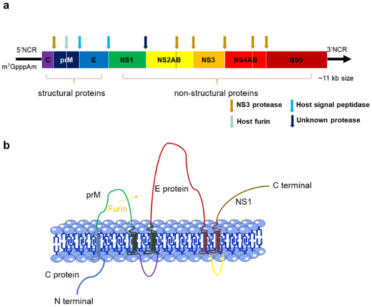
Flaviviruses are still a hidden threat to global human safety, as we are reminded by recent reports of dengue virus infections in Singapore and African-lineage-like Zika virus infections in Brazil. Therapeutic drugs or vaccines for flavivirus infections are in urgent need but are not well developed. The Flaviviridae family comprises a large group of enveloped viruses with a single-strand RNA genome of positive polarity. The genome of flavivirus encodes ten proteins, and each of them plays a different and important role in viral infection. In this review, we briefly summarized the major information of flavivirus and further introduced some strategies for the design and development of vaccines and anti-flavivirus compound drugs based on the structure of the viral proteins. There is no doubt that in the past few years, studies of antiviral drugs have achieved solid progress based on better understanding of the flavivirus biology. However, currently, there are no fully effective antiviral drugs or vaccines for most flaviviruses. We hope that this review may provide useful information for future development of anti-flavivirus drugs and vaccines.
Keywords: drug target; epidemics; flavivirus; immunology; protein structure; vaccine.
Conflict of interest statement
The authors declare that they have no competing interest in this article.
Figures
Similar articles
-
Structure and function of capsid protein in flavivirus infection and its applications in the development of vaccines and therapeutics.Vet Res. 2021 Jun 30;52(1):98. doi: 10.1186/s13567-021-00966-2. Vet Res. 2021. PMID: 34193256 Free PMC article. Review.
-
Establishment and Application of Flavivirus Replicons.Adv Exp Med Biol. 2018;1062:165-173. doi: 10.1007/978-981-10-8727-1_12. Adv Exp Med Biol. 2018. PMID: 29845532 Review.
-
Zika Virus Subgenomic Flavivirus RNA Generation Requires Cooperativity between Duplicated RNA Structures That Are Essential for Productive Infection in Human Cells.J Virol. 2020 Aug 31;94(18):e00343-20. doi: 10.1128/JVI.00343-20. Print 2020 Aug 31. J Virol. 2020. PMID: 32581095 Free PMC article.
-
An Envelope-Modified Tetravalent Dengue Virus-Like-Particle Vaccine Has Implications for Flavivirus Vaccine Design.J Virol. 2017 Nov 14;91(23):e01181-17. doi: 10.1128/JVI.01181-17. Print 2017 Dec 1. J Virol. 2017. PMID: 28956764 Free PMC article.
-
Zika virus structural biology and progress in vaccine development.Biotechnol Adv. 2018 Jan-Feb;36(1):47-53. doi: 10.1016/j.biotechadv.2017.09.004. Epub 2017 Sep 12. Biotechnol Adv. 2018. PMID: 28916391 Review.
Cited by
-
Single-cell RNA sequencing to understand host-virus interactions.Virol Sin. 2024 Feb;39(1):1-8. doi: 10.1016/j.virs.2023.11.009. Epub 2023 Nov 25. Virol Sin. 2024. PMID: 38008383 Free PMC article. Review.
-
Therapeutic Plasma Exchange for Murray Valley Encephalitis: A Case Report and Review of Clinical Features, Diagnosis, and Challenges in Management.Cureus. 2024 Sep 19;16(9):e69696. doi: 10.7759/cureus.69696. eCollection 2024 Sep. Cureus. 2024. PMID: 39429370 Free PMC article.
-
Antiviral Agents against Flavivirus Protease: Prospect and Future Direction.Pathogens. 2022 Feb 25;11(3):293. doi: 10.3390/pathogens11030293. Pathogens. 2022. PMID: 35335617 Free PMC article. Review.
-
Exploring Rigid and Flexible Scaffolds to Develop Potent Glucuronic Acid Glycodendrimers for Dengue Virus Inhibition.Bioconjug Chem. 2024 Jan 17;35(1):34-42. doi: 10.1021/acs.bioconjchem.3c00309. Epub 2023 Nov 15. Bioconjug Chem. 2024. PMID: 37964742 Free PMC article.
-
Repurposing of FDA-approved drugs against oligomerization domain of dengue virus NS1 protein: a computational approach.Mol Divers. 2024 Jul 17. doi: 10.1007/s11030-024-10936-3. Online ahead of print. Mol Divers. 2024. PMID: 39017952
References
-
- Dethoff E.A., Boerneke M.A., Gokhale N.S., Muhire B.M., Martin D.P., Sacco M.T., McFadden M.J., Weinstein J.B., Messer W.B., Horner S.M., et al. Pervasive tertiary structure in the dengue virus RNA genome. Proc. Natl. Acad. Sci. USA. 2018;115:11513–11518. doi: 10.1073/pnas.1716689115. - DOI - PMC - PubMed
-
- Kummerer B.M. Establishment and application of flavivirus replicons. Adv. Exp. Med. Biol. 2018;1062:165–173. - PubMed
-
- Martin E., Borucki M.K., Thissen J., Garcia-Luna S., Hwang M., de Valdez M.W., Jaing C.J., Hamer G.L., Frank M. Mosquito-Borne viruses and insect-specific viruses revealed in field-collected mosquitoes by a monitoring tool adapted from a microbial detection array. Appl. Environ. Microb. 2019;85:e01202-19. doi: 10.1128/AEM.01202-19. - DOI - PMC - PubMed
Publication types
Grants and funding
LinkOut - more resources
Full Text Sources
Miscellaneous
