Review
. 2021 Jun 23;22(13):6714.
doi: 10.3390/ijms22136714.

NOD-Like Receptors: Guards of Cellular Homeostasis Perturbation during Infection

Affiliations
Review

NOD-Like Receptors: Guards of Cellular Homeostasis Perturbation during Infection

Gang Pei et al. Int J Mol Sci. .

Abstract

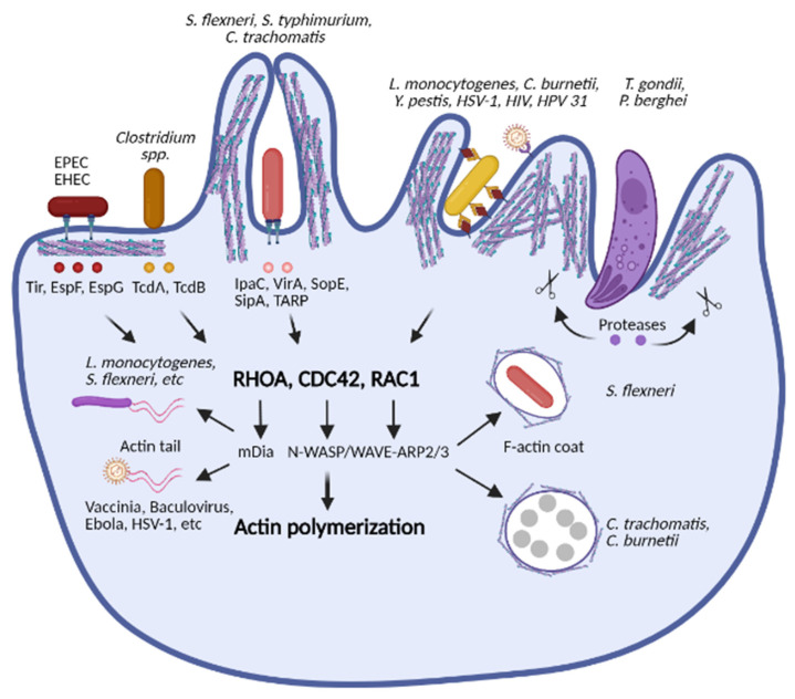
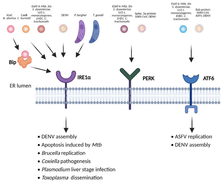
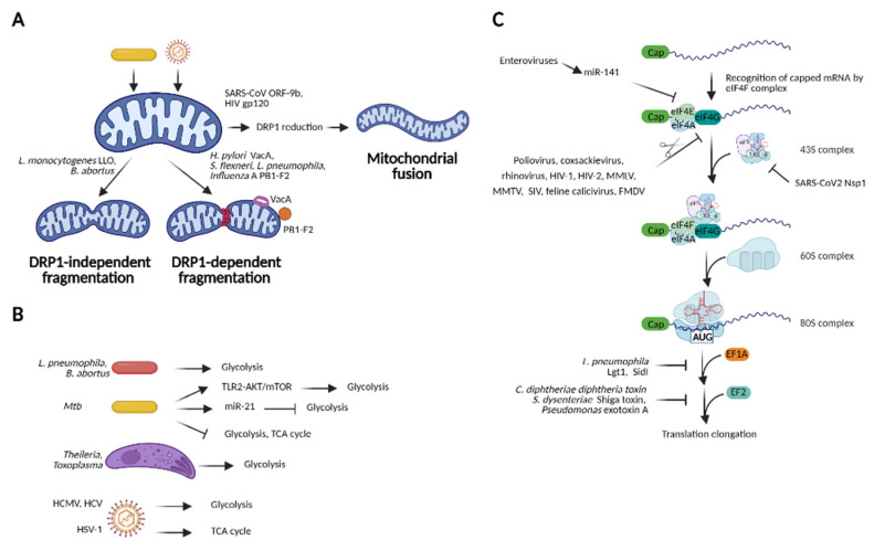
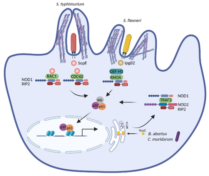
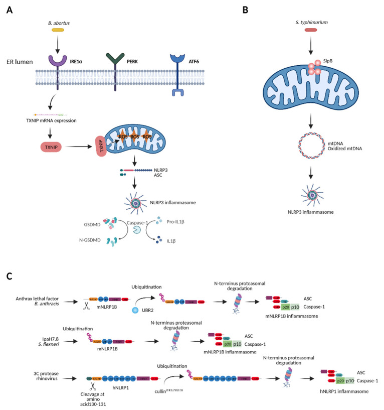
The innate immune system relies on families of pattern recognition receptors (PRRs) that detect distinct conserved molecular motifs from microbes to initiate antimicrobial responses. Activation of PRRs triggers a series of signaling cascades, leading to the release of pro-inflammatory cytokines, chemokines and antimicrobials, thereby contributing to the early host defense against microbes and regulating adaptive immunity. Additionally, PRRs can detect perturbation of cellular homeostasis caused by pathogens and fine-tune the immune responses. Among PRRs, nucleotide binding oligomerization domain (NOD)-like receptors (NLRs) have attracted particular interest in the context of cellular stress-induced inflammation during infection. Recently, mechanistic insights into the monitoring of cellular homeostasis perturbation by NLRs have been provided. We summarize the current knowledge about the disruption of cellular homeostasis by pathogens and focus on NLRs as innate immune sensors for its detection. We highlight the mechanisms employed by various pathogens to elicit cytoskeleton disruption, organelle stress as well as protein translation block, point out exemplary NLRs that guard cellular homeostasis during infection and introduce the concept of stress-associated molecular patterns (SAMPs). We postulate that integration of information about microbial patterns, danger signals, and SAMPs enables the innate immune system with adequate plasticity and precision in elaborating responses to microbes of variable virulence.

Keywords: NLRP1; NLRP3; NOD-like receptors; NOD1/2; cellular homeostasis; innate immunity; pathogens.


Conflict of interest statement

The authors declare no conflict of interest.

Figures

Figure 1
Figure 1
Pathogens cause cytoskeleton disruption. Bacteria and viruses subvert host cytoskeleton dynamics for their entry, intracellular survival and dissemination. They employ various effector molecules, toxins, or viral proteins to induce actin remodeling by manipulating Rho GTPases (RHOA, CDC42, and RAC1), which are the central regulators of the actin polymerization. Parasites such as Toxoplasma gondii and Plasmodium berghei also induce actin rearrangement by binding or cleaving actin-associated factors. Abbreviations: RHOA, Ras homolog family member A; CDC42, cell division control protein 42 homolog; RAC1, Ras-related C3 botulinum toxin substrate 1; mDia, mammalian homolog of Diaphanous; N-WASP, neuronal Wiskott-Aldrich Syndrome protein; WAVE, Wiskott-Aldrich syndrome protein family verprolin-homologous; ARP2/3, actin related protein 2/3; TirA, translocated intimin receptor A; EspF, enteropathogenic E. coli effector protein F; EspG, enteropathogenic E. coli effector protein G; TcdA, Clostridium difficile toxin A; TcdB, C. difficile toxin B; IpaC, invasion plasmid antigen C; VirA, virulence factor A; SopE, salmonella outer protein E; SipA, salmonella invasion protein A; TARP, type III secretion system actin-recruiting effector. Image created with BioRender.com.
Figure 2
Figure 2
Various pathogens cause ER stress to modulate host cell death or facilitate their replication and dissemination. Unfolded protein responses (UPR) are mediated by the ER stress sensors protein kinase R-like ER kinase (PERK), inositol-requiring enzyme 1α (IRE1α) and activating transcription factor 6 (ATF6). The 6 kDa early secretory antigenic target (ESAT-6) from M. tuberculosis (Mtb), shiga toxin 1 (Stx) from S. dysenteriae, listeriolysin O (LLO) from L. monocytogenes, dengue virus (DENV), etc. induce all three branches of UPR. VceC, a T4SS effector from B. abortus, binds to the ER chaperon binding immunoglobulin protein (BiP) and triggers IRE1α signaling. CaeB (C. burnetii anti-apoptotic effector B) from C. burnetii stimulates IRE1α signaling, facilitating its pathogenesis. The severe acute respiratory syndrome coronavirus (SARS-CoV) induces PERK and ATF6 signaling and the African Swine Fever Virus (ASFV) activates only ATF6. Image created with BioRender.com.
Figure 3
Figure 3
Mitochondrial dysfunction and blockade of host translation induced by pathogens. (A) Manipulation of mitochondrial dynamics by pathogens. Pathogenic viruses and bacteria cause mitochondrial fragmentation or mitochondrial fusion for replication, persistence in host cell or dissemination; (B) Modulation of host metabolism by pathogens. To acquire the nutrient from the host, pathogens trigger glycolysis or modulate the TCA cycle dependent on their metabolic requirements; (C) Inhibition of host translation by pathogens. To shut off host defense, many viruses such as poliovirus, coxsackievirus and rhinovirus, HIV-1, enteroviruses, and SARS-CoV2 target the eIF4F complex to inhibit translation initiation, while some bacterial pathogens utilize effector proteins or toxins to inactivate translation elongation factors-EF1A and EF2. Abbreviations: DRP1, dynamin related protein 1; VacA, vacuolating cytotoxin A; LLO, listeriolysin O; TLR2, toll-like receptor 2; AKT, protein kinase B; mTOR, mechanistic target of Rapamycin; TCA cycle, tricarboxylic acid cycle; HCMV, Human Cytomegalovirus; HCV, Hepatitis C Virus; HSV-1, Herpes simplex virus 1; HIV, Human immunodeficiency virus; MMLV, Moloney murine leukemia virus; MMTV, Mouse mammary tumor virus; SIV, Simian immunodeficiency virus; FMDV, Foot-and-mouth disease virus; eIF, eukaryotic translation initiation factor; EF1A/1B, eukaryotic translation elongation factor 1A/1B; Lgt1, Legionella glucosyltransferase 1; SidI, substrate I of Icm/Dot transporter; Nsp1, nonstructural protein 1. Image created with BioRender.com.
Figure 4
Figure 4
NOD1 and NOD2 trigger NF-κB activation by detecting pathogen-induced cytoskeleton disruption or pathogen-induced ER stress. Virulence factors from S. typhimurium and S. flexneri induce aberrant actin polymerization, subsequently triggering NOD1-mediated NF-κB activation. B. abortus and C. muridarum elicit ER stress, which is further sensed by NOD1 and NOD2 to enable NF-κB activation. Abbreviations: RHOA, Ras homolog family member A; CDC42, cell division control protein 42 homolog; RAC1, Ras-related C3 botulinum toxin substrate 1; GEF-H1, guanine nucleotide exchange factor H1; TRAF2, TNF receptor associated factor 2; RIP2, receptor-interacting-serine/threonine-protein kinase 2; IKB, inhibitor of κB; IRE1α, inositol-requiring enzyme-1a; IpgB2, invasion plasmid gene B2; SopE, Salmonella outer protein E; VceC, virB-coregulated effector C. Image created with BioRender.com.
Figure 5
Figure 5
Distinct NLRP inflammasomes are activated by ER stress, mitochondrial dysfunction or directly by pathogens. (A) B. abortus induces ER stress, which causes NLRP3 inflammasome activation. (B) S. typhimurium targets mitochondria to trigger NLRP3 inflammasome activation. (C) Murine and human NLRP1 (mNLRP1B, hNLRP1) are targeted by pathogen virulence factors. Abbreviations: PERK, PKR-like ER kinase; IRE1α, inositol-requiring enzyme 1α; ATF6, activating transcription factor 6; TXNIP, thioredoxin-interacting protein; ASC, apoptosis-associated Speck-like protein containing a CARD; GSDMD, Gasdermin D; UBR2, ubiquitin protein ligase E3 component N-Recognin 2; ROS, reactive oxygen species; mtDNA, mitochondrial DNA; SipB, Salmonella invasion protein B; IpaH7.8, invasion plasmid antigen H7.8. Image created with BioRender.com.

Similar articles

Cited by

References

    1. Kanneganti T.D., Lamkanfi M., Nunez G. Intracellular NOD-like receptors in host defense and disease. Immunity. 2007;27:549–559. doi: 10.1016/j.immuni.2007.10.002. - DOI - PubMed
    1. Motta V., Soares F., Sun T., Philpott D.J. NOD-like receptors: Versatile cytosolic sentinels. Physiol. Rev. 2015;95:149–178. doi: 10.1152/physrev.00009.2014. - DOI - PubMed
    1. Martin B.K., Chin K.C., Olsen J.C., Skinner C.A., Dey A., Ozato K., Ting J.P. Induction of MHC class I expression by the MHC class II transactivator CIITA. Immunity. 1997;6:591–600. doi: 10.1016/S1074-7613(00)80347-7. - DOI - PubMed
    1. Masternak K., Muhlethaler-Mottet A., Villard J., Zufferey M., Steimle V., Reith W. CIITA is a transcriptional coactivator that is recruited to MHC class II promoters by multiple synergistic interactions with an enhanceosome complex. Genes Dev. 2000;14:1156–1166. - PMC - PubMed
    1. Yang J., Zhao Y., Shi J., Shao F. Human NAIP and mouse NAIP1 recognize bacterial type III secretion needle protein for inflammasome activation. Proc. Natl. Acad. Sci. USA. 2013;110:14408–14413. doi: 10.1073/pnas.1306376110. - DOI - PMC - PubMed

MeSH terms

Substances

LinkOut - more resources