. 2021 Jun 5;26(11):3430.
doi: 10.3390/molecules26113430.

In Vitro and In Silico Anti-Arboviral Activities of Dihalogenated Phenolic Derivates of L-Tyrosine

Affiliations

In Vitro and In Silico Anti-Arboviral Activities of Dihalogenated Phenolic Derivates of L-Tyrosine

Vanessa Loaiza-Cano et al. Molecules. .

Abstract

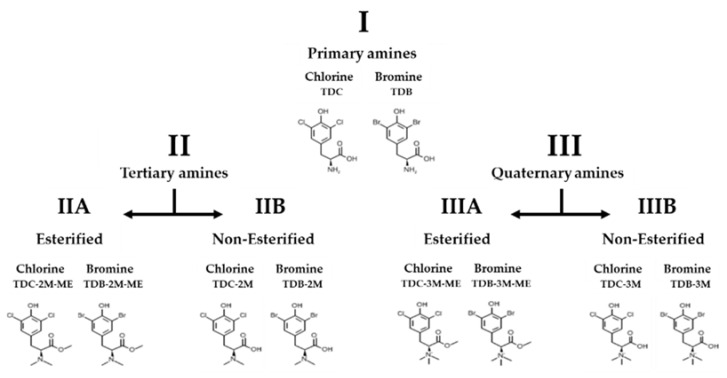
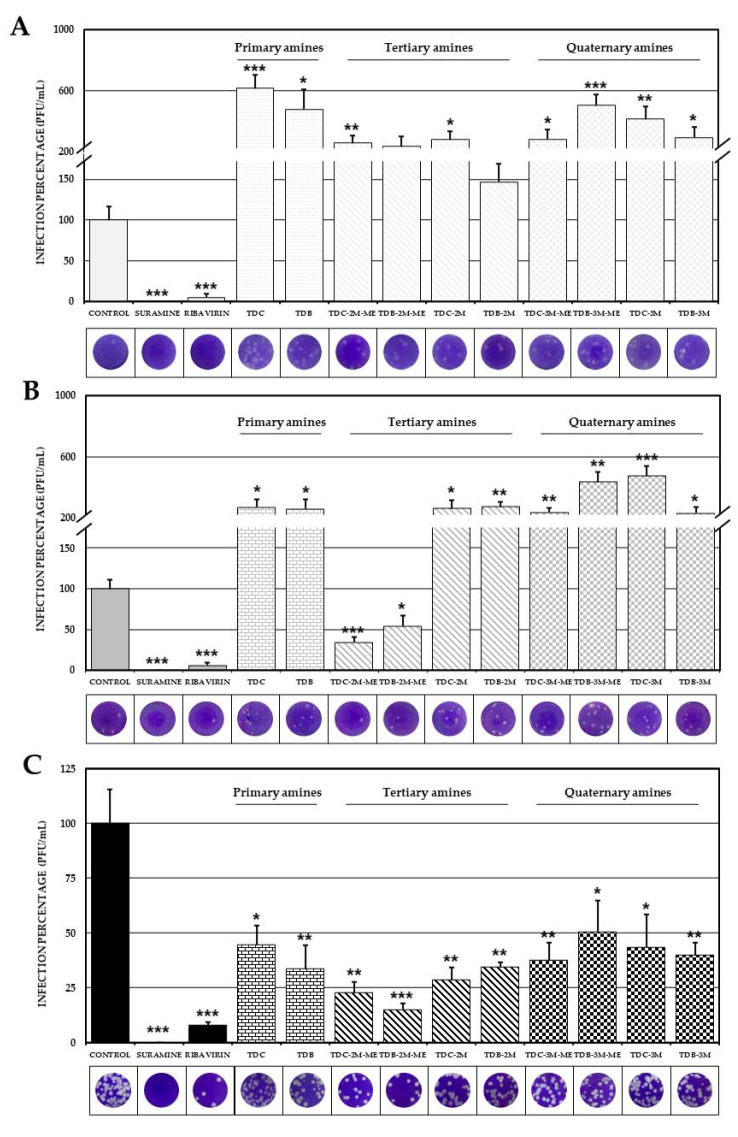
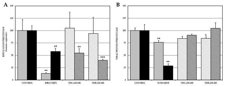
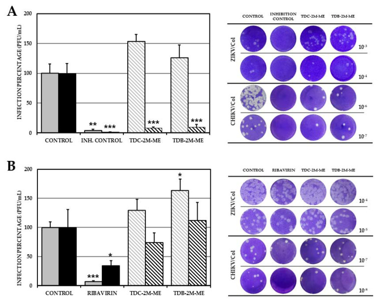
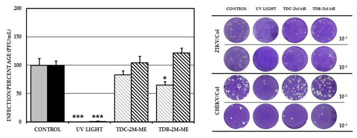
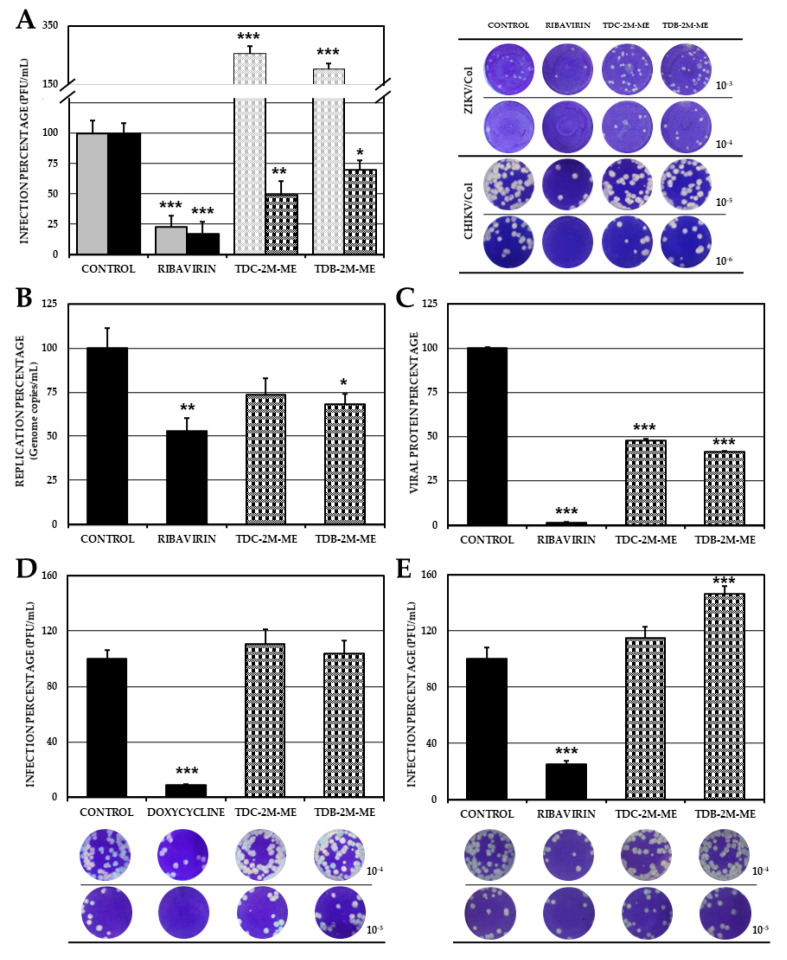
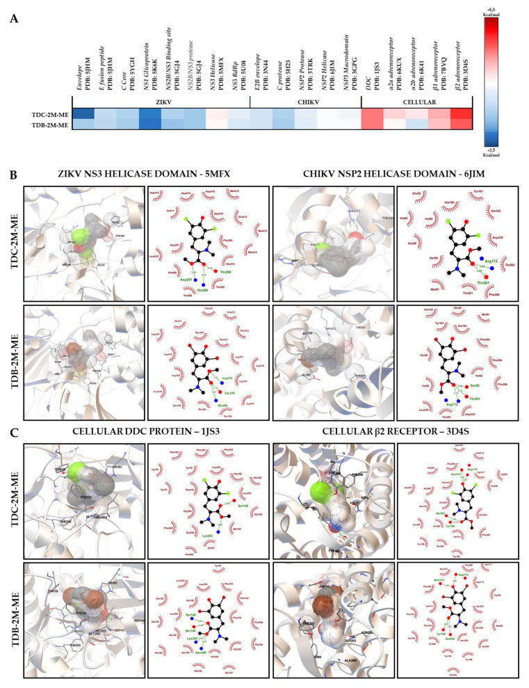
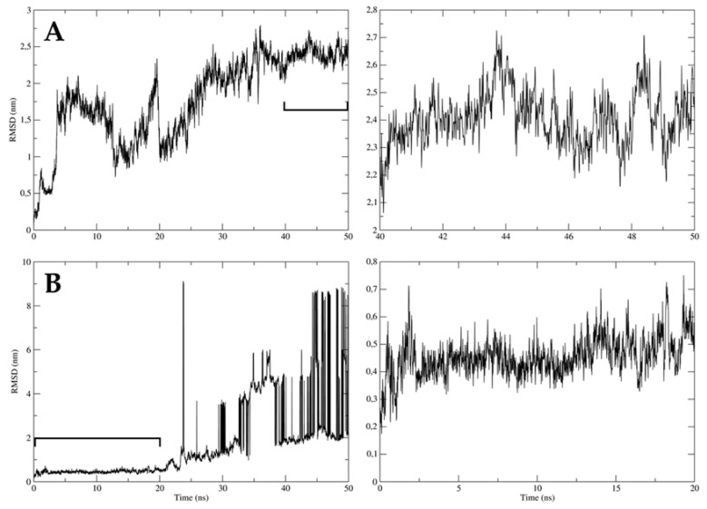
Despite the serious public health problem represented by the diseases caused by dengue (DENV), Zika (ZIKV) and chikungunya (CHIKV) viruses, there are still no specific licensed antivirals available for their treatment. Here, we examined the potential anti-arbovirus activity of ten di-halogenated compounds derived from L-tyrosine with modifications in amine and carboxyl groups. The activity of compounds on VERO cell line infection and the possible mechanism of action of the most promising compounds were evaluated. Finally, molecular docking between the compounds and viral and cellular proteins was evaluated in silico with Autodock Vina®, and the molecular dynamic with Gromacs®. Only two compounds (TDC-2M-ME and TDB-2M-ME) inhibited both ZIKV and CHIKV. Within the possible mechanism, in CHIKV, the two compounds decreased the number of genome copies and in the pre-treatment strategy the infectious viral particles. In the ZIKV model, only TDB-2M-ME inhibited the viral protein and demonstrate a virucidal effect. Moreover, in the U937 cell line infected with CHIKV, both compounds inhibited the viral protein and TDB-2M-ME inhibited the viral genome too. Finally, the in silico results showed a favorable binding energy between the compounds and the helicases of both viral models, the NSP3 of CHIKV and cellular proteins DDC and β2 adrenoreceptor.

Keywords: Zika virus; antiviral agents; chikungunya virus; computational biology; dengue virus; tyrosine.


Conflict of interest statement

The authors declare no conflict of interest.

Figures

Figure 1
Figure 1
Classification of evaluated compounds. Ten phenolic dihalogenated compounds derived from L-tyrosine were synthetized, then were classified into three groups as per the substitution in their amine group: I (Primary amines), II (Tertiary amines) and III (Quaternary amines). Finally, groups II and III were divided into sub-groups according to the carboxyl substitution with a methyl group (A or B, with or without esterification, respectively). Each sub-group has one di-chlorinated compound and one di-brominated compound.
Figure 2
Figure 2
Effect of compounds on the production of infectious viral particles. Percentages of infection calculated according to the results obtained by plaque assay (PFU/ml) of the supernatants collected from the antiviral screening of the compounds in VERO cells infected with DENV-2/S16803 (A), ZIKV/Col (B) or CHIKV/Col (C). In all cases, the control without treatment was assumed as 100% infection. The asterisks indicate statistically significant differences with respect to the control without compound (* p < 0.05, ** p < 0.01 and *** p < 0.001; t-Student) and error bars indicate standard error of the mean; n: 6. Moreover, are shown representative plaques of titration on VERO cells corresponding to each experimental condition.
Figure 3
Figure 3
Effect of the compounds TDC-2M-ME and TDB-2M-ME on viral replication and translation in VERO cells. Percentage of viral genome replication obtained by real-time PCR (genome copies/mL) in VERO cells infected with ZIKV/Col (gray bars) or CHIKV/Col (black bars) at MOI 5, assuming the control without treatment as 100% infection (A). Percentage of viral protein of ZIKV/Col (gray) and CHIKV/Col (black) evaluated by Cell-ELISA (absorbance) (B). The asterisks indicate statistically significant differences with respect to the control without compound (* p <0.05, ** p <0.01 and *** p <0.001; t-Student) and error bars indicate standard error of the mean; n: 4.
Figure 4
Figure 4
Effect of the compounds TDC-2M-ME and TDB-2M-ME on the production of infectious viral particles according to the treatment strategy in VERO cells. Percentages of infection calculated according to the results obtained by plaque assay of the supernatants collected from VERO cells treated before (A) or after (B) the infection with ZIKV/Col (gray bars) or CHIKV/Col (black bars). The control without treatment was assumed as 100% infection. The asterisks indicate statistically significant differences with respect to the control without compound (* p < 0.05, ** p <0.01 and *** p <0.001; t-Student) and error bars indicate standard error of the mean; n: 6. Moreover, are shown representative plaques of titration on VERO cells corresponding to each experimental condition.
Figure 5
Figure 5
Virucidal effect of the compounds TDC-2M-ME and TDB-2M-ME on the production of infectious viral particles. Percentages of infection calculated according to the results obtained by plaque assay of VERO cells treated and simultaneously infected with ZIKV/Col (gray bars) or CHIKV/Col (black bars). The control without treatment was assumed as 100% infection. The asterisks indicate statistically significant differences with respect to the control without compound (* p < 0.05, and *** p < 0.001; t-Student) and error bars indicate standard error of the mean; n: 6. Moreover, are shown representative plaques of titration on VERO cells corresponding to each experimental condition.
Figure 6
Figure 6
Antiviral effects of the compounds TDC-2M-ME and TDB-2M-ME in U937 cells. Infection percentages of cell cultures treated by combined strategy were calculated by plaque assay (PFU/mL) of the supernatants (A), real-time PCR (genome copies/mL) (B) and Cell-ELISA (absorbance) (C) in U937 monolayers. Also, the supernatants obtained of pre-treatment (D) and post-treatment (E) antiviral strategies were evaluated by plaque assay (PFU/mL). Evaluation on ZIKV/Col or CHIKV/Col infected cells are shown in gray and black, respectively. The control without treatment was assumed as 100% infection. The asterisks indicate statistically significant differences with respect to the control without compound (* p < 0.05, ** p < 0.01 and *** p < 0.001; t-Student) and error bars indicate standard error of the mean; n: 4. Moreover, are shown representative plaques of titration on VERO cells corresponding to each experimental condition.
Figure 7
Figure 7
In silico interactions between viral and cellular proteins and the antiviral compounds. Heat map of binding energies. Free binding energies were obtained by molecular docking with AutodockVina® of TDC-2M-ME and TDB-2M-ME compounds with viral proteins of ZIKV and CHIKV and cellular proteins related with L-tyrosine derivatives. Less than 0 kcal/mol were considered favorable energies. The averages were graphed in color scale from blue to red. Blue scale shows less favorable energies (>−6 kcal/mol), and red scale the best binding energies (≤−6 kcal/mol). The limit was established by the concordance of the results obtained from Monsalve-Escudero et al., 2021, where the in vitro (antiviral and virucidal activity) and in silico (molecular docking and molecular dynamic) results were concordant. Each complex was evaluated by triplicate (A). Most relevant interactions were obtained after molecular docking between the promissory antivirals and the proteins with which the best binding energies were obtained, viral helicases (B) and cellular proteins DDC and β2 receptor (C). Both graphics evidence the interactions showed by two different software PMV (left) and LigPlot® (Right). Hydrogen bonds are shown in green in both software, hydrophobic interactions in red in LigPlot® and π–π interactions in yellow in PMV software.
Figure 8
Figure 8
Stability of TDB-2M-ME and ZIKV envelope proteins complexes. The stability of the complexes formed by the only virucidal compound, TDB-2M-ME, and ZIKV envelope proteins, DIII (A) and fusion peptide (B), were evaluated; DIII-E ZIKV (A). The left plots indicate the complete simulation (50 ns) and the right plot indicates the timescale with the lowest oscillation of each simulation, 40 to 50 ns (A) and 0 to 20 ns (B). The y-axis represents the root mean square deviation (RMSD) in nanometers (nm), and the x-axis the timescale in nanoseconds (ns). The complex is considered stable if the oscillation is below 0.3 nm.

Similar articles

Cited by

References

    1. Paixão E.S., Teixeira M.G., Rodrigues L.C. Zika, chikungunya and dengue: The causes and threats of new and re-emerging arboviral diseases. BMJ Glob. Health. 2018;3:e000530. doi: 10.1136/bmjgh-2017-000530. - DOI - PMC - PubMed
    1. Jones R., Kulkarni M.A., Davidson T.M., Team R.-L.R., Talbot B. Arbovirus vectors of epidemiological concern in the Americas: A scoping review of entomological studies on Zika, dengue and chikungunya virus vectors. PLoS ONE. 2020;15:e0220753. doi: 10.1371/journal.pone.0220753. - DOI - PMC - PubMed
    1. Epelboin Y., Talaga S., Epelboin L., Dusfour I. Zika virus: An updated review of competent or naturally infected mosquitoes. PLoS Negl. Trop. Dis. 2017;11:e0005933. doi: 10.1371/journal.pntd.0005933. - DOI - PMC - PubMed
    1. Carrillo-Hernández M.Y., Ruiz-Saenz J., Villamizar L.J., Gómez-Rangel S.Y., Martínez-Gutierrez M. Co-circulation and simultaneous co-infection of dengue, chikungunya, and zika viruses in patients with febrile syndrome at the Colombian-Venezuelan border. BMC Infect. Dis. 2018;18:61. doi: 10.1186/s12879-018-2976-1. - DOI - PMC - PubMed
    1. Cabral-Castro M.J., Cavalcanti M.G., Peralta R.H.S., Peralta J.M. Molecular and serological techniques to detect co-circulation of DENV, ZIKV and CHIKV in suspected dengue-like syndrome patients. J. Clin. Virol. 2016;82:108–111. doi: 10.1016/j.jcv.2016.07.017. - DOI - PubMed

MeSH terms