. 2021 Jul 15;81(14):2887-2900.e5.
doi: 10.1016/j.molcel.2021.06.002. Epub 2021 Jun 24.

Structural insights into the functional divergence of WhiB-like proteins in Mycobacterium tuberculosis

Affiliations

Structural insights into the functional divergence of WhiB-like proteins in Mycobacterium tuberculosis

Tao Wan et al. Mol Cell. .

Abstract

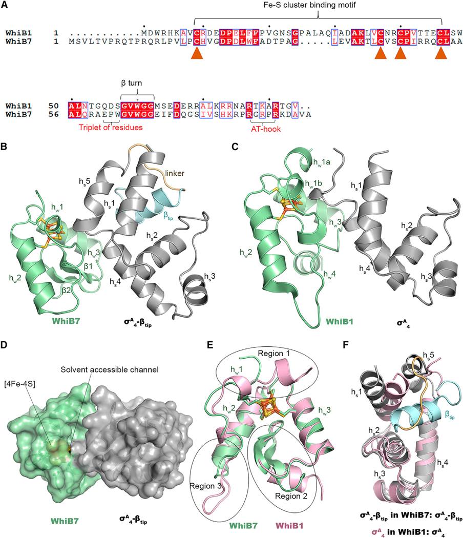
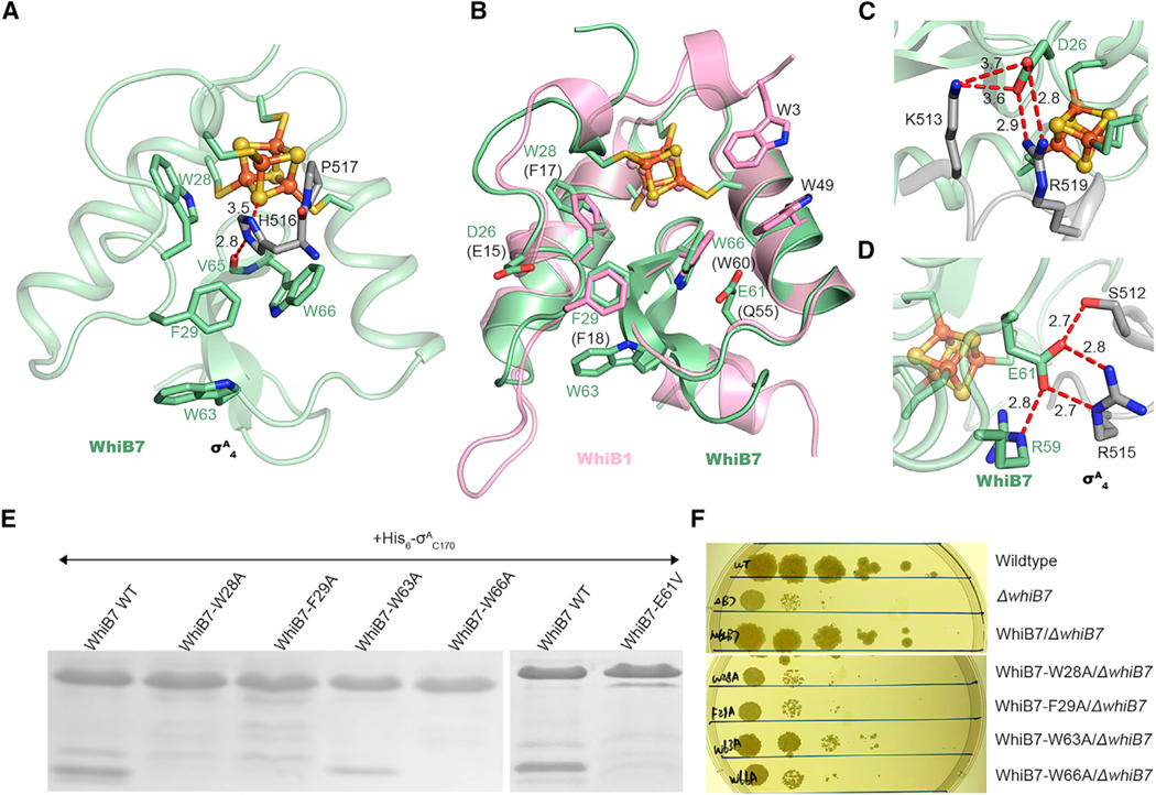
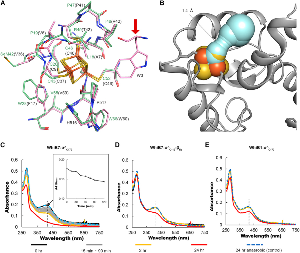
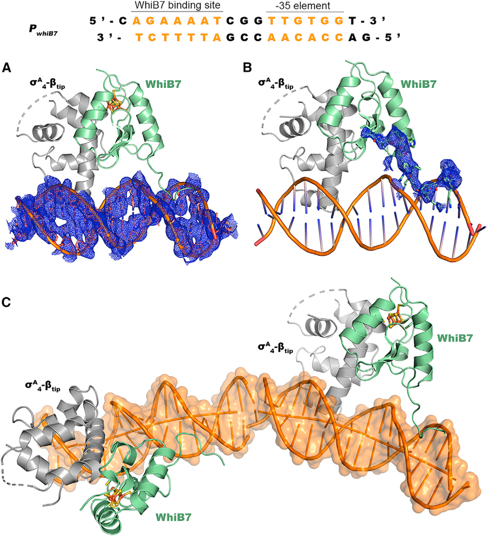
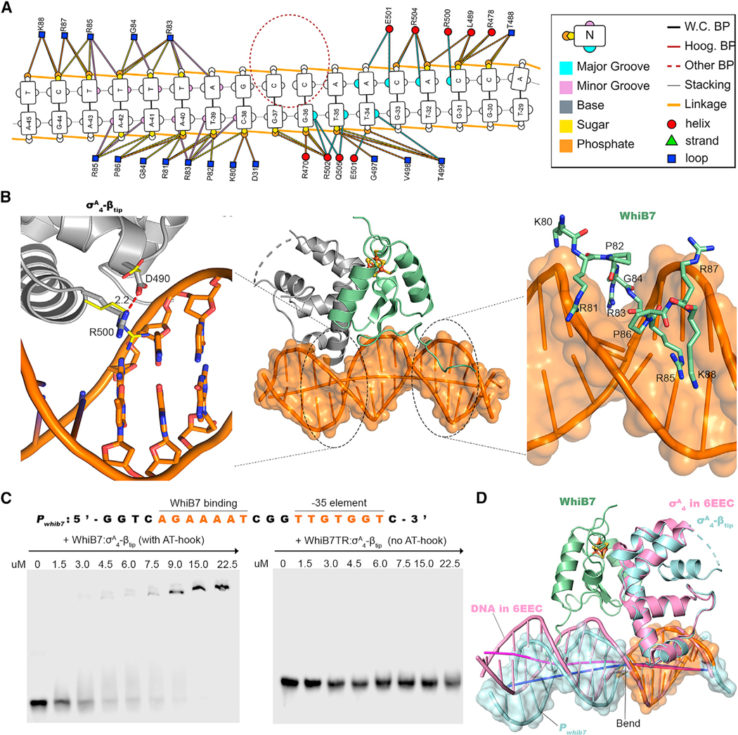
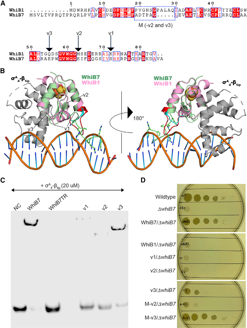
WhiB7 represents a distinct subclass of transcription factors in the WhiB-Like (Wbl) family, a unique group of iron-sulfur (4Fe-4S] cluster-containing proteins exclusive to the phylum of Actinobacteria. In Mycobacterium tuberculosis (Mtb), WhiB7 interacts with domain 4 of the primary sigma factor (σA4) in the RNA polymerase holoenzyme and activates genes involved in multiple drug resistance and redox homeostasis. Here, we report crystal structures of the WhiB7:σA4 complex alone and bound to its target promoter DNA at 1.55-Å and 2.6-Å resolution, respectively. These structures show how WhiB7 regulates gene expression by interacting with both σA4 and the AT-rich sequence upstream of the -35 promoter DNA via its C-terminal DNA-binding motif, the AT-hook. By combining comparative structural analysis of the two high-resolution σA4-bound Wbl structures with molecular and biochemical approaches, we identify the structural basis of the functional divergence between the two distinct subclasses of Wbl proteins in Mtb.

Keywords: AT-hook; Wbl family; WhiB1; WhiB7; X-ray crystallography; antibiotic resistance; iron-sulfur cluster; transcription factor; σ(A)(4).


Conflict of interest statement

Declaration of interests The authors declare no competing interests.

Figures

Figure 1.
Figure 1.. Comparison of the overall structures between the WhiB7TR:σA4-βtip complex and the WhiB1:σA4 complex
(A) Sequence alignment of Mtb WhiB1 and WhiB7. The conserved cysteines in the Fe-S cluster binding motif are indicated by solid brown triangles. The WhiB7 subclass signature motifs (the triplet-peptide sequence and the AT-hook) are indicated with red fonts. (B and C) Cartoon representations of (B) WhiB7TR:σA4tip and (C) WhiB1:σA4 (PDB: 6ONO) for side-by-side comparison. (D) Surface representation of the WhiB7TR:σA4tip structure, with a water-accessible channel from the complex surface toward the [4Fe-4S] cluster indicated by lines. (E) Overlay of WhiB1 (pink) and WhiB7 (pale green) in the two Wbl complexes. Three regions (regions 1–3) with significant differences between the two structures are indicated by circles. (F) 3D structural comparison of σA4 in WhiB7TR:σA4tip and WhiB1:σA4. In all structures, the [4Fe-4S] cluster is shown in ball-and-stick representation, with Fe atoms represented in orange and S atoms represented in yellow.
Figure 2.
Figure 2.. Molecular interface of WhiB7 and σA4 in comparison to WhiB1
(A) Close-up view of the molecular interface between WhiB7 (pale green) and σA4 (gray) in the [4Fe-4S] cluster binding pocket, with H516 and P517 of σA4 protruded into the cluster binding pocket and surrounded by the highly conserved aromatic residues, W28, F29, and W66, within 4 Å. W63, the only aromatic residue at the molecular interface but outside of the cluster binding site, is also shown for comparison. The H516-centered hydrogen bonding network is highlighted by red dashed lines. (B) Structural overlay of the key residues in WhiB7 (pale green) and WhiB1 (pink), respectively, at the interaction interface with σA4. (C and D) Highlights of the two clusters of polar contacts between WhiB7 (pale green) and σA4 (gray) centered on D26 (C) and E61 (D), respectively. (E) SDS-PAGE analyses of the samples from the co-expression and affinity purification of tagless WhiB7 (wild-type [WT] and mutants as indicated) and His6AC170. Full gel images and the UV-visible spectra of these samples are shown in Figure S3. (F) Spectinomycin sensitivity test by spotted dilutions (10—1–10—5) of Msm WT and the whiB7 deletion mutant (ΔwhiB7) alone or complemented by Mtb whiB7 (WT or mutant as indicated).
Figure 3.
Figure 3.. Comparison of the local environment and stability of the [4Fe-4S] cluster in the σA4-bound Wbl complexes
(A) Comparison of the residues within 4 Å of the [4Fe-4S] cluster in the σA4-bound WhiB7 (pale green) and WhiB1 (gray). The residues surrounding the clusters are well conserved between the two complexes, except for W3 in WhiB1 (indicated by a red arrow). (B) The solvent-accessible path from the complex surface toward the [4Fe-4S] cluster in WhiB7TR: σA4tip calculated by CAVER using a shell radius of 1.4 Å as the radius cutoff for solvent molecules. (C–E) Monitoring the [4Fe-4S] cluster degradation in (C) the σA4-bound WhiB7, (D) the σA4tip-bound WhiB7, and (E) the σA4-bound WhiB1 in the air O2-saturated Tris buffer by UV-visible spectroscopy. A decrease in the intensity of the absorption peak around 410 nm, as highlighted by the black dashed lines, is indicative of the cluster loss. As a control, the UV-visible spectra were collected on the same protein samples kept under anaerobic conditions for 24 h (24-h anaerobic) to rule out the possible difference in thermal stability of the complexes.
Figure 4.
Figure 4.. Structural characterization of the WhiB7:σA4-βtip:PwhiB7 complex
(A and B) Simulated-annealing 2Fo-Fc composite omit maps around the PwhiB7 DNA helix and the AT-hook in WhiB7, respectively, contoured at 1σ. (C) Head-to-tail packing of the symmetry-related WhiB7:σA4tip:PwhiB7 complexes in two neighboring asymmetric units. The joint of the two DNA helixes is indicated by a blue circle.
Figure 5.
Figure 5.. Interactions between the WhiB7:σA4-βtip complex and PwhiB7
(A) Overview of the DNA contacts by the AT-hook of WhiB7 and the DNA binding motif σA4. Nucleotide numbering is based on the position of –35 element. The nucleotides of the DNA helix and the residues of the protein complex at the protein:DNA interface are indicated in color, using the scheme shown in the color key on the right and are described in detail in the legend of Figure S5. All the base pairs in PwhiB7 are Watson-Crick base pairs (W.C. BP) without any Hoogsteen base pairs (Hoog. BP). A break of base-base stacking is indicated by a red circle, correlated with the starting point of DNA bending shown in (D). (B) Close-up view of interactions between the WhiB7:σA4tip complex and PwhiB7, with a focus on the DNA-binding motif of σA4 on the left and the AT-hook of WhiB7on the right. Residues R500 and D490 of σA4 are indicated with sticks, while the same pair of residues in the RNAP holoenzyme-DNA complex (PDB: 6EEC) are indicated with thin lines, with the carbon atoms represented in yellow. (C) The AT-hook-dependent interaction between the WhiB7: σA4tip complex and PwhiB7 (sequence as indicated) as analyzed by the EMSAs. (D) AT-hook of WhiB7-induced DNA bending indicated by overlaying of σA4 in the WhiB7: σA4tip:PwhiB7 structure with the the RNAP holoenzyme-DNA complex (PDB: 6EEC).
Figure 6.
Figure 6.. Identification of the key structural motifs in WhiB7 for transcriptional activation
(A and B) Illustration of the WhiB1-WhiB7 chimera design in the aligned protein sequences and the superimposed 3D structures of WhiB1 (pink) and WhiB7 (pale green), respectively, in complex with σA4 (gray). The residues in the AT-hook motif are indicated with sticks. σA4 in the WhiB1: σA4 complex is not shown in the figure for the clarity. Chimera v1 includes the N-terminal WhiB1 (aa 1–71) and the C-terminal AT-hook of WhiB7 (aa 78–92). Chimera v2 includes the N-terminal WhiB1 (aa 1–62) and the C-terminal peptide of WhiB7 corresponding to the second strand of the b hairpin and the AT-hook of WhiB7 (aa 69–92). Chimera v3 includes the N-terminal WhiB1 (aa 1–54) and the C-terminal peptide of WhiB7 corresponding to the intact β hairpin and the AT-hook of WhiB7 (aa 61–92). Chimeras M-v2 and M-v3 include an additional modification on chimeras v2 and v3, respectively, with a replacement of the peptide in WhiB1, highlighted in red in (B), with the corresponding peptide in WhiB7 (cyan), as indicated in the blue frame in (A). This modification is made based on the structural difference identified in region 3 of Figure 1E. (C) EMSAs of the σA4tip-bound WhiB1-WhiB7 chimeras. The PwhiB7 DNA sequence used in the assays is shown in Figure 5C. All the proteins used in the assay are in complex with His6- σAC112tip. The wild-type WhiB7 is used as the reference. No protein sample was added in the negative control (NC). (D) Spectinomycin sensitivity test by spotted dilutions (10–1–10–5) of Msm wild-type, the whiB7 deletion mutant (ΔwhiB7) alone or complemented by MtbWhiB7 (wild-type), MtbWhiB1 (wild-type), or a chimera as indicated.

Similar articles

Cited by

References

    1. Adams PD, Afonine PV, Bunkóczi G, Chen VB, Davis IW, Echols N, Headd JJ, Hung LW, Kapral GJ, Grosse-Kunstleve RW, et al. (2010). PHENIX: a comprehensive Python-based system for macromolecular structure solution. Acta Crystallogr. D Biol. Crystallogr 66, 213–221. - PMC - PubMed
    1. Alam MS, Garg SK, and Agrawal P. (2009). Studies on structural and functional divergence among seven WhiB proteins of Mycobacterium tuberculosis H37Rv. FEBS J. 276, 76–93. - PubMed
    1. Aravind L, and Landsman D. (1998). AT-hook motifs identified in a wide variety of DNA-binding proteins. Nucleic Acids Res. 26, 4413–4421. - PMC - PubMed
    1. Bagga R, Michalowski S, Sabnis R, Griffith JD, and Emerson BM (2000). HMG I/Y regulates long-range enhancer-dependent transcription on DNA and chromatin by changes in DNA topology. Nucleic Acids Res. 28, 2541–2550. - PMC - PubMed
    1. Blanco AG, Canals A, Bernués J, Solà M, and Coll M. (2011). The structure of a transcription activation subcomplex reveals how σ70 is recruited to PhoB promoters. EMBO J. 30, 3776–3785. - PMC - PubMed

Publication types

MeSH terms