

Liposomes and Extracellular Vesicles as Drug Delivery Systems: A Comparison of Composition, Pharmacokinetics, and Functionalization
- PMID: 34165909
- PMCID: PMC11468589
- DOI: 10.1002/adhm.202100639
Liposomes and Extracellular Vesicles as Drug Delivery Systems: A Comparison of Composition, Pharmacokinetics, and Functionalization
Abstract
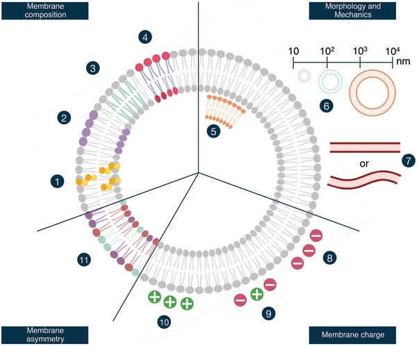
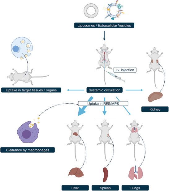
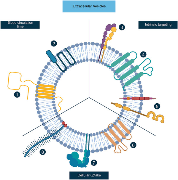
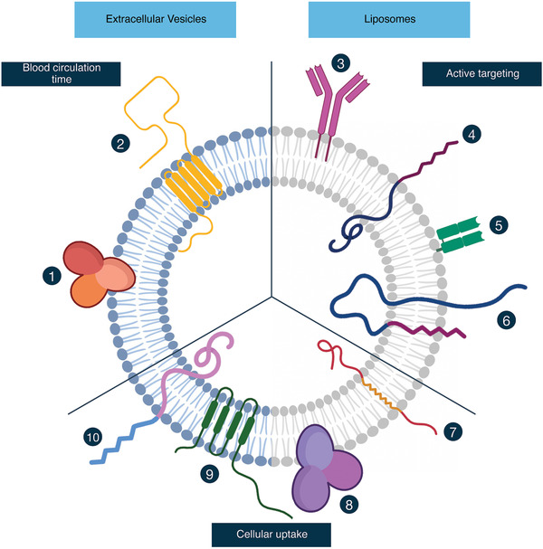
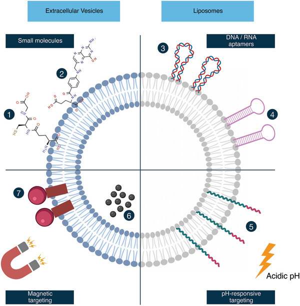
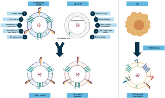
Over the past decades, lipid-based nanoparticle drug delivery systems (DDS) have caught the attention of researchers worldwide, encouraging the field to rapidly develop improved ways for effective drug delivery. One of the most prominent examples is liposomes, which are spherical shaped artificial vesicles composed of lipid bilayers and able to encapsulate both hydrophilic and hydrophobic materials. At the same time, biological nanoparticles naturally secreted by cells, called extracellular vesicles (EVs), have emerged as promising more complex biocompatible DDS. In this review paper, the differences and similarities in the composition of both vesicles are evaluated, and critical mediators that affect their pharmacokinetics are elucidate. Different strategies that have been assessed to tweak the pharmacokinetics of both liposomes and EVs are explored, detailing the effects on circulation time, targeting capacity, and cytoplasmic delivery of therapeutic cargo. Finally, whether a hybrid system, consisting of a combination of only the critical constituents of both vesicles, could offer the best of both worlds is discussed. Through these topics, novel leads for further research are provided and, more importantly, gain insight in what the liposome field and the EV field can learn from each other.
Keywords: biodistribution; cellular uptake; targeting moiety incorporation; therapeutic cargo delivery; vesicle functionalization.
© 2021 The Authors. Advanced Healthcare Materials published by Wiley-VCH GmbH.
Conflict of interest statement
The authors declare no conflict of interest.
Figures
Similar articles
-
A Comparison of Cellular Uptake Mechanisms, Delivery Efficacy, and Intracellular Fate between Liposomes and Extracellular Vesicles.Adv Healthc Mater. 2023 Oct;12(25):e2300319. doi: 10.1002/adhm.202300319. Epub 2023 Jul 9. Adv Healthc Mater. 2023. PMID: 37384827 Free PMC article. Review.
-
Modification of Extracellular Vesicles by Fusion with Liposomes for the Design of Personalized Biogenic Drug Delivery Systems.ACS Nano. 2018 Jul 24;12(7):6830-6842. doi: 10.1021/acsnano.8b02053. Epub 2018 Jul 10. ACS Nano. 2018. PMID: 29975503
-
Extracellular vesicles as drug delivery systems: lessons from the liposome field.J Control Release. 2014 Dec 10;195:72-85. doi: 10.1016/j.jconrel.2014.07.049. Epub 2014 Aug 2. J Control Release. 2014. PMID: 25094032 Review.
-
Systematic review of targeted extracellular vesicles for drug delivery - Considerations on methodological and biological heterogeneity.J Control Release. 2019 Jul 28;306:108-120. doi: 10.1016/j.jconrel.2019.06.006. Epub 2019 Jun 5. J Control Release. 2019. PMID: 31175896
-
Functional siRNA Delivery by Extracellular Vesicle-Liposome Hybrid Nanoparticles.Adv Healthc Mater. 2022 Mar;11(5):e2101202. doi: 10.1002/adhm.202101202. Epub 2021 Aug 11. Adv Healthc Mater. 2022. PMID: 34382360 Free PMC article.
Cited by
-
Therapeutics of the future: Navigating the pitfalls of extracellular vesicles research from an osteoarthritis perspective.J Extracell Vesicles. 2024 Jul;13(7):e12435. doi: 10.1002/jev2.12435. J Extracell Vesicles. 2024. PMID: 38943211 Free PMC article. Review.
-
Therapeutic potential and pharmacological significance of extracellular vesicles derived from traditional medicinal plants.Front Pharmacol. 2023 Dec 1;14:1272241. doi: 10.3389/fphar.2023.1272241. eCollection 2023. Front Pharmacol. 2023. PMID: 38108066 Free PMC article. Review.
-
Nanoscale Phytosomes as an Emerging Modality for Cancer Therapy.Cells. 2023 Aug 4;12(15):1999. doi: 10.3390/cells12151999. Cells. 2023. PMID: 37566078 Free PMC article. Review.
-
NIR-Mediated drug release and tumor theranostics using melanin-loaded liposomes.Biomater Res. 2022 Jun 3;26(1):22. doi: 10.1186/s40824-022-00270-w. Biomater Res. 2022. PMID: 35659113 Free PMC article.
-
Polymer Conjugate as the New Promising Drug Delivery System for Combination Therapy against Cancer.Curr Top Med Chem. 2024;24(13):1101-1119. doi: 10.2174/0115680266280603240321064308. Curr Top Med Chem. 2024. PMID: 39005059 Review.
References
-
- Doyle L. M., Wang M. Z., Cells 2019, 8, 727. - PubMed
-
- a) Thery C., Witwer K. W., Aikawa E., Alcaraz M. J., Anderson J. D., Andriantsitohaina R., Antoniou A., Arab T., Archer F., Atkin‐Smith G. K., Ayre D. C., Bach J. M., Bachurski D., Baharvand H., Balaj L., Baldacchino S., Bauer N. N., Baxter A. A., Bebawy M., Beckham C., Zavec A. B., Benmoussa A., Berardi A. C., Bergese P., Bielska E., Blenkiron C., Bobis‐Wozowicz S., Boilard E., Boireau W., Bongiovanni A., et al., J. Extracell. Vesicles 2018, 7, 1535750; - PMC - PubMed
- b) Witwer K. W., Thery C., J. Extracell. Vesicles 2019, 8, 1648167. - PMC - PubMed
-
- Elsharkasy O. M., Nordin J. Z., Hagey D. W., de Jong O. G., Schiffelers R. M., Andaloussi S. E., Vader P., Adv. Drug Delivery Rev. 2020, 159, 332. - PubMed
Publication types
MeSH terms
Substances
LinkOut - more resources
Full Text Sources
Miscellaneous
